Molecular Biology of the Cell - Volume 2 - Alberts B., Bray D., Lewis J., Raff M., Roberts K., Watson J. 1993
Intercellular Signaling
Mechanisms of signal transduction involving cell surface receptor proteins
All Water-soluble signaling molecules (including Neurotransmitters, Peptide Hormones, and growth factors), as well as some lipid-soluble ones, bind to specific protein receptors On the surface of target Cells. Surface receptors bind the signaling molecule (Ligand) with high affinity, and this extracellular event triggers an intracellular signal that alters Cell behavior.
Using ligands labeled with radioactive atoms, fluorescent Dyes, or electron-dense particles (such as colloidal gold), one can study the distribution of receptors on The Cell surface. It has been shown that the number of receptors for a specific ligand can range from 500 to over 100,000 per cell, and they are either randomly distributed across the membrane or concentrated in specific areas. Like other Membrane Proteins, cell-surface receptors are difficult to isolate in pure form and study, particularly because they account for less than 0.1% of the total Plasma Membrane protein mass. Techniques for cloning DNA sequences encoding surface receptors have helped overcome many of these challenges and fundamentally transformed our understanding of receptor Structure and function.
Unlike intracellular receptors for steroid and THYROID HORMONES, cell-surface receptors do not directly regulate Gene Expression. They merely transmit the signal across The Plasma Membrane, and their effect on processes in the Cytosol or nucleus is mediated by the generation of new intracellular signals. One might assume that cell-surface receptors simply transport external signaling molecules across the membrane into the cytosol, converting them into an intracellular signal, but this is not the case. Admittedly, many protein signaling molecules, such as Insulin, are indeed taken up by the cell via receptor-mediated endocytosis (Section 12.5.1), but they do not escape from endosomal or lysosomal vesicles into the cytosol. Apparently, The Role of the external ligand is simply to induce a conformational change in the receptor protein located on the cell surface. In fact, the action of a normal ligand can often be mimicked by receptor-binding Antibodies; this phenomenon underlies A number of pathological conditions. For example, a very common cause of hyperthyroidism—overactivity of The Thyroid Gland—in humans is the abnormal production of antibodies that bind to thyrotropin receptors; these receptors are thereby activated and cause Hyperfunction of the gland.
In this section, we will examine how a conformational change in a cell-surface protein receptor, induced by extracellular ligand binding, enables the receptor to directly or indirectly convert an external signal into an intracellular one.
12.3.1. At least three classes of cell-surface protein receptors are known: channel-linked, G-protein-linked, and catalytic [9]
Most cell-surface protein receptors can be assigned to one of three classes depending on the mechanism they use to transduce signals. Channel-linked receptors are neurotransmitter-Gated Ion Channels involved primarily in rapid synaptic signaling between electrically excitable cells. A small number of neurotransmitters are used to control such channels, briefly opening or closing the receptor-gated channel and thereby altering the ion permeability of the plasma membrane, and consequently the excitability of the postsynaptic cell. DNA sequence analysis of these receptors has shown that they belong to a single family of homologous, multipass transmembrane proteins. These receptors are discussed in Chapter 19 (Section 19.3) and will not be considered further here.
Catalytic receptors function as Enzymes upon ligand activation. Most known catalytic receptors are transmembrane proteins with a cytosolic domain possessing Tyrosine-specific protein kinase activity.
G-protein-linked receptors indirectly activate or inhibit specific enzymes or ion channels associated with the plasma membrane. The interaction between the receptor and the enzyme or ion channel is mediated by a third protein known as a GTP-binding regulatory protein (or G protein). Receptors linked to G proteins typically trigger a cascade of events that alter the concentration of one or more small Intracellular Signaling molecules, often called intracellular second messengers or mediators. These molecules, in turn, act by modifying The behavior of other target proteins within the cell. The two most important messengers are cyclic AMP (cAMP) and Ca2+. The signals they convey are generated via different pathways (both involving G proteins) and are utilized in all animal cells (see Fig. 12-13). We will examine these pathways before returning to catalytic receptors with tyrosine-specific protein kinase activity; we begin with the experiments that led to the discovery of cAMP and paved the way for our current understanding of how intracellular messengers are generated in response to an extracellular signal.
Class="center">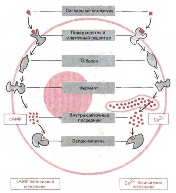
Fig. 12-13. Two main mechanisms by which G-protein-linked cell-surface receptors trigger The formation of intracellular messengers. In both cases, the binding of an extracellular ligand alters the conformation of the receptor's cytosolic domain, enabling it to bind to a G protein, which then activates (or inhibits) a specific plasma membrane enzyme. In some cases, the G protein interacts with an ion channel rather than an enzyme. In the cAMP pathway, the enzyme activated by the Gs protein synthesizes cyclic AMP. In the Ca2+ pathway, an enzyme generates a soluble messenger that releases Ca2+ ions from intracellular stores. Both cAMP and Ca2+ bind to other specific proteins within the cell, altering their activity.
12.3.2. Cyclic AMP is a ubiquitous messenger in animal cells [10]
In Muscle or Liver cells, exposure to adrenaline stimulates The breakdown of Glycogen stores. It was found that adrenaline activates the enzyme Glycogen phosphorylase, which catalyzes this breakdown. Further investigation revealed that treating isolated liver cell membranes with adrenaline (in the presence of ATP) causes The production of a low-molecular-weight, heat-stable factor capable of replacing the hormone and activating phosphorylase in a cell-free extract of the same cells. In 1959, this mediator was identified as cyclic AMP (cAMP, Fig. 12-14), which was later shown to regulate numerous intracellular reactions in all prokaryotic and animal cells studied to date.
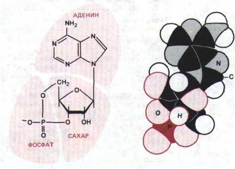
Fig. 12-14. Chemical structure and space-filling model of cyclic AMP (C, H, N, O, and P represent carbon, hydrogen, nitrogen, oxygen, and phosphorus atoms, respectively).
The identification of cyclic AMP led to The Study of the enzymes involved in its Synthesis and degradation. For cAMP to function as an intracellular messenger, its concentration within the cell (typically < 10-6 M) must undergo rapid fluctuations in response to specific extracellular signals (hormonal stimulation can cause a 5-fold increase within seconds). As we will see (Section 12.4.7), this requires rapid synthesis to be balanced by rapid degradation or removal. Cyclic AMP is synthesized from ATP by adenylyl cyclase, an enzyme bound to the cell's plasma membrane, but it is rapidly broken down by one or more enzymes—cAMP phosphodiesterases—which hydrolyze it to adenosine-5'-monophosphate (5'-AMP) (Fig. 12-15).
12-16
12.3.3. The receptor and adenylyl cyclase are separate proteins that functionally interact within the plasma membrane
Many hormones and local chemical mediators act by altering cAMP concentrations, and they do so by activating (or in some cases inhibiting) adenylyl cyclase rather than by affecting phosphodiesterase activity. Just as the same steroid hormone exerts distinct effects on various target cells, different cells respond very differently to changes in intracellular cAMP concentration (Table 12-2). Any ligand that activates adenylyl cyclase in a given cell typically elicits the same response. For example, at least four different hormones activate adenylyl cyclase in fat cells, and all of them induce the breakdown of triglycerides (fat reserves) into Fatty acids (Table 12-2). It appears that different receptors for these hormones activate a common pool of adenylyl cyclase molecules. That receptors and adenylyl cyclase are separate molecules was demonstrated in receptor "transplantation" experiments. For example, adrenaline receptors extracted from detergent-solubilized Plasma Membranes possess no adenylyl cyclase activity; however, when incorporated into the plasma membrane of other cells that lack their own adrenaline receptors, these "transplanted" receptors, upon activation by the hormone, are capable of functionally interacting with the recipient cell's adenylyl cyclase (Fig. 12-16).
Table 12-2. Some cellular responses to hormones mediated by cyclic AMP
|
Target tissue |
Hormone |
Major response |
|
Thyroid gland |
Thyroid-stimulating hormone (TSH) |
Synthesis and Secretion of thyroxine |
|
Adrenal cortex |
Adrenocorticotropic hormone (ACTH) |
Secretion of cortisol |
|
Luteinizing hormone |
Secretion of progesterone |
|
|
Muscle, liver |
Adrenaline |
Glycogen breakdown |
|
Parathyroid hormone |
Bone resorption |
|
|
Adrenaline |
Increase in heart rate and contraction force |
|
|
Vasopressin |
Water reabsorption |
|
|
Adipose tissue |
Adrenaline, ACTH, Glucagon, TSH |
Breakdown of triacylglycerols |

Fig. 12-15. Synthesis and degradation of cAMP. Pyrophosphatase drives the synthesis of cAMP to completion by hydrolyzing the released pyrophosphate.
12.3.4. Receptors activate adenylyl cyclase via a stimulatory G protein (Gs) [12]
In Bacteria, receptors and adenylyl cyclase molecules are directly coupled, but in animal cells, this coupling is mediated by an additional protein. The existence of such an indirect mechanism was first suggested by the finding that GTP is required for the hormonal activation of adenylyl cyclase in lysed cells. Subsequently, mutant cell lines were isolated in which adrenaline binding fails to activate adenylyl cyclase, despite the presence of normal amounts of both receptors and the cyclase itself. By mixing plasma membrane preparations from such "uncoupled" cells with detergent extracts of membrane from other cells, it was possible to reconstitute a hormone-sensitive adenylyl cyclase system that requires GTP for its operation. These extracts were found to contain a specialized membrane-bound GTP-binding protein (G protein) that is absent in the "uncoupled" mutant cells. Because this protein participates in enzyme activation, it was named the stimulatory G protein (Gs). Individuals with an inherited deficiency of Gs respond poorly to many hormones and therefore suffer from growth defects, delayed Puberty, mental retardation, and a variety of metabolic abnormalities. More recently, the adrenaline-activatable adenylyl cyclase system has been reconstituted from purified components (adrenaline receptor, Gs protein, and the catalytic component of adenylyl cyclase) by incorporating them into synthetic phospholipid Liposomes (Section 6.2.2). These experiments demonstrated that these three proteins are sufficient for the hormonal activation of adenylyl cyclase.
For the Gs protein to transmit a signal from the receptor to adenylyl cyclase, it must undergo a structural change upon receiving the signal. GTP is utilized for this purpose. When Gs is activated by the hormone-receptor complex, it simultaneously binds a GTP molecule, thereby acquiring The ability to activate an adenylyl cyclase molecule. Gs maintains adenylyl cyclase in an active state as long as the GTP molecule remains intact. Once Gs, which acts as a GTPase, hydrolyzes GTP to GDP, cyclase activation ceases.
12-17
12.3.5 Gs protein is a heterotrimer that dissociates into subunits upon activation [13]
The function of the Gs protein critically depends on its subunit structure. It is composed of three Polypeptides: the a-chain (Gsa), which binds and hydrolyzes GTP and activates adenylate cyclase, and a tight complex of the ß- and y-chains (Gβy), which anchors Gs to the inner leaflet of the cytoplasmic membrane. Fig. 12-17 illustrates the current model of receptor activation coupling to adenylate cyclase activation via Gs. In its inactive state, the Gs protein exists as a trimer with GDP bound to its a-subunit. Upon activation by the hormone-receptor complex, the guanine nucleotide binding site on Gs undergoes a conformational change, allowing GTP to bind in place of GDP. It is believed that GTP binding causes Gsa to dissociate from Gßy and tightly bind to an adenylate cyclase molecule, thereby activating it to synthesize cAMP. In less than a minute, Gsa hydrolyzes its bound GTP to GDP, which leads to the detachment of Gsa from adenylate cyclase (rendering the enzyme inactive) and the reassociation of Gsa with Gßy, regenerating the inactive Gs molecule.
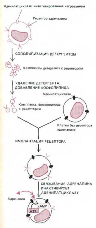
Fig. 12-16. Functionally active adrenergic receptors can be extracted from cells in which adenylate cyclase has been inactivated by heat, and incorporated into the plasma membrane of cells lacking such receptors. Upon activation by adrenaline, these "transplanted" receptors activate adenylate cyclase in the plasma membrane of recipient cells (the G-protein coupling the receptors to the cyclase is not shown in the diagram). There are at least Three types of adrenergic receptors (also called adrenoceptors)—a1, a2, and ß; only ß-receptors activate adenylate cyclase.

Fig. 12-17. Modern model illustrating how protein receptors can functionally couple to adenylate cyclase via the stimulatory G protein (Gs). As long as the signaling ligand remains bound, the receptor protein can continually activate new molecules of the Gs protein, thereby amplifying the response. An additional Amplification mechanism (which is more prominent in certain signaling systems) involves retaining bound GTP on the Gs protein α-subunit for many seconds, keeping adenylate cyclase activated throughout this period. According to an alternative model, adenylate cyclase remains associated with Gs in both its active and inactive states.
The bacterial adenylate cyclase system lacks the Gs intermediate. Why, then, have animal cells evolved such a complex, multi-step signal Transduction mechanism involving a G protein inserted between the receptor and the activated enzyme? One reason may be The Need for signal amplification (see Section 12.4.6), while another is the requirement for additional control checkpoints.
Gs allows for Two Types of amplification. In the simplest case, a single activated protein receptor can theoretically collide with and activate multiple Gs protein molecules, which in turn trigger the activation of numerous adenylate cyclase molecules. In some instances, however, an extracellular ligand may not remain bound to its receptor long enough for this amplification mechanism to operate; for example, certain ligands dissociate from their receptors in less than a second. Meanwhile, Gs itself is believed to remain active for over 10-15 seconds before the bound GTP is hydrolyzed. Thus, it keeps adenylate cyclase in an active state for a considerable time even after the extracellular ligand has dissociated. This effect can be dramatically demonstrated by adding a non-hydrolyzable GTP analogue to lysed cells: subsequent hormone Treatment leads to a prolonged synthesis of cAMP.
G proteins not only amplify the signal but also serve as a crucial regulatory hub where the entire activation process can be modulated. In principle, the efficiency of receptor-enzyme interaction can be altered by Covalent Modification of the G protein or by changes in its concentration within the plasma membrane. The most striking illustration of this is the action of the bacterial toxin responsible for cholera symptoms. Cholera toxin is an enzyme that catalyzes The transfer of ADP-ribose from intracellular NAD to the α-subunit of the Gs protein, causing the latter to lose its ability to hydrolyze bound GTP. Adenylate cyclase activated by such a modified Gs α-subunit remains active indefinitely. As a result, a sustained elevation of cAMP levels in intestinal epithelial cells triggers a massive efflux of water and sodium from these cells into the intestinal lumen, leading to severe diarrhea—the hallmark symptom of cholera.
Gs is just one member of a large family of G proteins that couple receptors to various enzymes and ion channels in Introduction/5.html">Eukaryotic Cell membranes. As we will see shortly, another member of this family does not activate, but rather inhibits, adenylate cyclase.
12.3.6 Receptors Inhibit Adenylate Cyclase Activity via an Inhibitory G Protein (Gi) [13]
The same signaling molecule can either increase or decrease the intracellular concentration of cAMP depending on the type of receptor it binds to. For example, there are several types of adrenergic receptors: ß-adrenergic receptors activate adenylate cyclase, whereas a2-adrenergic receptors inhibit it. The different final outcomes are determined by the G proteins that couple these receptors to adenylate cyclase: ß-receptors act via Gs, whereas a2-receptors act via an inhibitory G protein (Gi) that contains the same ßy-complex as Gs, but a different a-subunit (Gia). Once activated, the a2-adrenergic receptor interacts with Gßy, leading to the replacement of GDP with GTP at the guanine nucleotide-binding site on the a-subunit. In this process, Gia is believed to dissociate from Gβy, and both subunits participate in inhibiting adenylate cyclase: Gia directly suppresses adenylate cyclase activity, whereas the action of Gβy stems from binding the free Gsа-subunit and, consequently, abolishing its stimulatory effect on adenylate cyclase.
Just as cholera toxin increases cAMP levels by ADP-ribosylating the Gsa subunit and inactivating its GTPase activity, pertussis toxin—a product of the bacteria that cause whooping cough—produces the same effect by ADP-ribosylating Gia. In this case, however, the Modification of the G protein prevents it from interacting with receptors; therefore, upon receptor activation, adenylate cyclase is not inhibited.
Although G proteins were originally discovered through their effects on adenylate cyclase, they can also function through other pathways (see Table 12-3). Specifically, by activating phospholipase C (Section 12.3.9), certain G proteins can link receptor activation to changes in cytosolic Ca2+ concentration, and Ca2+ serves as an intracellular messenger even more widely than cAMP.
Table 12-3. Some GTP-binding regulatory proteins involved in cellular signal transduction
|
G protein type |
a-Subunit1 |
Function |
Modifying bacterial toxin |
|
Gs |
as |
Activation of adenylate cyclase |
Cholera |
|
Gi |
ai |
Inhibition of adenylate cyclase |
Pertussis |
|
Gp |
? |
Activation of phosphoinositide-specific phospholipase C |
Pertussis (in some cells only) |
|
GO |
a0 |
Major G protein of the Brain; may regulate ion channels |
Pertussis |
|
Ta |
Activation of cGMP phosphodiesterase in vertebrate retinal rods (see Section 12.3.12) |
Pertussis and cholera |
|
|
ras proteins |
2 |
Involved in The stimulation of Cell Division by growth factors (see Sections 12.3.11 and 13.4.5) |
1 Except for ras proteins (and Gp, whose structure is unclear), G proteins are heterotrimers in which the a-subunit is loosely bound to the ßy dimer. All known a-subunits (mol. wt. 40,000–50,000) are homologous, and most of them share identical (or very similar) ß-subunits (mol. wt. 35,000) and y-subunits (mol. wt. 8,000).
2 ras proteins are single polypeptides (mol. wt. 21,000) with minimal Homology to the subunits of other G proteins; it remains unknown whether they couple receptors to effector proteins in the same manner as other G proteins.
12.3.7 Ca2+ ions are stored in a specialized intracellular compartment [14]
The concentration of free Ca2+ ions in the cell cytosol is very low (on the order of 10-7 M), whereas their concentration in the extracellular fluid (exceeding 10-3 M) and in specialized intracellular Ca2+-storing compartments is quite high. Such a significant Ca2+ gradient tends to "drive" these ions into the cytosol across the plasma membrane and the membranes of intracellular storage Organelles. When a signal transiently opens Ca2+ channels in these membranes, Ca2+ ions rush into the cytosol, sharply increasing their local concentration and activating calcium-sensitive cellular mechanisms.

Fig. 12-18. The principal mechanisms enabling the cell to maintain a very low cytosolic concentration of free Ca2+ ions despite their high concentration in the extracellular fluid. Ca2+ is actively pumped out of the cytosol into the extracellular space (A) and into membrane-enclosed intracellular organelles that store Ca2+ (B). In addition, free Calcium Ions are tightly bound by various intracellular molecules. Mitochondria are also capable of pumping Ca2+ out of the cytosol, but they do so effectively only at very high Ca2+ concentrations, which typically occur during cell damage.
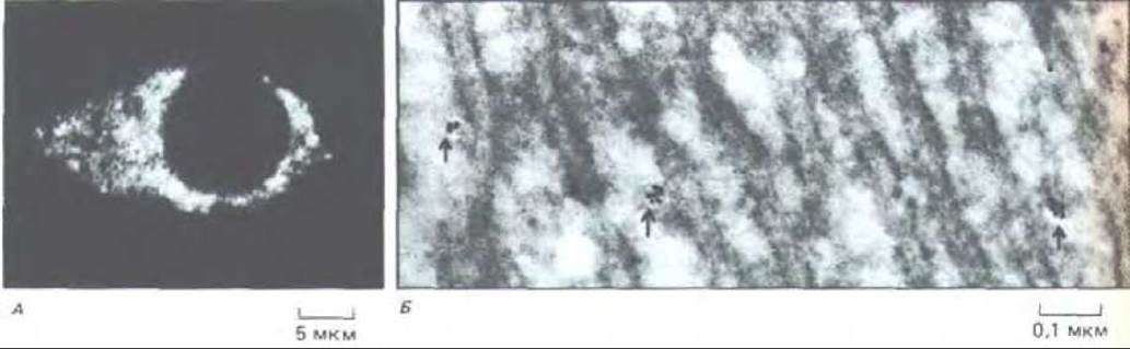
Fig. 12-19. Intracellular stores for Ca2+ ions, visualized using antibodies against the Ca2+-binding protein calsequestrin. A.
Immunofluorescence micrograph of a cultured rat nerve cell; calcium-binding sites are visible throughout the Cytoplasm. B. Electron micrograph of a frozen thin section of rat liver. Judging by the distribution of colloidal gold-labeled antibodies (indicated by the arrow), Ca2+ ions are not stored in the granular Endoplasmic reticulum, although they may be present in certain elements of the smooth reticulum. Quantitative analysis of such electron micrographs made it possible to estimate the volume of the storage organelles, which accounts for less than 1% of the cell volume. (A - P. Volpe et al., Proc. Natl. Acad. Sci. USA 85: 1091-1095, 1988; B — kindly provided by J. Meldolesi.)
For this signaling mechanism to function properly, a low cytosolic concentration of Ca2+ must be maintained, which is achieved through several pathways (Fig. 12-18). All Eukaryotic cells possess a Ca2+-dependent ATPase in their plasma membrane, which utilizes the energy of ATP Hydrolysis to pump Ca2+ ions out of the cytosol. In muscle and Nerve Cells, which rely heavily on Ca2+-mediated signaling, the plasma membranes contain an additional calcium pump that couples Ca2+ efflux to the influx of Na+ into the cell. This Na+-Ca2+ exchanger has a relatively low affinity for Ca2+ and therefore only becomes fully operational when the normal Ca2+ level is exceeded tenfold, a condition that occurs during repeated stimulation of a muscle or nerve cell.
The calcium pump in the membranes of specialized organelles also plays a crucial role in keeping cytosolic Ca2+ concentrations low: this Ca2+-ATPase is capable of pumping large quantities of calcium ions from the cytosol into organelles against a steep concentration gradient, even when cytosolic levels are low. Ca2+ ions are stored within the lumen of the storage organelle in loose association with calsequestrin, a Ca2+-binding protein that features a low affinity for Ca2+ (Kd = 103 L/mol) but a high capacity (~50 Ca2+ ions per molecule). When cells are stained with antibodies against calsequestrin and the Ca2+-ATPase, the same membrane-bounded organelles are revealed, which are smaller in size and distinct from the granular endoplasmic reticulum (Fig. 12-19). These organelles are homologous to the sarcoplasmic reticulum in muscle cells (Section 11.1.11) and, similarly, are specialized for the storage and release of Ca2+. Hereinafter, we will refer to them as Ca2+-storage organelles.
Typically, the concentration of free calcium ions in the cytosol ranges from approximately 10-7 M at rest to 5×10⁻⁵ M upon activation by an external signal. However, if a cell is damaged and fails to effectively pump Ca2+ ions out of the cytosol, their concentration can reach dangerously high levels (> 10-5 M). Under these circumstances, a high-capacity, low-affinity calcium pump located in The inner mitochondrial membrane comes into play, utilizing the energy of the electrochemical gradient generated across this membrane by electron transport during Oxidative Phosphorylation (Section 7.1.8).
12.3.8 The Ca2+ Ion Acts as a Ubiquitous Intracellular Mediator [15]
The first evidence regarding the role of the Ca2+ ion as an intracellular mediator was obtained in 1947, when it was discovered that injecting a small amount of Ca2+ into a Skeletal Muscle cell induces contraction. In recent years, it has become clear that Ca2+ serves as an intracellular mediator in a remarkably diverse array of cellular responses, including secretory processes and proliferation. Two types of signal transduction involving Ca2+ ions have been identified (Fig. 12-20): one operates predominantly in electrically active cells, while the other Functions in almost all eukaryotic cells. The first of these pathways has been thoroughly investigated in nerve cells, where plasma membrane depolarization triggers Ca2+ uptake by the nerve terminal, thereby initiating neurotransmitter secretion; Ca2+ enters through voltage-gated calcium channels that open upon depolarization of the nerve terminal plasma membrane during the arrival of an Action Potential (Section 19.3.2). In the second, universally distributed mechanism, the binding of a signaling molecule to a cell-surface receptor leads to the release of Ca2+ from intracellular stores; events at the cell surface are coupled to the opening of calcium channels in the intracellular membrane via another intracellular mediator, Inositol trisphosphate. The latter is generated in the plasma membrane through the rapid hydrolysis of minor Phospholipids (inositol phospholipids).
12-14
12.3.9 Inositol Trisphosphate (InsP3) Couples Receptor Activation to Ca2+ Release from Intracellular Stores [16]
The hypothesis regarding the role of inositol phospholipids (phosphoinositides) in signal transduction first emerged in 1953, when it was discovered that certain extracellular signals stimulate the incorporation of radioactive phosphate into phosphatidylinositol (PI), a minor phospholipid of The cell membrane. It was later demonstrated that this incorporation results from Cleavage and resynthesis reactions of inositol phospholipids triggered by a receptor that activates the enzyme phosphoinositide-specific phospholipase C. Among the inositol phospholipids, the two phosphorylated derivatives of PI—PI phosphate (PIP) and PI bisphosphate (PIP2)—play the most critical role in signal transduction and are believed to reside predominantly in the inner leaflet of the plasma membrane lipid bilayer (Fig. 12-21). Although PIP2 is present in animal cell membranes in much smaller quantities than PI, its hydrolysis is of paramount importance.
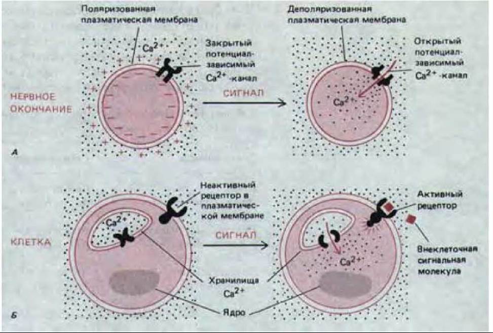
Fig. 12-20. Two primary pathways by which Ca2+ can enter the cytosol to function as an intracellular mediator for extracellular signals. In case A, calcium enters the nerve terminal from the extracellular fluid through voltage-gated calcium channels when the nerve terminal membrane is depolarized by an action potential. In case B, the binding of an extracellular signaling molecule to a surface receptor stimulates the release of Ca2+ from its intracellular stores.
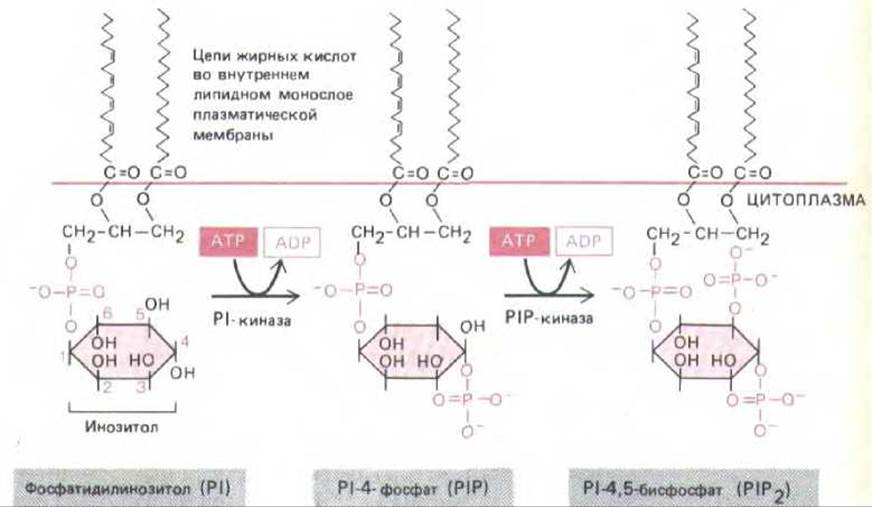
Fig. 12-21. Inositol phospholipids (phosphoinositides) in mammalian cells account for less than 10% of all cell membrane phospholipids. Polyphosphoinositides (PIP and PIP2) are phosphorylation products of phosphatidylinositol (PI). Although all three inositol phospholipids can be cleaved in response to an external signal, PIP2 hydrolysis plays The most significant role, despite accounting for less than 10% of total inositol phospholipids and less than 1% of all cellular phospholipids.
The chain of events linking an external signal to an internal response via PIP2 hydrolysis begins with the attachment of a signaling molecule to its receptor in the plasma membrane. It is now established that inositol phospholipid hydrolysis is involved in signal transduction from more than 25 different cell-surface receptors (Table 12-4). Although the details of this activation are less clear than in the case of adenylate cyclase, accumulating evidence indicates that a similar multi-step mechanism operates here in the plasma membrane as well. Presumably, the activated receptor stimulates a G protein (provisionally designated as Gp), which in turn activates phospholipase C. In less than a second, this enzyme converts PIP2 into two products: inositol trisphosphate and diacylglycerol (Fig. 12-22). At this point, the signal transduction pathway splits into two branches. Since both molecules play a critical role in the subsequent Development of the response, we will examine them separately.
Table 12-4. Selected cellular responses mediated via the phosphoinositide cascade.
|
Target tissue |
Signaling molecule |
Major response |
|
Liver |
Vasopressin |
Glycogen breakdown |
|
Acetylcholine |
Amylase secretion |
|
|
Smooth muscle |
Acetylcholine |
Contraction |
|
Pancreatic ß-cells |
Acetylcholine |
Insulin secretion |
|
Mast cells |
Antigen |
Histamine secretion |
|
Platelets |
Serotonin and PDGF secretion |

Fig. 12-22. The hydrolysis of PIP2 yields inositol trisphosphate (InsP3), which diffuses through the cytosol and releases Ca2+ from Ca2+-storage organelles. The other hydrolysis product, diacylglycerol, also plays an essential role in intracellular signal transduction by activating protein kinase C (see text below).
Inositol trisphosphate (InsP3) is a small, water-soluble molecule that releases Ca2+ ions from their intracellular stores (Fig.
12-22). When InsP3 is added to permeabilized cells (whose membranes have been rendered permeable via special treatment) or to isolated intracellular vesicles, it induces Ca2+ efflux into the medium. Apparently, the binding of InsP3 to a receptor on the cytoplasmic surface of the intracellular organelle opens calcium channels in its membrane. Two mechanisms ensure that the calcium release is transient: 1) Ca2+ ions entering the cytosol are rapidly pumped out, primarily to the extracellular space (later, once the stimulus is removed, the cell gradually replenishes its Ca2+ reserve by taking it up across the plasma membrane); 2) a fraction of InsP3 is rapidly dephosphorylated (and thereby inactivated) by a specific phosphatase. However, not all InsP3 is dephosphorylated; on the contrary, a portion of it is phosphorylated to inositol 1,3,4,5-tetrakisphosphate (InsP4), which may mediate slower and more prolonged cellular responses. In some cells, Ca2+ release occurs in a series of "pulses," each lasting 10 seconds or longer.
12.3.10 Diacylglycerol, Generated via PIP2 Hydrolysis, Activates Protein Kinase C [17]
While InsP3 produced by PIP2 hydrolysis elevates cytosolic Ca2+ concentrations, the other cleavage product of PIP2—diacylglycerol—produces entirely different effects. It serves two potential "signaling" roles: it can be further degraded to yield arachidonic acid, which is required for the synthesis of Prostaglandins and related lipid mediators (see Fig. 12-8), or, more importantly, it can activate a specific protein kinase that subsequently phosphorylates a range of proteins with diverse functions in the target cell.
The enzyme activated by diacylglycerol is called protein kinase C, or C-kinase, because its activity depends on Ca2+. Diacylglycerol—a product of receptor activation—together with phosphatidylserine, a phospholipid of the inner plasma membrane leaflet, binds to protein kinase C, greatly increasing its affinity for Ca2+ so that it becomes active even at low cytosolic Ca2+ concentrations. In many cells, however, C-kinase is normally activated, presumably through the combined action of diacylglycerol and Ca2+ ions, the concentration of which rises in the cytosol under METABOLISM/18.html">The Influence of InsP3. The activation of C-kinase is short-lived, as diacylglycerol is phosphorylated to phosphatidic acid or degraded to release arachidonic acid within a few seconds.
C-kinase, activated by diacylglycerol and Ca2+, transfers a terminal phosphate group from ATP to specific Serine or Threonine residues on target proteins, which vary depending on the cell type. For example, in many animal cells, C-kinase appears to phosphorylate and thereby activate the plasma membrane Na+/H+ exchanger, which regulates intracellular pH (Section 6.4.10); an elevation in intracellular pH can promote cell proliferation. C-kinase concentrations are highest in the brain, where, among other functions, it phosphorylates neuronal ion channels, thereby altering their properties and cellular excitability (Section 19.5). In some cells, C-kinase activation enhances the Transcription of specific genes. The promoters of at least some of these genes contain a common enhancer sequence recognized by a regulatory protein whose activity increases upon C-kinase activation (see Table 10-1). However, it remains unclear how C-kinase activates this protein—whether by directly phosphorylating (and thus activating) it, or indirectly through a protein kinase cascade.
Each of the two phosphoinositide signaling pathways can be mimicked by adding appropriate pharmacological agents to intact cells. The effects of InsP3 are mimicked by calcium ionophores, such as A23187 or ionomycin, which allow Ca2+ ions to enter the cytosol from the extracellular space (Section 6.4.19). The action of diacylglycerol can be mimicked by monoacyl derivatives of diacylglycerol or by phorbol esters—plant-derived substances that bind to C-kinase and directly activate it (Fig. 12-23). Using these Reagents, it has been demonstrated that achieving a full cellular response often requires both branches to act in concert. For instance, the proliferation of many cell types in culture can be stimulated by simultaneously applying a calcium ionophore and a C-kinase activator, whereas these reagents are ineffective individually.
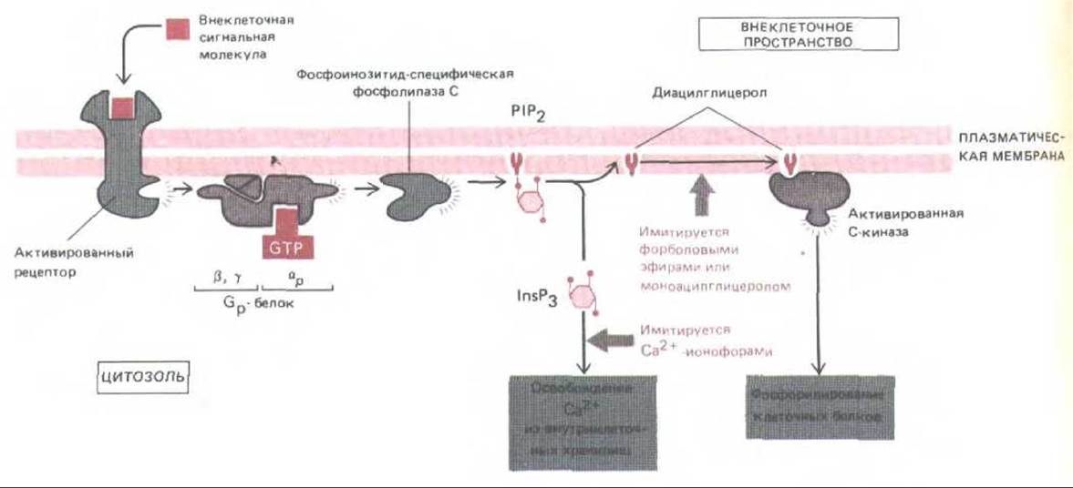
Fig. 12-23. The two Branches of the inositol phospholipid pathway. The activated receptor is thought to bind to a specific G protein (Gp), causing its a-subunit to dissociate and activate phospholipase C, which cleaves P1P2 to yield InsP3 and diacylglycerol. The true Protein Structure remains unclear. Diacylglycerol (with the participation of associated Ca2+ ions and phosphatidylserine, not shown in the diagram) activates protein kinase C. Both enzymes—phospholipase C and protein kinase C—are present in cells in both soluble and membrane-bound forms; upon activation, one or both of them translocate from the cytosol to the inner surface of the plasma membrane. The Effect of InsP3 can be mimicked experimentally in intact cells by treatment with a Ca2+ ionophore, whereas the effect of diacylglycerol is mimicked by treatment with monoacylglycerol derivatives or phorbol esters, which bind to and activate protein kinase C.
When cells begin to proliferate excessively for some reason, it naturally attracts attention. It was precisely for this reason that the effects of phorbol esters on cell proliferation were first discovered through their action as "tumor promoters" in animal organisms. Tumor promoters are substances that do not cause Cancer on their own, but can induce tumor growth in an animal after exposure to a small dose of a carcinogen that is insufficient to cause cancer by itself. When it was later discovered that phorbol esters directly activate protein kinase C, this helped to elucidate one of the intracellular mechanisms of cellular response to growth signals. In general, fundamental research into the causes of cancer, particularly animal Oncogenic Viruses, has provided a wealth of valuable insights into intracellular signal transduction pathways.
12.3.11. The so-called ras oncogenes encode a novel class of G proteins involved in the Regulation of Cell division
Apparently, several different signaling pathways are utilized to regulate Cell Growth and Division. Some genes normally encoding proteins that are components of these pathways were occasionally captured at random by Retroviruses and integrated into their genomes, often in a modified form (these retroviruses are also known as oncogenic RNA viruses). Such viral genes "stolen" from the cell in some cases impart to viruses the ability to cause uncontrolled proliferation of infected cells and thus trigger tumor development. These genes were named Viral Oncogenes. The study of tumors led to the discovery of tumor-causing viruses, then oncogenes, and this in turn led to the discovery of normal cellular Proto-oncogenes from which the viral oncogenes originated (Section 13.4.2).
A large class of oncogenes and proto-oncogenes discovered in this way was named the ras genes (since they were first found in viruses causing rat Sarcoma). These genes encode G proteins that reside on the inner surface of the plasma membrane, where they bind and hydrolyze GTP. These ras proteins, encoded by viral ras oncogenes, differ from normal ras proteins (encoded by proto-oncogenes) by an amino acid substitution at one of two positions. This is typically sufficient to disrupt the GTPase activity, and along with it, the intrinsic inactivation mechanism of the G protein (Section 12.3.4). If cultured cells are microinjected with antibodies against ras proto-oncogene products, these cells lose the ability to divide in response to growth factors. On this basis, it is believed that ras proteins are somehow involved in coupling growth factor receptors to intracellular effector proteins. The Nature of the effector proteins and the coupling mechanism remain elusive, although accumulating evidence suggests that effector proteins may regulate the phosphoinositide signaling pathway—at least as one of their functions. Because ras proteins are single polypeptides, the hypothesis that the Gp protein coupling receptors to phospholipase C is encoded by a ras proto-oncogene is highly unlikely: in that case, Gp would differ too much from all other G proteins—the homologous heterotrimers discussed in this chapter (see Table 12-3). It is far more plausible that the ras proto-oncogene family, comprising at least three members, encodes a distinct group of G proteins with novel functions. Despite several unresolved questions, the study of oncogenes has opened up an important new approach to elucidating normal G protein-mediated cell signaling mechanisms, as well as other cellular signaling pathways.
12.3.12. G protein activators form a large family of homologous Glycoproteins that span the membrane seven times [19]
Signaling systems utilizing G proteins are remarkably diverse. We have seen that some G proteins (Gs and Gi) couple receptors to adenylate cyclase, while others (Gp) couple them to phospholipase C. In the vertebrate eye, a G protein called transducin couples the absorption of a photon by a rhodopsin molecule to the activation of the enzyme phosphodiesterase, which hydrolyzes cGMP (Section 12.4.5); a drop in the concentration of this intracellular messenger leads to electrical Changes in the photoreceptor cell. All these G proteins can indirectly alter the state of ion channels in one way or another, but some G proteins can interact with them directly. For example, the binding of acetylcholine to receptors on heart muscle cells activates a Gi-like protein that directly activates a potassium channel in the plasma membrane. (These receptors, sensitive to the toadstool alkaloid muscarinic, are called muscarinic acetylcholine receptors, in contrast to others—nicotinic acetylcholine receptors—which are channel-forming receptors of skeletal muscle; see Section 6.4.17.)
All G proteins characterized so far, operating in these vastly different systems, are evolutionarily related. They share a similar subunit structure and Amino Acid Sequence: the a-subunits of transducin and Gi, for instance, are 65% identical in sequence. It is not surprising that many, if not all, receptors interacting with these G proteins are also homologous to one another, as became clear after the sequencing of their cDNAs. Generalization of the primary structures of this rapidly growing class of receptors revealed a common architecture consisting of a single polypeptide chain that spans The Lipid Bilayer seven times. This family of seven-transmembrane receptor proteins includes ß-adrenergic receptors (Fig. 12-24), muscarinic acetylcholine receptors, several neuropeptide receptors, and even rhodopsin. It appears that all these glycoproteins belong to a very large family of evolutionarily related receptors. This general structural "motif" likely arose early in evolution, as it is already characteristic of Bacteriorhodopsin (a light-driven proton pump that, however, does not operate via a G protein; see Section 6.27) and receptor proteins involved in The regulation of Yeast mating (Section 10.3.2). However, not all cell surface receptors span the membrane multiple times, and we now turn to another class of receptors—the family of tyrosine-specific protein Kinases.
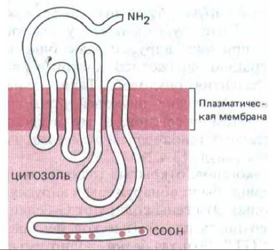
Fig. 12-24. Schematic diagram of the putative transmembrane orientation of the ß-adrenergic receptor in the plasma membrane. Regions of the cytoplasmic "tail" highlighted in red indicate the positions of serine residues that serve as potential targets for phosphorylation during receptor desensitization (Section 15.5.2). It has been established that in other G protein-coupled receptors, the polypeptide chain also spans the membrane seven times. (Based on R. A. Dixon et al., Nature 321: 75–79, 1986.)
12.3.13. Many catalytic receptors are single-pass transmembrane glycoproteins with tyrosine-specific protein kinase activity [20]
A vast group of surface receptors transduces extracellular signals into intracellular ones via G proteins, but There are also receptors that act through a more direct pathway. These are catalytic receptor proteins, the best-studied of which are single-pass transmembrane tyrosine-specific protein kinases, whose catalytic domain resides on the inner side of the plasma membrane. Upon ligand binding, they are activated and transfer a terminal phosphate group from ATP to the hydroxyl group of a tyrosine residue in specific target cell proteins. This protein kinase family includes the receptors for insulin, many growth factors including platelet-derived growth factor (PDGF; see Section 13.3.4), and epidermal growth factor (EGF), which stimulates the division of epidermal and many other cells (see Fig. 12-25). Most other protein kinases phosphorylate serine or, less commonly, threonine residues in proteins, meaning that phosphotyrosine accounts for less than 0.1% of all phosphorylated cellular proteins. In all cases studied, the receptor protein with tyrosine kinase activity also phosphorylates itself upon activation. In the case of the Insulin Receptor, this autophosphorylation enhances kinase activity via a positive feedback loop.
How does ligand binding to the extracellular domain of a receptor activate the catalytic domain on the opposite side of the plasma membrane? It is difficult to imagine how conformational changes could be transmitted across the lipid bilayer through a single transmembrane a-helix. In the case of the EGF receptor, ligand binding induces Conformational Changes in the extracellular domain, leading to receptor dimerization. It is possible that the interaction between two adjacent cytoplasmic domains confers catalytic activity upon the dimer.
There is substantial evidence that the kinase function of receptors plays a crucial role in the signaling process. For instance, cells harboring a mutant insulin receptor with a single amino acid substitution that selectively abolishes kinase activity fail to respond to insulin. However, identifying the key substrates phosphorylated alongside the receptor itself is extremely difficult, and consequently, the precise role of tyrosine phosphorylation in signal transduction remains elusive. In the case of the PDGF receptor, one of the substrates appears to be a kinase that phosphorylates phosphatidylinositol (PI kinase, see Fig. 12-21). This may explain the paradoxical fact that PDGF causes a slow activation of the phosphoinositide signaling pathway, even though its receptor is not coupled to Gp1.
Following ligand binding, many catalytic receptors are internalized via endocytosis in coated pits containing receptor-ligand complexes (Section 6.5.7). In some cases, such endocytosis appears to be driven by receptor autophosphorylation. This process may play an important role both in the degradation of signaling molecules and in the regulation of receptor density on the target cell surface (Section 12.5). Furthermore, the concomitant relocation of the tyrosine kinase domain to a new cellular locale may also be essential for signaling, although this remains to be proven.
1 Recently, evidence has emerged that the receptors for at least some growth factors can also phosphorylate phospholipase C upon ligand binding, thereby enhancing the hydrolysis of P1P2. — Transl.
12.3.14. Products of certain oncogenes are abnormal catalytic receptors with dysregulated kinase activity [21]
The first tyrosine protein kinase was discovered in 1979. It was not a cell surface receptor, but rather an intracellular viral oncogene product—a protein named pp60 v-src (Section 13.4.2). The first receptor found to possess tyrosine kinase activity (in 1982) was the EGF receptor. A few years later, it was revealed that the viral oncogene erbB encodes a truncated variant of the EGF receptor. This truncated protein has lost the extracellular EGF-binding domain but retained the intracellular tyrosine kinase domain, with the result that cells bearing such defective receptors behave as though they are continuously driven by a proliferation signal. Later, it was discovered that the neu oncogene, active in certain chemically induced rat Nervous system tumors, encodes an abnormal receptor that functions as a tyrosine kinase, although the identity of the ligand (presumably a growth factor) for the normal receptor has not been established. In this case, the abnormal and normal receptors differ by only a single amino acid residue in the protein's sole transmembrane segment. This modification alone is sufficient to render the tyrosine kinase constitutively active. These studies underscore the critical role of tyrosine kinases in the control of cell proliferation.
Numerous other links between oncogenes and normal proliferative signaling pathways have been uncovered. The sis oncogene, for instance, encodes a functionally active subunit of PDGF, whereas the erbA oncogene encodes an altered form of the thyroid hormone receptor. As we will discuss in more detail in Chapter 13, the investigation of oncogenes opens up a promising avenue for identifying and understanding the full spectrum of mechanisms by which proliferative signals achieve their targets.
The products of roughly half of all oncogenes discovered to date are protein kinases that phosphorylate target proteins on tyrosine, serine, or threonine residues. This is not surprising, since phosphorylation plays a vital role in signal transduction processes triggered by both catalytic and G protein-coupled receptors, and a remarkably large family of protein kinases exists to carry it out. Over 70 protein kinases are already known, and all of them presumably descend from a common precursor, as their catalytic domains are homologous (Fig. 12-25). In fact, it is now possible to predict whether a protein is a kinase and, if so, which residues—serine, threonine, or tyrosine—it will phosphorylate simply from its amino acid sequence. In the next section, we will see that the two major intracellular messengers—cAMP and Ca2+—mediate many of their effects by activating serine/threonine-specific protein kinases.
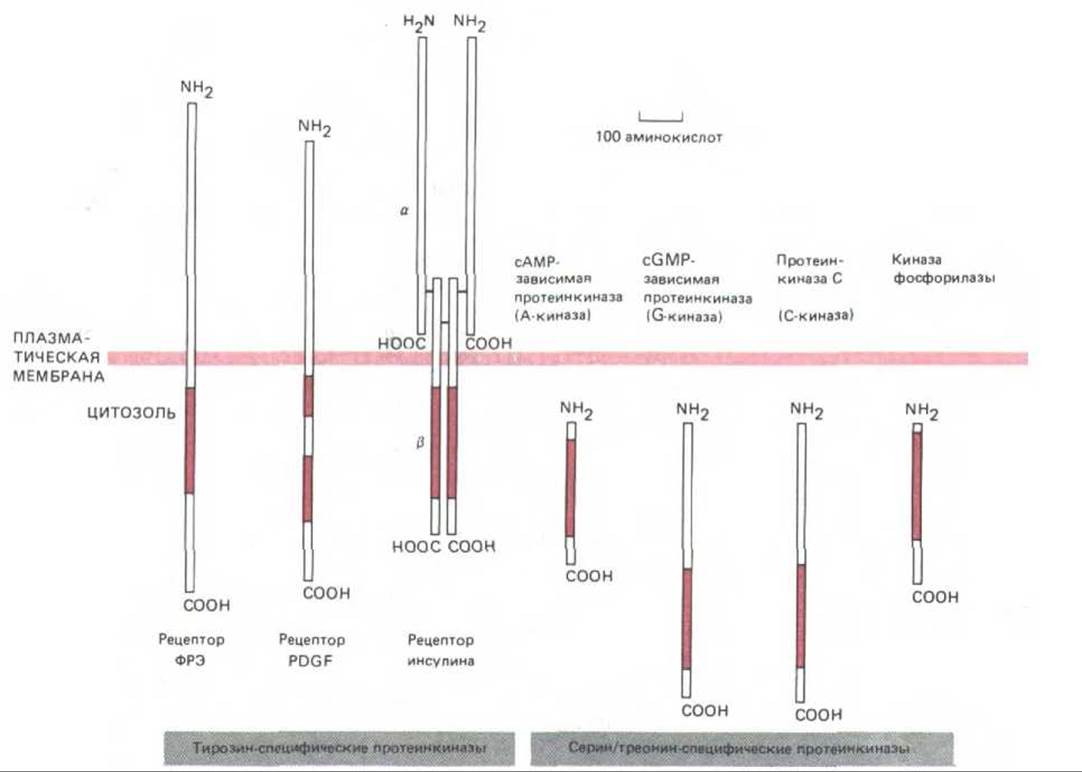
Fig. 12-25. Sizes and localization of the catalytic domains of several protein kinases discussed in this chapter. In all cases, the catalytic domain (highlighted in color) consists of approximately 250 amino acid residues and shares a conserved amino acid sequence, suggesting that they all evolved from a common precursor. The three tyrosine-specific kinases shown here are transmembrane receptor proteins that are activated upon binding a specific extracellular ligand and phosphorylate a set of intracellular proteins (including themselves) on tyrosine residues. Both chains of the insulin receptor are encoded by a single gene whose product—a precursor protein—is cleaved into two chains linked by Disulfide Bonds. The extracellular portion of the PDGF receptor is apparently folded into five immunoglobulin-like domains—suggesting that this protein belongs to the immunoglobulin superfamily (Section 18.6.20). The Regulatory Subunits of A-kinase (see Fig. 12-27) and phosphorylase kinase (see Fig. 12-31), which are normally associated with these kinases, are omitted from the diagram for simplicity.
Summary
Three main families of cell surface receptors are known to transmit extracellular signals via distinct mechanisms. Channel-linked receptors are neurotransmitter-gated ion channels that rapidly open or close in response to neurotransmitter binding, thereby altering the electrical excitability of the cell. Catalytic receptors are predominantly tyrosine-specific kinases that directly phosphorylate specific target cell proteins on tyrosine residues. G protein-coupled receptors indirectly activate or inhibit plasma membrane-bound enzymes or ion channels via regulatory GTP-binding proteins (G proteins). These G proteins turn themselves off by slowly hydrolyzing the bound GTP. Some of these receptors activate or inhibit adenylate cyclase, thereby altering the concentration of the intracellular messenger cAMP. Others activate phosphoinositide-specific phospholipase C, which hydrolyzes phosphatidylinositol bisphosphate (P1P2) to generate two intracellular messengers: (1) inositol trisphosphate (InsP3), which releases Ca2+ ions from intracellular stores and increases their concentration in the cytosol; and (2) diacylglycerol, which remains in the plasma membrane and activates protein kinase C, leading to the phosphorylation of various cellular proteins. The diverse responses elicited by the activation of G protein-coupled receptors are rapidly terminated once the extracellular signaling ligand is removed. Concurrently, the G proteins inactivate themselves, InsP3 is rapidly dephosphorylated by a phosphatase, diacylglycerol is converted to phosphatidic acid or broken down to yield arachidonic acid, cAMP is hydrolyzed by phosphodiesterase, and Ca2+ ions are swiftly pumped out of the cytosol.
Last update: 12/08/2026
Editorial and Educational Adaptation: This material has been compiled based on the primary/original source text. The project team performed an editorial review, corrected technical inaccuracies, structured sections, and adapted the content for an educational format.
What was processed:
- elimination of formatting defects (OCR errors, structural breaks, corrupted characters);
- editorial organization of content;
- standardization of terminology in accordance with academic sources;
- verification of factual statements against the original source text.
All mentions of the author, publication year, and origin of the primary text have been preserved in accordance with the source.