Molecular Biology of the Cell - Volume 2 - Alberts B., Bray D., Lewis J., Raff M., Roberts K., Watson J. 1993
Intercellular signaling
Signaling involving intracellular receptors: mechanisms of steroid hormone action
A multitude of ontogenetic and physiological processes across a wide range of organisms—from Fungi to humans—are regulated by a small number of Steroid Hormones synthesized from Cholesterol. Being relatively small, hydrophobic molecules (with a molecular mass of around 300), these hormones readily cross Cell/30.html">The Plasma Membrane via simple diffusion. Once inside the target cell, each type of steroid hormone binds tightly yet reversibly to its specific receptor protein. This hormone binding induces an allosteric conformational change in the receptor protein (a process known as receptor activation), which enhances the receptor's affinity for DNA. This enables the receptor to bind to specific genes within The Nucleus and regulate their METABOLISM/31.html">Transcription. THYROID HORMONES act in a very similar fashion, binding to their own receptors, which share a striking structural similarity with steroid receptors.
12-10
12.2.1. Steroid Hormone-Receptor Complexes Bind to Specific DNA Sequences and Regulate Gene Transcription
A typical target cell contains approximately 10,000 steroid Hormone Receptors. Each receptor can reversibly bind a single molecule of a specific hormone with high affinity (with an affinity constant Ka ranging from 108 to 1010 L/mol). Because receptors account for less than 0.01% of the total cellular protein mass, purifying and characterizing them has proven extremely difficult. Recently, however, the DNA sequences encoding several vertebrate steroid hormone receptors (their cDNAs) have been cloned and sequenced, revealing that these receptor Proteins share a highly conserved Structure. Their polypeptide chains, consisting of about 800 amino acid residues, fold into three distinct domains: a C-terminal hormone-binding domain, a central DNA-binding domain, and an N-terminal domain responsible for activating gene transcription (Fig. 12-9).
Class="center">
Fig. 12-9. Model of a steroid hormone receptor protein. In its inactive state, the receptor is believed to be associated with an inhibitory protein that blocks its DNA-binding domain. Hormone binding triggers the dissociation of this inhibitory protein, thereby activating the receptor. This model was originally derived from the cortisol (glucocorticoid) receptor, but receptors for estrogens, testosterone, progesterone, aldosterone, thyroid hormone, retinoic acid, and vitamin D share a remarkably similar structure (see Fig. 10-25); together, these proteins form the steroid hormone receptor superfamily. In the case of the cortisol and estrogen receptors, the inhibitory protein is the heat Shock protein hsp90, which has a molecular mass of approximately 90,000 (Section 8.2.7).
Certain types of steroid hormone receptors reside in the Cytosol in the absence of the hormone, whereas others are located in the nucleus. In both cases, hormone binding increases the receptor's affinity for DNA, allowing it to bind tightly to specific nucleotide sequences within the gene regulated by that hormone. The binding of the hormone-receptor complex to these specific gene regions activates (or, in some cases, represses) the Transcription of the gene.
Obtaining direct proof that activated steroid hormone receptors bind to specific genes was exceptionally challenging and was only achieved in 1983 with the advent of Recombinant DNA technology. This technology made it possible to clone steroid-regulated genes and isolate specific DNA sequences in large quantities. It also required the purification of the receptor proteins themselves—a notoriously laborious and time-consuming Procedure. Once purified receptors became available, their target DNA sequences were mapped in vitro using the DNase footprinting assay (Section 4.6.6); these experiments demonstrated that receptor binding protects a specific set of DNA nucleotide sequences from mild Cleavage by Nucleases or chemical Reagents. Deleting these short recognition sequences from the gene abolishes the steroid hormone's ability to activate its transcription. Furthermore, if a short DNA fragment containing this recognition sequence is fused to a reporter gene and introduced into a cell expressing the receptor protein, the corresponding steroid hormone will activate the transcription of the reporter gene. These experiments indicate that the DNA sequences recognized in vitro by activated steroid hormone receptors truly mediate the hormone's action within The Cell. Genes sensitive to steroid hormones typically contain multiple clusters of these recognition sequences, usually located upstream (and occasionally downstream) of the coding region or even within the gene itself (Fig. 12-10). Given the high degree of structural Homology among various steroid hormone receptors, the striking similarity in the sequences they recognize is hardly surprising.
Nevertheless, only a small fraction of genes in any given target cell fall under the direct influence of steroid hormones. For instance, 30 minutes after adding cortisol to a culture of rat Liver Cells, out of the thousands of proteins resolvable by two-dimensional gel Electrophoresis, only seven were affected: the levels of six increased, while one decreased. This effect was fully reversible: upon hormone withdrawal, the synthesis rates of these proteins returned to baseline. Because this method is estimated to detect roughly 10% of cellular proteins, the binding of cortisol to all 10,000 of its receptor molecules in a liver cell likely influences the transcription of only about fifty genes—far fewer than would be predicted based on the number of potential DNA binding sites. This implies that many activated receptors bind to DNA sites where their presence exerts no discernible effect.

Fig. 12-10. Localization of DNA sequences within the mouse mammary tumor virus gene that are recognized by activated cortisol receptors. Each colored bar represents a single receptor-binding site (8 NUCLEOTIDES in length). [After K. R. Yamamoto. In: Transfer and Expression of Eukaryotic Genes (H. Ginsberg, H. J. Vogel, eds.), pp. 79-92. New York: Academic Press, 1984.]
How does the binding of a hormone-receptor complex to a gene activate its transcription? Studies have shown that hormone-responsive DNA elements can stimulate transcription even when located thousands of Base Pairs away from the promoter where RNA Synthesis initiates. The MECHANISM OF ACTION of such DNA elements, known as transcriptional enhancers, is discussed in Chapter 10 (Section 10.2.11).
12-12
12.2.2. Steroid Hormones Often Trigger Both Primary and secondary Responses [7]
In many instances, the cellular response to a steroid hormone unfolds in two distinct stages. The direct transcriptional induction of a set of specific genes is termed the primary response. Subsequently, the protein products of these genes can, in turn, activate other genes, eliciting a delayed secondary response. Thus, a simple hormonal trigger is capable of orchestrating highly complex shifts in Gene Expression patterns.
A striking example of this phenomenon is found in the fruit fly Drosophila. Within just 5 to 10 minutes following an injection of the molting hormone ecdysone, six new sites of intense RNA synthesis appear as puffs on the giant polytene Chromosomes of the Salivary Glands (see Section 9.2.6). After a certain time interval, some of the proteins synthesized during the primary response induce RNA synthesis at approximately a hundred new sites, leading to The production of a large cohort of secondary-response proteins. One or more of these primary-response proteins ultimately regulate the entire sequence by exerting feedback control to shut down the transcription of the genes that participated in the primary response (Fig. 12-11). It is highly probable that similar mechanisms mediate the Amplification and regulation of many hormonal responses in mammalian cells as well.
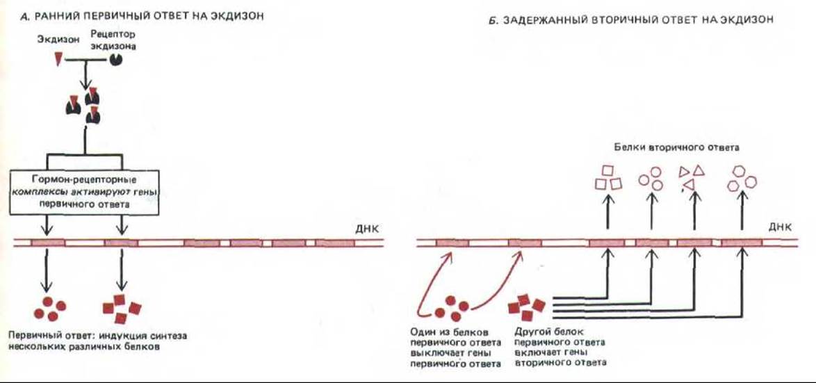
Fig. 12-11. Schematic diagram of the early primary (A) and delayed secondary (B) responses of Drosophila cells to ecdysone. Some of the proteins synthesized during the primary response turn on the genes responsible for the secondary response, whereas others turn off the genes involved in the primary response. In reality, both responses involve a greater number of genes than depicted in this simplified diagram.
12.2.3. Steroid Hormones Regulate Different Sets of Genes in Different Target Cells [8]
The cellular response to steroid hormones, much like hormones in general, is determined not only by The Nature of the hormone itself, but equally by the identity of the target cell (see Fig. 12-5). In principle, this can be explained in two ways: either different cell types possess distinct receptors for the same hormone, or the receptors are identical, but they activate different genes. Current evidence strongly Supports the latter explanation.
This evidence stems from Molecular Genetics experiments demonstrating that the receptor proteins for estradiol, cortisol, and progesterone are each encoded by a unique, single gene, as well as from studies of mammalian mutants with defective receptors for the male sex hormone testosterone. All mammals that fail to experience testosterone signaling during embryonic development naturally follow the female developmental pathway. Genetically male mutants possess normal Testes that actively secrete testosterone, yet their Tissues fail to respond to the hormone due to defective receptors. Consequently, these genetic males develop a full set of female secondary sexual characteristics, and their testes fail to descend into the Scrotum, remaining instead in the Abdominal cavity. This syndrome, known as testicular feminization, occurs in mice, rats, cattle, and humans alike. Although only the gene encoding the testosterone receptor is mutated, every diverse cell type that normally responds to this hormone is affected (Fig. 12-12).
Why does the same steroid hormone activate different sets of genes in different cell types? As discussed in Chapter 10, the Activation of a eukaryotic gene typically requires the Cooperative binding of multiple regulatory proteins (Section 10.1.5). Therefore, a steroid hormone receptor can activate a gene only in the presence of the appropriate combination of regulatory proteins, some of which are strictly tissue-specific.

Fig. 12-12. Different target cells respond differently to testosterone despite containing identical receptor proteins. In testicular feminization syndrome, a mutation in a single gene that alters the testosterone receptor causes all target cells to lose their ability to respond to the hormone.
Thus, each steroid hormone elicits a characteristic spectrum of responses because 1) receptors for that hormone are present only in specific cell types, and 2) the cells of each type contain distinct combinations of other tissue-specific regulatory proteins that act in concert with the activated steroid receptor to influence the transcription of specific sets of genes.
Steroid hormones are small, hydrophobic molecules derived from cholesterol. In the bloodstream, they circulate as Water-soluble complexes bound to specific carrier proteins. Upon dissociation from the carrier, they diffuse across the plasma membrane of target cells and reversibly bind to specific receptor proteins in the Cytoplasm or nucleus. Upon hormone binding, the receptor exhibits an increased affinity for specific DNA sequences that function as enhancers, thereby stimulating the transcription of several neighboring genes. The products of some of these genes may, in turn, activate other genes and elicit a delayed secondary response, thus amplifying the hormone's action. Each steroid hormone is recognized by its own unique receptor belonging to a family of homologous proteins. The same receptor regulates different sets of genes in different target cells, likely because the transcription of specific genes also requires other DNA-binding proteins that vary among cell types.
Last update: 12/08/2026
Editorial and Educational Adaptation: This material has been compiled based on the primary/original source text. The project team performed an editorial review, corrected technical inaccuracies, structured sections, and adapted the content for an educational format.
What was processed:
- elimination of formatting defects (OCR errors, structural breaks, corrupted characters);
- editorial organization of content;
- standardization of terminology in accordance with academic sources;
- verification of factual statements against the original source text.
All mentions of the author, publication year, and origin of the primary text have been preserved in accordance with the source.