Review of Medical Physiology - William F. Ganong 2002
Respiration
Pulmonary Functions
Gas Exchange in the Lungs
Sampling of Alveolar Air
Theoretically, all gas except for the initial 150 mL exhaled with each breath originates from the alveoli (alveolar air); however, some gas mixing occurs at the interface between the dead space and the alveolar air (see Fig. 34-17). Therefore, the final portion of the expired air is typically collected for analysis. Modern equipment featuring a custom automated valve allows for the collection of the last 10 mL of gas exhaled during resting ventilation. Figure 34-18 compares the composition of alveolar gas with that of inspired and expired air.
The alveolar gas equation can also be used to calculate PAO2:
Class="center">
where FiO2 is the fractional concentration of dry gas; PiO2 is the fractional concentration in inspired air; and R is the respiratory exchange ratio (see Chapter 17).
Composition of Alveolar Air
Oxygen continuously diffuses from the alveolar gas into the bloodstream, while CO2 diffuses from the Blood into the alveoli. At equilibrium, the inspired air mixes with the alveolar gas, replenishing the O2 taken up by the blood and diluting the CO2 added from the alveoli. A portion of this mixture is then exhaled. Consequently, the O2 content in alveolar gas decreases while the CO2 content increases until the next breath. Given that the alveolar gas volume is approximately 2 L at the end of expiration (functional residual capacity, see Fig. 34-7), each 350 mL increment of inspired or expired air has a relatively minor effect on PO2 and PCO2. Furthermore, the composition of alveolar gas remains remarkably stable even outside of resting conditions.
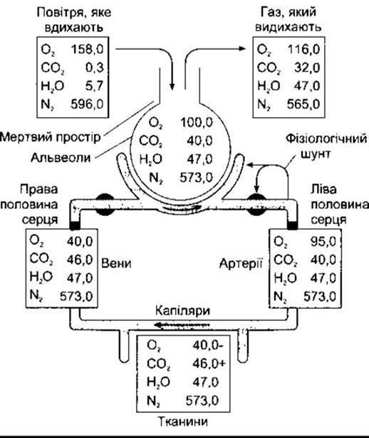
Fig. 34-18. Partial pressures of gases (mmHg) in various Regions of the Respiratory system and Circulation.
Diffusion Across the Alveolocapillary Membrane
Gases diffuse between the alveoli and the pulmonary capillary blood in both directions across the ultrathin alveolocapillary membrane, which consists of the pulmonary epithelium, capillary endothelium, and their fused basement membranes (see Fig. 34-3). Solutes pass from the alveoli into the capillary blood, reaching equilibrium within 0.75 s—the typical transit time of blood through pulmonary capillaries at rest—though this also depends on the chemical reaction rates of the solutes with blood components. For instance, nitrous oxide (used as an anesthetic gas) does not bind to blood constituents, and N2O reaches equilibrium within 0.1 s (Fig. 34-19). In this case, N2O uptake is not diffusion-limited, but rather perfusion-limited (dependent entirely on pulmonary capillary blood flow). Conversely, erythrocyte Hemoglobin takes up carbon monoxide so avidly that the partial pressure of CO in the capillaries remains extremely low, preventing equilibrium from being reached within the 0.75 s capillary transit time. Therefore, CO transfer is not perfusion-limited at rest, but is strictly diffusion-limited. Oxygen lies intermediate between N2O and CO; it is bound by hemoglobin but has a lower affinity than CO, reaching equilibrium with capillary blood in approximately 0.3 s. Consequently, its uptake is also perfusion-limited.

Fig. 34-19. Uptake of various gases during their transit through pulmonary capillaries (over 0.75 s); N2O does not bind in the blood, so its partial pressure rises rapidly to match alveolar levels. In contrast, CO is rapidly sequestered by erythrocytes, keeping its partial pressure well below that in the alveoli. Values for O2 fall intermediately.
The diffusing capacity of the Lungs for an inspired gas is directly proportional to the surface area of the alveolocapillary membrane and inversely proportional to its thickness. Diffusing capacity for CO (DLCO) is commonly measured as an index of overall pulmonary diffusing capacity because its uptake is diffusion-limited. The value of DLCO is proportional to The rate of CO uptake into the blood (VCO) divided by the partial pressure of CO in the blood entering the pulmonary capillaries. An exception applies to chronic smokers: their back-pressure of CO is nearly zero and thus negligible, simplifying the equation to:

The normal resting value for DLCO is 25 mL/min/mmHg. It increases approximately threefold during physical exertion due to capillary dilatation and the recruitment of additional perfused capillaries. In normal alveolar gas, PO2 is typically 100 mmHg (see Fig. 34-18), whereas the PO2 of mixed venous blood entering the capillaries is 40 mmHg. The diffusing capacity for O2 at rest, comparable to that of CO, is 25 mL/min/mmHg, raising capillary blood PO2 to 97 mmHg—slightly below the alveolar PO2. A further drop in aortic PO2 to 95 mmHg results from normal physiological shunting (see below). The value of DLO2 increases to 65 mL/min/mmHg or higher during exercise and declines in conditions such as sarcoidosis and berylliosis, which cause alveolar wall fibrosis. Another cause of pulmonary fibrosis is overexpression of PDGF (see Chapter 27) by alveolar macrophages, which ultimately stimulates neighboring mesenchymal Cells.
Mixed venous PCO2 is 46 mmHg, whereas alveolar PCO2 is 40 mmHg; thus, CO2 diffuses down its pressure gradient from the blood into the alveoli. The PCO2 of blood leaving the lungs drops to 40 mmHg. Because CO2 diffuses freely across Introduction/36.html">Biological Membranes, the diffusing capacity of the lungs for CO2 is significantly greater than that for O2. Consequently, CO2 retention is rarely a clinical issue in patients with alveolar fibrosis, whereas impaired O2 diffusing capacity presents a major physiological challenge.
Last update: 10/08/2026
Editorial and Educational Adaptation: This material has been compiled based on the primary/original source text. The project team performed an editorial review, corrected technical inaccuracies, structured sections, and adapted the content for an educational format.
What was processed:
- elimination of formatting defects (OCR errors, structural breaks, corrupted characters);
- editorial organization of content;
- standardization of terminology in accordance with academic sources;
- verification of factual statements against the original source text.
All mentions of the author, publication year, and origin of the primary text have been preserved in accordance with the source.