Antibiotics (Properties, Application, Interaction) - M.P. Cherenko 1999
Anesthesiology and Intensive Care
ANESTHESIOLOGY
Anesthesiology (from Greek an – negation, absence + aistesis – sensation + logos – word, science) is the science that studies Methods of eliminating sensations. It is the science of pain relief and Other Methods of protecting the body from excessive stimuli caused by surgical intervention. Protection of the body from surgical trauma must begin even before surgery, continue during its performance, and throughout the early postoperative period. Prevention of the undesirable effects of surgical aggression can be achieved through local or general anesthesia methods.
Local anesthesia (regional pain control) is the abolition of sensation in specific areas of the body while full consciousness is maintained.
General anesthesia, or narcosis, is an artificially induced, reversible impairment of Central Nervous system function accompanied by loss of consciousness, a reduction in various types of sensitivity, and suppression of reflex activity. Deep narcosis is typically also accompanied by Muscle relaxation, blockade of neurovegetative and neuroendocrine reactions, and marked depression of the body's vital Functions. Modern anesthesiological care makes it possible to purposefully influence specific links of The Nervous System without exerting a pronounced negative impact on the body. The objectives of anesthesiological management during surgical intervention include: elimination of negative emotions, pain sensations, muscle tension, pathological neuroendocrine and neurovegetative reactions, as well as The regulation of basic vital functions during surgery and in the early postoperative period.
The first objective of anesthesiological management—reducing psychological trauma and negative emotions—can be achieved using general anesthetic agents and tranquilizers. The abolition of pain sensitivity (analgesia) is achieved through local and general anesthetic agents, as well as narcotic and non-narcotic analgesics.
The blockade of undesirable neurovegetative and neuroendocrine reactions can be accomplished not only with general anesthetic agents, but also with pharmacological preparations of highly targeted action. For this purpose, anticholinergic (cholinolytic) and antiadrenergic (adrenolytic) agents are most frequently used. They help mitigate the NEGATIVE IMPACT OF not only surgical trauma, but also certain factors associated with anesthesia, preventing the excessive activation and subsequent exhaustion of neurovegetative and neuroendocrine mechanisms.
The suppression of protective motor reactions and the prevention of increased muscle tone in response to painful stimuli are achieved through The Use of anesthetic agents and muscle relaxants. The latter, by blocking the transmission of nerve impulses at the neuromuscular synapse, cause a reversible paralysis of Cytology/practical/58.html">Striated Skeletal Muscle tissue. This makes it possible to achieve muscle relaxation (myoplegia) at a light level of anesthesia, before its toxic effects on the body manifest. Since muscle relaxants paralyze striated Muscles, including Respiratory Muscles, their application requires the use of mechanical ventilation.
Artificial pulmonary ventilation (APV) has made it possible to eliminate one of the main drawbacks of general anesthetic agents—the depression of external respiration.
An important objective of anesthesiological management during surgery is also the maintenance of adequate Blood Circulation. An example of targeted regulation of vascular tone is induced hypotension.
Only adequate correction of Water-electrolyte, protein, and other METABOLISM/4.html">Types of Metabolism can ensure a favorable course in the postoperative period.
Thus, modern anesthesia possesses a vast arsenal of methods and agents for maintaining adequate pain relief and regulating the body's primary vital functions during surgery and in the first hours thereafter.
HISTORY OF The Development of ANESTHESIOLOGY
The earliest records of pain relief during surgery and painful medical Procedures (using wine, mandrake ROOT, opium, Indian hemp, and thornapple) are found in the Ebers Papyrus (3rd–2nd millennia BC). Later, these remedies came to be used in Egypt, China, Greece, and Rome. Hippocrates (459–377 BC) noted that "to relieve pain is a divine work."
In Assyria, during short-term surgeries, patients' neck vessels were compressed, thereby inducing fainting (an unconscious state). Many of these methods were also used in Kievan Rus.
Attempts to achieve pain relief in specific areas of the body were also made in ancient times. Initially, the vessels of a limb were compressed for this purpose, and later cold began to be used. These methods were mentioned by Avicenna (980–1037). The aforementioned methods of pain relief were largely ineffective or dangerous to the patient's life.
In the 19th century, the Discovery of the narcotic properties of A number of chemical compounds ushered in a new era in the development of pain relief. In 1200, R. Lullius discovered ether. It was first used clinically by C. W. Long. In January 1842, under ether anesthesia, he extracted a tooth from a patient, and in March he removed a tumor located in the occipital region. However, this publication did not appear in print until 1852. Consequently, W.T.G. Morton is considered the pioneer of ether anesthesia; in October 1846, he publicly administered ether anesthesia (using a specially constructed apparatus) during the removal of a submandibular tumor. News of the successful use of ether spread around the world. As early as February 1847 in Russia, it was used by F.I. Inozemtsev, N.I. Pirogov, H.A. Vantsetti, and V.A. Karavaev.
Almost simultaneously, the effects of nitrous oxide (laughing gas) were discovered. In December 1844, dentist H. Wells tested its effects on himself during the extraction of a diseased tooth.
Nitrous oxide anesthesia combined with oxygen remains one of the most common types of general anesthesia to this day.
In November 1847, gynecologist J. Simpson reported the successful use of chloroform for pain relief. Twenty days later, it was used by N.I. Pirogov at the 1st Military Land Hospital in St. Petersburg. However, the high incidence of complications and high mortality rate associated with chloroform subsequently forced its abandonment.
In 1911, Lehmann used trichloroethylene for experimental anesthesia. It was first used clinically for pain relief during surgery by E. Piper.
In 1934, Waters used cyclopropane for pain relief in clinical practice. Neither of these drugs found widespread application. In 1945 in England, Suckling synthesized halothane (fluothane).
Today, halothane is one of the most widely used inhalational anesthetic agents, employed both for mononarcosis and in combination with nitrous oxide and ether (azeotropic mixture).
Non-inhalational narcosis came into widespread clinical practice much later than inhalational anesthesia, although as early as 1847 N.I. Pirogov used ether experimentally for rectal and intravenous narcosis.
Russian pharmacologist N.P. Kravkov in 1902 proposed using hebaral/hedonal for intravenous narcosis, and in 1909 S.P. Fedorov used it clinically. However, the drug did not find wide application due to poor solubility.
In 1932, Weese used a barbituric acid derivative—evipal sodium (hexenal)—for intravenous narcosis, and starting in 1936, J. Lundy began using thiopental sodium. Hexenal and thiopental sodium are used for intravenous narcosis, and in children they are administered rectally.
In 1941, H. Selye discovered the ability of Steroid Hormones (progesterone, desoxycorticosterone) to suppress the central nervous system.
In 1955, a steroid preparation, hydroxydione, was synthesized, which exhibited pronounced narcotic properties while lacking hormonal activity. It was released under the trade name "Predione for Injection" (Viadril G). It is most commonly used in combination with other anesthetic agents.
In 1960, H. Nabori and co-workers synthesized a narcotic substance structurally close to natural metabolites—sodium oxybutyrate, which has been used in our practice since 1961 for intravenous anesthesia and intensive care.
In 1964, a new ultra-short-acting anesthetic agent, propanidid (Epontol, Sombrevin), was synthesized. Over time, it was established that mortality and complication rates following the use of propanidid were higher than those associated with barbiturate anesthesia.
Nowadays, ultra-short-acting intravenous anesthetics such as etomidate and propofol are used effectively.
Parallel to the Introduction of general anesthetics into clinical practice, methods for local anesthesia were being developed. The impetus for this development came from the work of V.K. Anrep (1880), who published the results of his study on the pharmacological effects of cocaine. These findings were utilized by Koller, N.A. Karatsupov, and Z.A. Zakharyevsky, who applied cocaine in ophthalmology. Halsted (1885) used it for nerve block anesthesia, a year later Oberst and A.I. Lukashevich used it for regional anesthesia, and in 1897,
A. Bier used it for spinal anesthesia. In 1901, Sicard and Cathelin (using cocaine) performed a variation of epidural anesthesia—sacral blockade. Since this drug had toxic systemic effects at high concentrations, Reclus (1889) and Schleich (1892) suggested infiltrating the site of the future incision with weak cocaine solutions. With this technique, the anesthetic acts directly on sensory nerve endings within the operative field. This marked the beginning of the development of local infiltration anesthesia. Local anesthesia became particularly widespread after Einhorn synthesized novocaine in 1905, and O.V. Vishnevsky developed and introduced infiltration anesthesia using novocaine via the "tight creeping infiltrate" method, "cross-extremity" anesthesia, and various blockade techniques. In recent years, one of the variants of regional anesthesia—epidural anesthesia, the theoretical prerequisites for which were developed back in 1885 by Corning—has gained widespread acceptance.
It should be noted that neither local nor general anesthesia using a single anesthetic agent (mononarcosis) could satisfy all the requirements demanded of pain relief. Combining several agents appeared more promising in this regard. The practice of combining anesthetics began long ago in an effort to reduce chloroform doses. For this purpose, multiple agents were administered simultaneously, most frequently chloroform and ether, or chloroform and scopolamine.
A significant drawback of this type of anesthesia is that, when using a combination of various agents—much like with mononarcosis—loss of consciousness, analgesia, and muscle relaxation are achieved only under conditions of deep anesthesia, which has a negative impact on the body.
A new era in anesthesiology began with the introduction of muscle-relaxing agents (muscle relaxants) that have minimal effects on other Organs and systems. The introduction of muscle relaxants into clinical anesthesiology is associated with the names of Canadian scientists Griffith and Johnson, who in 1942 used intocostrin, a purified preparation of curare. Since then, relaxants have become widely adopted. Myoplegia caused by the administration of muscle relaxants leads to the depression or complete cessation of breathing. This circumstance necessitated the development of artificial lung ventilation methods. To perform this, it was necessary to seal the "lung-machine" system, which was successfully achieved through tracheal intubation. This made the widespread use of endotracheal anesthesia possible.
The use of muscle relaxants and tracheal intubation contributed to a new stage in the development of anesthesiology. Endotracheal anesthesia combined with muscle relaxants and mechanical ventilation began to be used during the most complex surgical interventions in patients with severe impairments of vital functions.
The evolution of anesthesiology is also characterized by an intensive search for new methods of pain relief that eliminate The Need for general anesthetic agents. This gave rise to various types of neuroleptanalgesia, ataralgesia, ketamine anesthesia, and central analgesia using morphine, fentanyl, and other analgesic drugs.
THEORIES OF ANESTHESIA
Ever since the discovery of anesthesia, numerous attempts have been made to explain the pharmacological action of various anesthetic agents. The mechanism by which the anesthetic state develops is usually explained by physico-chemical or physiological phenomena. In chronological order, the main theories can be outlined as follows: 1) Claude Bernard's coagulation theory (1875); 2) Meyer-Overton lipid theory (1899–1901); 3) Verworn's nerve Cell depression theory (1912); 4) Traube's adsorption (surface tension) theory (1904–1913), supported by Warburg (1914–1918); 5) Pauling's hydrate microcrystal theory (1961).
In recent years, the membrane theory of the MECHANISM OF ACTION of general anesthetics at the subcellular and molecular levels has gained widespread recognition. It explains the onset of anesthesia through The Effect of these agents on the polarization and depolarization MECHANISMS OF CELL membranes.
By dissolving in The Cell membrane, anesthetic agents impair its permeability to sodium ions, disrupt the generation of excitation, and reduce the Action Potential.
In the early 1950s, The Role of the reticular formation in maintaining wakefulness was established. It was hypothesized that the loss of consciousness stems from a primary blocking action of the anesthetic agent on the reticular formation, followed by deafferentation of the Cerebral Cortex due to the elimination of ascending activating influences. However, recent studies have demonstrated that conduction blockade at the reticular level is not the triggering mechanism for all types of anesthesia. It is characteristic only of barbiturate and propanidid anesthesia. Ether and cyclopropane primarily affect the cerebral cortex while having virtually no effect on the reticular formation, whereas ketamine predominantly affects and excites the limbic systems of the Brain.
In light of the above, it can be concluded that a unified theory of anesthesia is unattainable not only at the systemic level, but even at the CELLULAR AND MOLECULAR levels. Today, it is difficult to isolate any universal neurophysiological mechanisms of anesthesia. It is likely more accurate to assume that each anesthetic agent possesses its own distinct Specificity and sequence of action.
INHALATION ANESTHESIA
Inhalation anesthesia is achieved by the inhalation of vapors from various volatile anesthetic agents or anesthetic gases. Depending on the route of delivery into the Lungs, anesthesia is termed mask anesthesia when administered via a facemask; nasopharyngeal anesthesia when delivered through a nasopharyngeal tube; intubation anesthesia when introduced directly into the lung via an endotracheal tube; endotracheal anesthesia when delivered directly into the Trachea; and endobronchial anesthesia when directed into the main Bronchi.
Inhalation anesthesia is the most common form of general anesthesia. Its primary advantage is excellent controllability.
The rate of induction, safety profile, and recovery time vary among different inhalation anesthetics. These parameters depend on the Specific characteristics of their tissue uptake and distribution throughout the body.
Currently, nitrous oxide, halothane, and ether are the most commonly used agents in anesthetic practice, while trichloroethylene and others are used less frequently.
Based on their potency, inhalation anesthetics can be ranked in the following descending order: trichloroethylene, chloroform, methoxyflurane, halothane, cyclopropane, ether, and nitrous oxide.
ETHER ANESTHESIA
Ether exerts a pronounced local and resorptive effect. The local action manifests as mucous membrane hyperemia, reflex laryngospasm, vomiting, and increased bronchial secretion, while the resorptive action leads to the development of characteristic stages of anesthesia.
First stage (analgesia). In this stage, pain sensitivity is blunted or abolished while consciousness is preserved to varying degrees.
These symptoms are caused by the body's reflex response to the irritation of the respiratory mucous membranes by the anesthetic agent. With a gradual increase in concentration, the irritating effect of ether may sometimes be unnoticeable, allowing anesthesia to transition smoothly into the Second Stage.
It should be remembered that in The first stage, consciousness is merely clouded, and the patient may catch the meaning of a conversation; tactile and Temperature sensitivity, as well as memory for past and present events, are preserved, and Hearing becomes acute. Any noise at this time seems like a roar.
Second stage (excitement). This varies among individuals: in debilitated, exhausted patients, or following the administration of sedatives, it tends to be brief and indistinct. In excitable, physically robust individuals who abuse alcohol, the excitation stage is particularly pronounced. Respiration at this time is irregular and loud, Teeth are clenched, lacrimation and salivation are increased, and all forms of reflex activity are heightened. For this reason, vomiting most commonly occurs during the second stage, and ventricular fibrillation may ensue. Since any stimulation intensifies excitation, one must not attempt to insert an airway or initiate surgical intervention during this stage.
Pronounced changes in cardiovascular function are observed In the second stage. These manifest as tachycardia, elevated blood pressure, and Cardiac Arrhythmias. As anesthesia deepens, the symptoms of excitation gradually subside, muscles begin to relax, respiration normalizes, and the Third Stage of anesthesia is reached.
The third (surgical) stage of anesthesia is divided into 3 planes. The first plane (III1) — light anesthesia — is characterized by the onset of calm Sleep accompanied by deep and rhythmic respiration, in which the muscles and Diaphragm participate evenly; inspiration and expiration are of equal duration. The pupils are constricted, palpebral and swallowing Reflexes disappear, and the corneal reflex is slightly diminished. Pharyngeal and laryngeal reflexes are preserved, and any attempt to insert an endotracheal tube may cause coughing, vomiting, and laryngospasm. Pulse rate and blood pressure approach baseline levels, while the tone of skeletal muscles, extremities, and the lower abdominal wall decreases. There is no response to mild stimuli, but intense pain elicits motor and autonomic reactions. Therefore, this plane of anesthesia is unsuitable for prolonged and traumatic operations. It is used for abscess incision, drain insertion, dressings, and outpatient surgeries accompanied by significant pain. Intra-abdominal surgery using mask anesthesia is impossible due to the lack of abdominal muscle relaxation.
The second plane of the surgical stage (III2) — moderate anesthesia — is characterized by a further weakening of reflex responses to painful stimuli and can be used for most surgical interventions on the extremities and the upper and lower abdomen under mask anesthesia.
At this plane of anesthesia, Eyeball movements and corneal reflexes are absent, pupils are narrow with sluggish light reaction, and the cornea is moist. Lacrimation is reduced, and pharyngeal, cough, laryngeal, and tendon reflexes are absent. Respiration is even and rhythmic (slightly slower than in the first plane of anesthesia), with some weakening of thoracic respiration and a predominance of diaphragmatic breathing. Blood pressure and pulse rate remain unchanged.
Muscle tone in the lower anterior abdominal wall is decreased, although complete relaxation is not achieved.
The third plane of the surgical stage (III3) — deep anesthesia — is characterized by progressive depression of reflex activity and major vital functions, which can become life-threatening with prolonged use, especially in debilitated patients. This represents the extreme limit of permissible narcotic depression of the body's vital functions. Consequently, maximum vigilance by the anesthesiologist is required to prevent further deepening of anesthesia and overdosage. A sign of the Toxic Effect of ether on vital organs is excessive respiratory depression (absence of thoracic breathing and a sharp decrease in diaphragmatic breathing), leading to reduced alveolar ventilation. All reflex responses to painful stimuli disappear, and only the response to rectal sphincter stretching remains. The pupils dilate and do not react to light, lacrimation ceases (dry cornea), eyelids remain open, and the eyeballs are fixed in a central position. Signs of cardiovascular depression appear, including marked tachycardia, arterial hypotension, increased central venous pressure, suppressed renal function (up to anuria), and the development of metabolic and respiratory acidosis.
The Fourth Stage of anesthesia is the agonal stage. It results from an excessive depth of anesthesia and can lead to irreversible changes in central nervous system Cells if its duration exceeds 3—4 minutes. Cardiac arrest occurs 2—3 minutes after the cessation of respiration. Prior to this, pulse and blood pressure become undetectable, cold clammy sweat appears, the Skin acquires a grayish hue, sphincters gape, resulting in involuntary urination and defecation. Only prompt cardiopulmonary resuscitation measures initiated no later than 3—5 minutes after circulatory arrest can save the patient. The administration of the anesthetic agent must be stopped immediately, Artificial ventilation and closed-chest massage initiated, replacement solutions infused, and defibrillation performed if fibrillation is present.
During emergence from anesthesia, a patient passes through the same stages as during induction, but in reverse order. However, the boundaries between individual planes of anesthesia and even the stages themselves are indistinct, and the excitement stage is less pronounced.
The technique of mask anesthesia has certain specific features due to the prominence of the excitation stage. As a rule, ether anesthesia is currently administered using anesthesia machines. Ether vapor is inhaled along with oxygen. Anesthesia is most commonly performed using a semi-closed circuit. Prior to anesthesia, the patient breathes pure oxygen through a mask for 2—3 min. Afterward, the vaporizer flowmeter is set to 1—2 vol. %, and the patient adapts to the smell of ether over 3—4 min. Its delivery is then gradually increased to 10—20 vol. %. Once excitation begins, the administration of the anesthetic agent must not be interrupted. The mask is removed only briefly in the event of vomiting. After clearing the vomitus, anesthesia is resumed. Until consciousness, coughing, and swallowing reflexes are fully restored, the patient must not be left unattended, as vomiting, aspiration into the respiratory tract, hemodynamic instability, and impaired external respiration or gas exchange may occur. Even after complete recovery of consciousness, intensive monitoring of the patient should be continued for several hours.
Advantages of ether anesthesia:
1) a wide therapeutic index combined with relatively low toxicity;
2) absence of depression on hemodynamics and parenchymal organ functions;
3) bronchodilatory effect and respiratory stimulation at concentrations providing adequate anesthesia for the majority of surgical procedures;
4) pronounced analgesic effect and The ability to achieve good muscle relaxation;
5) the possibility of using air instead of oxygen.
Disadvantages:
1) irritation of the respiratory mucosa;
2) unpleasant odor;
3) risk of explosion;
4) prolonged induction period with a pronounced stage of excitement;
5) nausea and vomiting during the induction of anesthesia and in the postoperative period;
6) prolonged recovery period.
Indications: ether is currently rarely used for monoanesthesia via the mask method. It is sometimes employed in combination with other anesthetic agents.
Contraindications: acute inflammatory processes in the respiratory tract and lungs, myasthenia gravis, and surgeries requiring the use of an electrosurgical knife for electrocoagulation.
HALOTHANE ANESTHESIA
Halothane is a potent narcotic agent (4–5 times stronger than ether and 50 times stronger than nitrous oxide), non-irritating to the respiratory tract; it suppresses salivation and the secretion of salivary and bronchial glands, dilates the bronchi, and is rapidly eliminated from the body. It depresses laryngeal and swallowing reflexes, allowing for tracheal intubation without the use of muscle relaxants. Following emergence, tremors and chills are frequently observed, and vomiting is less common (in 2–6% of cases).
Halothane depresses respiration (due to a direct effect on the respiratory center and relaxation of the respiratory muscles). It exerts a direct depressive effect on myocardial function.
The drug lowers blood pressure, stimulates the Vagus nerve center, and reduces heart rate.
The technique for mask-induced halothane anesthesia is identical to that of any other inhalation anesthetic. Given the narrow therapeutic index of the drug, it should be administered only with specialized vaporizers located outside the gas circulation system.
Initially, oxygen is delivered to the patient through a mask, after which the halothane vaporizer is opened, and its concentration is gradually increased to 2–3 vol. % over 2–3 min. Once the patient enters the anesthetic state, the concentration is reduced to 1.0–1.5 vol. %. Induction is rapid (5–7 min), without any sensation of suffocation or similar discomfort.
The surgical stage of halothane anesthesia is divided into 3 levels.
Emergence from this type of anesthesia occurs without excitation, vomiting is rare, and consciousness is restored within 5–10 min.
Advantages:
1) non-explosiveness;
2) the ability to use high oxygen concentrations;
3) rapid and pleasant induction for the patient, as well as swift recovery;
4) the ability to provide good muscle relaxation;
5) suppression of salivary and bronchial gland secretion;
6) bronchodilator effect;
7) chemical stability.
Disadvantages:
1) narrow therapeutic margin;
2) inhibition of the sympathoadrenal system function;
3) pronounced depressive effect of the drug on the myocardium and respiration;
4) increased cardiac sensitivity to catecholamines and the propensity to induce arrhythmias;
5) impaired hepatic blood flow and potential Liver damage, especially upon repeated administration;
6) occurrence of chills and metabolic acidosis in the postoperative period.
Indications: for anesthesia during short-term surgical procedures on the limbs and Abdominal cavity; in minor purulent surgery and traumatology. It is widely used to potentiate the effects of nitrous oxide and is indicated for patients with concomitant Bronchial Asthma, pulmonary surgeries, and pheochromocytoma.
Contraindications: liver and Kidney diseases, hypovolemia, severe heart failure, adrenocortical insufficiency, and the absence of a specialized vaporizer.
NITROUS OXIDE ANESTHESIA
Nitrous oxide is an inert gas that is eliminated unchanged from the body primarily through the lungs. At concentrations not exceeding 80%, it has no adverse effect on the function of vital organs. The agent does not suppress hemodynamics or gas exchange, does not affect liver or kidney function, and only slightly stimulates the sympathoadrenal system.
Indications. Due to its insufficient anesthetic potency and the occurrence of excitation, single-agent nitrous oxide anesthesia is used for analgesia only during painful procedures in obstetrics, dentistry, and outpatient practice. Nitrous oxide has found widespread application as a component of balanced anesthesia in combination with analgesics, muscle relaxants, neuroleptanalgesic agents, and other general anesthetics.
Nitrous oxide anesthesia can be administered using any anesthesia machine equipped with flowmeters for nitrous oxide and oxygen. As a rule, a semiclosed or semiopen circuit is used. Gas pressure reduction occurs in the pressure regulator. As nitrous oxide passes through it, the small amounts of water vapor it contains condense, and the water may freeze, leading to a gradual cessation of nitrous oxide flow. This is because the evaporation of nitrous oxide is accompanied by heat absorption, and its temperature can drop to the freezing point of water. This can be prevented by using a special pressure regulator or by heating its connection point to the cylinder. To prevent freezing during centralized gas supply, it is advisable to open several cylinders. A carbon dioxide absorber can be used during nitrous oxide anesthesia.
Considering the risk of Hypoxia due to inaccurate dosing, it is not recommended to use nitrous oxide at concentrations exceeding 75%, i.e., at an oxygen ratio higher than 3:1 (e.g., 6 L of nitrous oxide and 2 L of oxygen). The mask should be tightly applied to the patient's face to prevent the anesthetic gas mixture from mixing with atmospheric air. Anesthesia is terminated by gradually decreasing the anesthetic concentration. After stopping the nitrous oxide flow, pure oxygen should be inhaled for 4–5 minutes, because if the oxygen supply is discontinued immediately, nitrous oxide rapidly diffuses from the blood into the alveoli and, mixing with atmospheric air, reduces the alveolar oxygen concentration below normal values. This results in so-called diffusion hypoxia.
The stage of analgesia is achieved by inhaling 50–66% nitrous oxide, i.e., using it with oxygen in a 1:1 or 2:1 ratio. In debilitated patients with a prolonged pathological process and in intoxicated patients, even such a concentration of the drug may lead to loss of consciousness. Many patients exhibit motor and verbal agitation (physically robust individuals, patients with labile psyches, and those who abuse alcohol).
By increasing the nitrous oxide concentration to 75% in debilitated patients following enhanced premedication, the first level of the surgical stage can be achieved, allowing for minor surgical procedures as well as painful therapeutic and diagnostic manipulations that do not require muscle relaxation.
The positive properties of nitrous oxide include:
1) non-inflammability;
2) absence of irritating effects on the respiratory tract;
3) rapid and pleasant induction of and emergence from anesthesia for the patient;
4) pronounced analgesic effect;
5) absence of adverse effects on major vital functions and parenchymal organs;
7) relatively low cost.
Disadvantages:
1) its low anesthetic potency and frequent inability to achieve the surgical stage of anesthesia and muscle relaxation;
2) presence of agitation, especially in children with labile psyches and individuals who abuse alcohol.
NON-INHALATION ANESTHESIA
The primary type of non-inhalation anesthesia is intravenous. Rectal, intramuscular, intraosseous, subcutaneous, hypnotic, and electronarcosis are used much less frequently. Some of these methods (subcutaneous) are of historical interest only, as they are no longer used today. Other types of anesthesia (rectal, intramuscular, hypnotic anesthesia) are used to a limited extent for specific indications, while others (electronarcosis) are still in the stage of development and study.
Currently, the three short-acting drugs most widely used are hexenal (a barbituric acid derivative), thiopental sodium (a thiobarbituric acid derivative), and ketamine.
BARBITURATE ANESTHESIA
Thiopental sodium (pentothal sodium, thiopentone) is a yellowish or yellowish-green crystalline powder with a faint garlic odor. Its aqueous solution has a distinctly basic reaction and tends to precipitate. A 1— 2 % solution is prepared immediately prior to intravenous administration. The total dose of the drug should not exceed 1 g of dry substance, most commonly administered at 7—10 mg/kg, although its anesthetic potency is slightly higher than that of hexenal. Loss of consciousness occurs within 1.5—2 minutes, and the duration of anesthetic sleep is 20—25 minutes. In case of accidental subcutaneous injection, thiopentone sodium can cause necrosis of adipose tissue, whereas intra-arterial injection leads to severe arterial spasm, which may result in Gangrene of the extremity.
Upon entering the bloodstream, the majority of barbiturates (75 %) bind to Proteins, and only a fraction exerts an anesthetic effect. Therefore, in hypoproteinemia, small doses of anesthetics produce a pronounced narcotic effect. The degree of barbiturate-protein binding depends on blood pH: it decreases in acidosis, which increases the body's sensitivity to barbiturates, and increases in alkalosis. Barbiturates are absorbed by well-perfused organs and Tissues (heart, Kidneys, central nervous system, liver, etc.). The liver plays a decisive role in the degradation of barbiturates. The degree of their inactivation in the liver largely depends on the intensity of Carbohydrate Metabolism therein. A liver deficient in Glycogen breaks down barbiturates significantly more slowly.
The anesthetic effect of barbiturates is associated with their suppression of the reticular Formation of the Brainstem. Barbituric acid derivatives possess narcotic properties, yet they do not provide an analgesic effect.
Barbiturates depress respiration, potentially leading to complete apnea. However, even during deep barbiturate anesthesia, swallowing and laryngeal reflexes are preserved, and bronchial muscle tone increases. Therefore, irritation of the Larynx and throat can cause coughing, hiccups, and laryngospasm. Increased vagal tone induced by barbiturates (especially thiopental sodium) may lead to bronchiolospasm or cardiac arrest during intubation or bronchoscopy.
Barbiturates significantly affect cardiovascular function, leading to the development of hypotension.
Thiopental sodium and hexenal do not increase The Heart's sensitivity to catecholamines. At usual doses, barbiturates do not exert a negative effect on a healthy liver. However, in cases of impaired liver function, these disorders may be exacerbated by their influence. Barbiturates have no direct effect on the kidneys.
Indications: for induction anesthesia, anesthesia for short surgical procedures, and performance of therapeutic and diagnostic procedures accompanied by significant pain.
Contraindications: history of allergic reactions to the drug, absence of an anesthesiologist and a mechanical ventilation device. Relative contraindications include: latent bronchiolospasm or bronchial asthma, Shock, collapse, marked hypotension, parenchymal liver disease, Addison's disease, severe hypoproteinemia and metabolic acidosis, and porphyrinuria.
Thiopental sodium and hexenal are administered intravenously slowly: initially 2—3 ml of a 1 % solution to determine whether there is hypersensitivity to the anesthetic or if the solution has been injected subcutaneously, and after 20—30 seconds, the remaining dose is administered at a rate of 5—10 ml/min. Indications for discontinuing the anesthetic administration include: disappearance of the ciliary reflex, loss of consciousness, pupillary constriction, alignment of the eyeballs along the orbital axis, and relaxation of the lower jaw muscles. Typically, this depth of anesthesia is achieved with a drug dose of 5—10 mg per 1 kg of body weight.
Surgical intervention under mono-anesthesia with barbiturates can be performed only when the protective reaction to pain disappears, muscle tone decreases, pupils constrict, and their reaction to light slows down. Respiration becomes shallow and infrequent, and ARTERIAL BLOOD PRESSURE begins to drop.
The positive qualities of intravenous barbiturate anesthesia include:
1) ease of administration;
2) rapid induction (without excitation, vomiting, or unpleasant sensations);
3) absence of respiratory tract irritation.
Disadvantages include:
1) relatively narrow therapeutic index;
2) lack of analgesic effect;
3) difficulty in controlling the depth of anesthesia;
4) vagotonic action, which increases the risk of laryngospasm;
5) pronounced depression of respiration and cardiovascular activity;
6) prolonged post-anesthetic depression;
7) insufficient muscle relaxation;
8) preservation of swallowing and laryngeal reflexes;
9) irritant effect in case of subcutaneous and intra-arterial administration of thiopental sodium.
ANESTHESIA WITH SODIUM OXYBUTYRATE
Sodium oxybutyrate is a derivative of gamma-aminobutyric acid, a natural body metabolite found in significant amounts in the brain. This explains why, unlike other anesthetic agents, it does not suppress cellular metabolism, oxidation, and phosphorylation processes. Sodium oxybutyrate exhibits pronounced sedative, specific narcotic, and mild analgesic effects while demonstrating virtually no toxicity to the body.
Mononarcosis using sodium oxybutyrate is rarely employed due to its insufficient analgesic activity. In surgery, it is used for induction and combined anesthesia. Combined with inhalation anesthetic agents (nitrous oxide, ether, halothane) or neuroleptanalgesia drugs, it can be used for pain relief in most surgical interventions. It is most advisable to use it in debilitated patients, those with heart defects, or impaired parenchymal organ functions accompanied by marked intoxication.
The drug is administered intravenously and slowly to prevent motor excitation and seizures, which may occur if administered rapidly. A sleep resembling physiological sleep occurs 5–10 minutes after administering the drug at a dose of 75–150 mg/kg and lasts for 30–40 minutes. Pain sensitivity and all reflexes are preserved; occasionally, muscle tremors, twitching, and irregular breathing with normal or even increased tidal volume may occur. Hemodynamic changes are negligible.
KETAMINE ANESTHESIA
Ketamine (ketalar) is a phencyclidine derivative, an anesthetic agent intended for intravenous and intramuscular administration. It is available as an aqueous solution in vials containing up to 20 ml of a 1% solution. A distinct feature of this drug is its pronounced ability to block pain conduction and perception with minimal impact on the Reticular Formation and the limbic System of the brain.
Mononarcosis with ketamine can be recommended for pain management during surgeries and procedures that do not require muscle relaxation. Given that ketamine does not depress blood circulation, it is indicated for pain relief in patients with high operative risk as one of the components of anesthesia. It is also used in oncological operations and abdominal surgery.
Ketamine is not used in patients with Hypertension, cerebrovascular diseases, thoracic and abdominal aortic aneurysms, alcoholism, Epilepsy, or psychiatric disorders.
The technique for pain management is the same as for other types of intravenous anesthesia. Premedication must necessarily include atropine sulfate and diazepam. For short-term surgical interventions not requiring relaxation, the drug is administered intramuscularly at a dose of 5–6 mg/kg; after 20–30 minutes, an additional dose of 3 mg/kg is administered to maintain anesthesia. Before prolonged traumatic surgical interventions, it is administered intravenously at 2–4 mg/kg over 2 minutes. After loss of consciousness, muscle relaxants are administered, and patients undergo artificial lung ventilation, using nitrous oxide (60–75 vol. %) to maintain anesthesia. After 20–30 minutes, ketamine is re-administered in maintenance doses of 0.5 mg/kg. The surgical stage of anesthesia occurs 1–2 minutes after intravenous and 6–8 minutes after intramuscular administration, lasting 10–15 and 30–40 minutes, respectively.
Complications associated with ketamine anesthesia include nausea, vomiting, increased muscle tone, involuntary movements, and rash. In the postoperative period, delirium, drowsiness, delusions, and hallucinations frequently occur.
The advantages of ketamine are:
1) the possibility of intramuscular administration;
2) the absence of a hypotensive effect;
3) relatively low toxicity (the toxic dose is 15–20 times higher than the narcotic dose);
4) pronounced analgesia.
The disadvantages of the drug are:
1) muscle rigidity;
2) the onset of postoperative hallucinations and delusions (these phenomena are eliminated by administering diazepam);
3) increased pharyngeal and laryngeal reflexes, enhanced salivation;
4) the ability to increase intracranial pressure;
5) stimulation of sympathoadrenal and adrenoreactive systems.
EQUIPMENT AND INSTRUMENTATION FOR ANESTHESIA
An anesthesia machine is a device designed to deliver concentrated volatile or gaseous anesthetic agents along with oxygen or air to a patient. A modern machine allows for precise dosing of gas mixtures and creates optimal conditions for maintaining Gas Exchange in the lungs, i.e., the delivery of oxygen to the lungs and the elimination of exhaled carbon dioxide.
A modern anesthesia machine (Fig. 13) consists of 4 main units: a cylinder with a pressure regulator, a gas flowmeter, an anesthetic vaporizer, and a breathing system.
Gas reservoirs are cylinders (oxygen cylinders are painted blue, nitrous oxide cylinders are gray, and cyclopropane cylinders are orange). From the cylinders, oxygen and nitrous oxide flow into the pressure regulator, where the gas pressure is reduced to 304–405.4 kPa (3–4 atm) to ensure a steady supply. From the cylinders, the gases pass through the regulator and hoses to gas flowmeters, which regulate the delivery of gases into the breathing system. The machines are equipped with rotameter-type flowmeters (rotameters) that monitor the volume of gas delivered per minute.
Ether, halothane, trichloroethylene, and other liquid anesthetic agents enter the patient's lungs during inhalation anesthesia in vapor form, for which they are previously vaporized in a special device—a vaporizer. In this device, the vapor of liquid anesthetic agents achieves the required concentration through the partial or complete movement of carrier gas passing through bypass channels. Thus, vapor-gas mixtures of a specific concentration are formed at the vaporizer outlet.
From the vaporizer, the anesthetic mixture enters the Respiratory system of the anesthesia machine, which comes in two types: non-rebreathing and rebreathing. In a rebreathing system, the gas-anesthetic mixture exhaled by the patient is partially or fully returned to the system, whereas in a non-rebreathing system, the exhaled mixture is released into the atmosphere. The non-rebreathing circuit can operate via an open or semi-open system. If the air for the respiratory mixture enters the machine from the atmosphere and the exhaled gas is completely vented into the atmosphere, such a system is called open. If the gas forming the respiratory mixture comes from cylinders while the exhaled air is vented into the atmosphere, it is a semi-open system.
Class="center">
Fig. 13. "Polsharkon-2" universal anesthesia machine: 1 — pressure regulator, 2 — vaporizers, 3 — corrugated breathing hose, 4 — mask, 5 — flowmeter unit
Open-drop anesthesia can be administered using a simple face mask. Most commonly, Schimmelbusch or Waud-Schimmelbusch (or Schimmelbusch/Vancouver) masks are used for this purpose. They consist of a metal frame covered with several layers of gauze, onto which liquid anesthetic (such as ether, halothane, or chloroform) is dropped. The patient inhales the anesthetic vapor along with atmospheric air and exhales directly into the atmosphere.
The advantages of this method include simplicity, minimal dead space, and low resistance to breathing. However, this method does not allow for precise dosing of anesthetic vapor concentration, and it results in excessive pollution of the operating room air as well as significant loss of moisture and heat. Furthermore, this type of anesthesia does not permit artificial or assisted pulmonary ventilation. It is also difficult to use during surgeries on the HEAD, neck, or when the patient is in the prone position. All of these factors limit the clinical utility of this anesthesia technique.
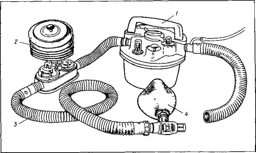
Fig. 14. Apparatus for administering open-drop anesthesia:
1 — vaporizer with thermocompressor, 2 — breathing attachment, 3 — corrugated hose, 4 — mask
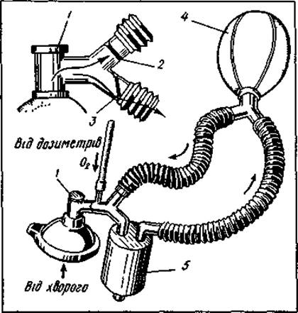
Fig. 15. Rebreathing circuit: 1 — adapter, 2,3 — Valves, 4 — breathing bag, 5 — CO2 absorber
Open-drop anesthesia using a simple anesthesia machine is somewhat safer (Fig. 14). Its main components are a vaporizer equipped with a thermal compensator and a breathing attachment with a bag, which can be used to perform artificial pulmonary ventilation. After inhalation, the air passes through the vaporizer, becomes saturated with anesthetic vapor, and enters the patient's lungs via a mask (or endotracheal tube). Exhalation occurs into the atmosphere through the expiratory valve. Open-drop anesthesia is particularly convenient for use in field or military-medical conditions.
Semi-open anesthesia has certain advantages over the open method because it allows the patient to be supplied with a high oxygen concentration mixture and makes it possible to use gaseous anesthetics. To perform anesthesia via a semi-open circuit, most modern anesthesia machines are equipped with special non-rebreathing valves that regulate the gas mixture flow. The gas mixture flows from the anesthesia machine into the patient's respiratory tract, while exhaled air is released into the atmosphere.
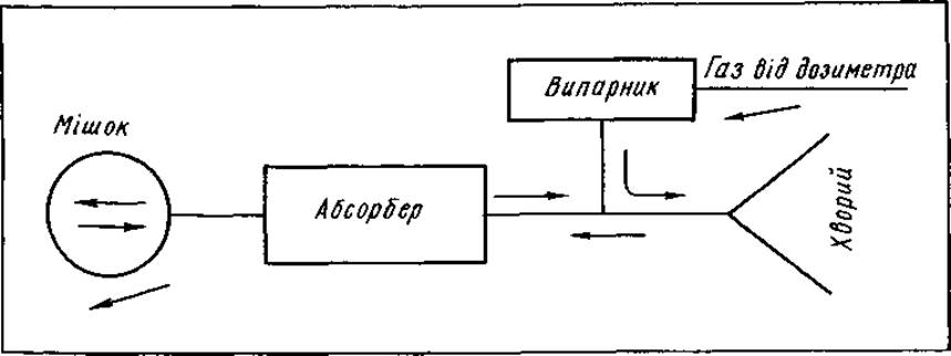
Fig. 16. Pendular gas rebreathing system
Gas rebreathing systems (rebreathing circuits) are divided into circular and pendular systems. In a circular system, the gas-anesthetic mixture circulates in a closed loop via valves: after inhalation, it enters the lungs; after exhalation, it enters the breathing bag (Fig. 15). To prevent hypercapnia, the gas-anesthetic mixture exhaled by the patient is passed through an absorber containing a chemical CO2 absorbent (HP-1 - GOST 6755-53).
Before each use of the system, the absorber must be refilled with fresh absorbent material stored in a hermetically sealed container.
The rebreathing system can be operated using either a closed or semi-closed circuit. In the former case, all of the exhaled gas mixture is directed back into the anesthesia machine and re-introduced into the patient's lungs. The gas flow in this setup must not exceed The amount of oxygen and anesthetics consumed by the lungs. In the second option, a larger volume of gas mixture is delivered to the lungs than is actually consumed, with the excess being vented out through the relief valve. A strictly closed circuit is practically difficult to maintain, so clinicians typically use a semi-closed circuit with varying degrees of gas rebreathing (Fig. 16). If a high gas flow (over 6 L/min) is used in a semi-closed circuit, the system begins to function as a non-rebreathing system, and the concentration of the inspired mixture is nearly identical to its concentration at the vaporizer outlet.
Special instruments and devices are used to perform mask and endotracheal anesthesia and to manage patients in the early postoperative period: masks, breathing bags, airways, dental props, Mouth gags, endotracheal tubes, stylets, connectors, anesthetic forceps, nebulizers, hose receivers, flexible rubber connecting tubes, flexible rubber breathing hoses, and others.
Anesthesia masks are used for Oxygen therapy, inhalation anesthesia, and assisted artificial pulmonary ventilation. Most commonly, they are placed over the MOUTH AND Nose; nasal masks are used less frequently. Anesthesia masks come in several sizes. The rim of the mask features an inflatable cuff that ensures a hermetic seal for the machine-patient system. A retaining strap is used to secure the mask to the face.
During deep anesthesia, muscle relaxation causes the Tongue to fall back, which obstructs the airway. To prevent this complication, airways are used (Fig. 17). They are manufactured from rigid rubber, plastics, metal, or combinations of these Materials.
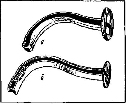
Fig. 17. Airways: a — rubber; b — metal
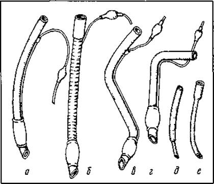
Fig. 18. Endotracheal tubes: a — Magill tube with cuff; b — reinforced tube; c — Kühn tube with cuff; d — angled tube with cuff; e, f — Cole tubes
Before lengthy and traumatic surgeries requiring muscle relaxation or the cessation of spontaneous breathing, tracheal intubation is performed; in cases of pulmonary pathology, main bronchial intubation may also be carried out. Endotracheal tubes are used for this purpose (Fig. 18). They are made of rigid rubber or plastic. Plastic tubes possess thermoplastic properties, which significantly reduces their adverse impact on the respiratory tract.
There are many types of endotracheal tubes. Some feature an inflatable cuff that, when inflated, isolates the trachea from the Oral Cavity and Esophagus. For nasal intubation, uncuffed tubes are more commonly used.
Fig. 19. Laryngoscopes (a, b)
As a rule, tracheal intubation is performed using a laryngoscope. It consists of two parts: a handle containing electrical batteries, and a blade with a light bulb. Blades come in three sizes and various shapes (Fig. 19).
Most commonly, tracheal intubation is performed under general anesthesia with or without muscle relaxants, and less frequently under local anesthesia. In the latter case, dicaine or lidocaine is used as a local anesthetic. In specific cases of nasotracheal intubation, anesthetic forceps and a stylet are used to help guide the endotracheal tube into the glottis.
MAIN STAGES OF ANESTHESIOLOGICAL MANAGEMENT IN SURGICAL INTERVENTIONS
The efficacy of Surgical Treatment is largely determined by the adequacy of its Anesthetic Management. It comprises the following stages: preoperative examination and Assessment of the patient's condition, preparation for surgery and anesthesia, administration of general anesthesia, and management of the early postoperative period.
During the initial interaction with the patient, attention should be paid to their mental state, and in cases of heightened excitability, the preoperative dose of sedatives should be increased. It is important to determine whether the patient has harmful habits (smoking, alcoholism, substance abuse), as these are frequently associated with resistance to general anesthetics.
Premedication Schemes
|
Time and route of administration |
Scheme I |
Scheme II |
|
Night before surgery, at bedtime — oral |
Phenobarbital or sodium etaminal 0.1 g, noxyron 0.2 g; chlordiazepoxide 10–15 mg, or trioxazine 0.3–0.5 g; suprastin 20–25 mg |
Phenobarbital or sodium etaminal 0.2 g; diprasin 25–30 mg, or diphenhydramine 0.1 g, diazepam 5–10 mg |
|
Morning, 2 hours before surgery — oral; 40 minutes before surgery — intramuscularly, or 5–10 minutes before — intravenously |
Atropine sulfate 0.3–0.7 mg, or metacin 0.5–0.7 mg, or scopolamine hydrobromide 0.25–0.5 mg, promedol 20–40 mg, or omnopon 20 mg, or morphine hydrochloride 10 mg |
Atropine sulfate 0.3–0.7 mg, or metacin 0.5–0.7 mg, or scopolamine hydrobromide 0.25–0.5 mg, promedol 20–40 mg, or omnopon 20 mg, or morphine hydrochloride 10 mg, or droperidol 2.5 mg, fentanyl 0.05 mg |
Any elective surgical intervention performed under general anesthesia requires a specific level of laboratory and clinical evaluation. If there is evidence of other comorbid conditions, a consultation with relevant specialists is required.
The results of the preoperative patient assessment determine the anesthesiologist's strategy before, during, and after surgery. Adequate anesthetic management of a surgical Procedure requires an objective understanding of the degree of operative risk. The risk level is assessed in points. The scope and nature of preoperative preparation are largely determined by the severity of the primary and concomitant diseases.
Before elective surgical interventions, the following measures are carried out: a Structure/133.html">Discussion between the anesthesiologist and the patient regarding anesthesia and surgery; restriction of food intake prior to surgery (at least 5–6 hours); a cleansing enema the evening before surgery and in the morning; emptying of the Urinary Bladder; removal of removable dentures; and medical preparation (premedication). Prior to emergency surgical interventions, gastric lavage is performed via a gastric tube.
Premedication is administered to calm the patient, ensure rest prior to surgery, relieve mental tension, and normalize metabolism. Premedication reduces the consumption of general anesthetics, prevents undesirable neurovegetative reactions and side effects of narcotic agents, and inhibits salivation, bronchial secretion, and perspiration. Agents with energy-potentiating properties are utilized. To improve sleep, hypnotics are prescribed, which also possess a certain sedative effect that is enhanced by the administration of tranquilizers. Narcotic analgesics act similarly; they significantly raise the pain threshold and reduce the required concentration of anesthetics by potentiating their effect. To a large extent, the sedative effect is supplemented by certain antihistamines with mild neuroleptic properties. All these drugs inhibit undesirable neuroreflex reactions, though they do not completely replace anticholinergic agents.
As a rule, 2–3 premedication regimens are used in clinical practice. The proposed scheme provides average doses of premedication agents most frequently used for patients with a body weight of 50–70 kg. For debilitated, elderly, and senile patients, the drug dosage is reduced accordingly. Premedication according to Scheme I is most frequently used. It is designed for calm, balanced patients undergoing relatively non-traumatic and short-term surgery. Premedication according to Scheme II is indicated for patients with increased nervous system excitability and intensive metabolic processes (thyrotoxicosis), as well as for prolonged and traumatic surgical interventions.
In cases of prolonged and traumatic surgical interventions, 30–40 minutes before surgery, patients are administered an intramuscular injection of 0.0025 g of droperidol and 0.0005 g of fentanyl along with atropine sulfate.
MASK ANESTHESIA
Mask anesthesia is used primarily in patients without pronounced Disorders of the respiratory and cardiovascular systems or parenchymal organ function. It can be employed when there is no need for muscle relaxation or positioning that would hinder pulmonary ventilation. Open-drop mask anesthesia is now extremely rarely used. It is most commonly administered via a semi-open circuit. Mask anesthesia has the following disadvantages:
1) an increase in dead space;
2) difficulty in maintaining airway patency due to potential laryngospasm during light anesthesia, tongue retraction, and aspiration of mucus or blood during deep anesthesia;
3) since muscle relaxation does not occur at a light level of anesthesia, it is necessary to deepen anesthesia to the second level of the surgical stage, which is accompanied by the depression of vital body functions;
4) difficulty in providing assisted and, especially, controlled mechanical ventilation, as mask anesthesia can cause gastric distension with air;
5) the impossibility of providing anesthesia for many otolaryngologic, ophthalmologic, and neurosurgical operations, as the anesthesiologist's manipulations may restrict the surgeon's access.
Modern balanced endotracheal anesthesia lacks many of these disadvantages.
ENDOTRACHEAL ANESTHESIA
Today, anesthesia for many prolonged surgical interventions is performed under endotracheal anesthesia. Compared to mask anesthesia, it offers several advantages:
1) it prevents vocal cord spasm, tongue retraction, and aspiration of vomit, blood, and mucus, thereby ensuring airway patency;
2) it creates optimal conditions for artificial pulmonary ventilation in any patient position;
3) it provides the opportunity for the extensive use of muscle relaxants, achieving muscle relaxation at a superficial level of anesthesia with minimal Toxic effects of the anesthetic on the body;
4) it reduces the dead space in the breathing system of the anesthesia machine compared to mask anesthesia;
5) it creates conditions for active suctioning of mucus, pus, blood, debris, and vomit from the Trachea and Bronchi;
6) it allows individual lung areas to be excluded from ventilation while maintaining it in other sections;
7) it makes it possible to perform surgical interventions in the Head and Neck region.
At the same time, endotracheal anesthesia also has certain disadvantages not observed with other methods of anesthesia:
1) complexity of the technique, which requires special equipment and personnel training;
2) irritating effect of the endotracheal tube on the tracheal mucosa;
3) potential spread of infection to Lower Respiratory Tract structures.
Indications: prior to thoracic surgery, prolonged (over 1 hour) and traumatic abdominal surgeries, neurosurgical interventions, as well as operations accompanied by massive blood loss, in cases of severe thyroid disorders, and when good muscle relaxation is required. An indication for this type of anesthesia is surgical interventions performed in a patient position on the operating table that disrupts the biomechanics of breathing and requires artificial pulmonary ventilation (Trendelenburg position, prone position, etc.).
Contraindications: acute infectious and Inflammatory Diseases of the Upper Respiratory Tract (tracheitis, laryngitis, pharyngitis, rhinitis, etc.).
Complications of intubation anesthesia frequently include tracheitis, laryngitis, or pharyngitis caused by trauma to the laryngeal and pharyngeal mucosa during intubation, tight packing of the oral Pharynx, and prolonged presence of the intubation tube in the trachea. These conditions are characterized by pain upon swallowing, hoarseness, and other unpleasant sensations. As a rule, they resolve within 2—3 days. Therapy for these complications consists of gargling with warm hypertonic and antiseptic solutions, and administering oil and steam inhalations.
In debilitated patients with organ and peripheral Circulatory Disorders, even short-term (1—2 hours) pressure from an inflated cuff on the tracheal mucosa leads to impaired blood circulation with the subsequent formation of pressure ulcers. To prevent them, one must avoid excessive cuff inflation and periodically (every 1—2 hours) release the air from it.
A later and relatively rare complication of endotracheal anesthesia is the development of vocal cord granulomas, which result from mucosal necrosis and fibrinous-suppurative inflammation in the area of the vocal cords and surrounding tissues.
COMBINED GENERAL ANESTHESIA
Combined anesthesia is defined as anesthesia achieved through the simultaneous or sequential use of various agents: general anesthetics, tranquilizers, analgesics, and muscle relaxants. This makes it possible to significantly reduce the concentration of anesthetic agents and, consequently, their toxic effect on the body. In anesthetic practice, nitrous oxide is most commonly combined with one of the strongest inhalation or non-inhalation anesthetic agents.
NEUROLEPTANALGESIA
Neuroleptanalgesia (NLA) is a type of combined pain relief in which a special state of the body—neurolepsis—is achieved through the combination of neuroleptic substances and narcotic analgesics. It is manifested by a decrease in mental and motor activity, a state of indifference bordering on catatonia and catalepsy, and a loss of sensation without loss of consciousness. The neuroleptic droperidol and the analgesic fentanyl are most frequently combined. A characteristic feature of NLA is the stability of cardiovascular function even during the most traumatic stages of surgery.
ATARALGESIA
The combination of the tranquilizer diazepam with narcotic analgesics (fentanyl, pentazocine) in anesthetic practice is termed ataralgesia. In terms of its effects on the body, this method shares much in common with NLA.
LOCAL ANESTHESIA
Local anesthesia holds a significant share in clinical practice. This is due to its ease of execution, relative safety, and the lack of need for special complex equipment. The most common types in clinical practice are local infiltration, surface (terminal) anesthesia, and Various Forms of nerve block anesthesia. Cryo-anesthesia, intraosseous anesthesia, intravenous regional anesthesia under a tourniquet, and acupuncture analgesia are used much less frequently. Pharmacological agents used for local pain relief belong to the group of local anesthetic substances.
The widespread recognition of this analgesia method was facilitated by the works of A.V. Vishnevsky, A.A. Vishnevsky, and their followers.
Today, local anesthesia holds a prominent place in outpatient and polyclinic practice for pain management during minor surgical procedures.
Table 1. Comparative characteristics of local anesthetic agents and their dosage According to the route of administration
|
Drug |
Anesthetic potency |
Toxicity |
Solution concentration and single dose for various methods of local anesthesia |
|||||||||
|
surface |
infiltration |
nerve block |
epidural |
spinal |
||||||||
|
% |
mg/kg |
% |
mg/ kg |
% |
mg/kg |
% |
mg/kg |
% |
mg/kg |
|||
|
Novocaine |
1 |
1 |
0,25 |
25 |
1 |
1,0 |
||||||
|
0,5 |
20 |
2 |
7.0 |
|||||||||
|
Trimecaine |
2,5- |
1,2- |
0,20 |
20 |
1 |
12,0 |
2 |
12 |
||||
|
3,5 |
1,4 |
0,5 |
15 |
2 |
10 |
3 |
10 |
|||||
|
Dicaine |
8-10 |
10-15 |
0,25 |
1 |
||||||||
|
2,0 |
1 |
|||||||||||
|
Xycaine |
3-4 |
1,5- |
5,0 |
15 |
0,25 |
15 |
1 |
15 |
2 |
10,0 |
||
|
2,0 |
0,5 |
10 |
||||||||||
Currently, anesthesiological practice primarily employs novocaine, trimecaine, xycaine, and dicaine (Table 1).
Novocaine is used mainly for local infiltration and nerve block anesthesia. Most commonly, it is used for local anesthesia in 0.25–0.5% solutions. Anesthesia lasts up to 1.5 hours in such cases. Up to 2 g of novocaine is permitted to be administered per 1 hour of surgery. Maximum single doses for adults: when using a 0.25% solution — 500 ml, 0.5% — 200 ml, 1% — 75 ml, 2% — 25 ml. The drug is frequently used in combination with adrenaline, which slows down its absorption rate.
Immediately before anesthesia, 2–3 drops of a 0.1% adrenaline solution are added per 100 ml of a 0.25%–0.5% novocaine solution. This allows the anesthetic dose to be reduced by 1.5–2 times and decreases its toxicity. The reduction in novocaine toxicity is due to the fact that, with slow absorption, it has time to be hydrolyzed by plasma pseudocholinesterase and does not accumulate.
Trimecaine is used for local infiltration anesthesia in the form of a 0.25–0.5% solution. Single doses generally do not exceed 20 mg/kg. Unlike novocaine, it is effective on scar-altered tissues. For nerve block anesthesia, a 1% (up to 100 ml) or 2% (up to 20 ml) solution is used, while for epidural anesthesia, a 2–3% solution (10–12 mg/kg) is used.
Xycaine (lidocaine) is one of the most stable anesthetic agents, retaining its properties under the action of acids, bases, and boiling. It rarely causes allergic reactions, is 4 times more potent than novocaine, and is twice as toxic. Its duration of action is 3–5 hours. Xycaine is used for infiltration anesthesia as a 0.25–0.5% solution, for nerve block as a 1% solution, for epidural anesthesia as a 2% solution, and for surface anesthesia as a 5% solution. The maximum single dose for adults is up to 200 ml in solution, and up to 500 mg when combined with adrenaline.
Dicaine is used in a 0.25–2% solution for surface anesthesia because it easily penetrates mucous membranes while failing to penetrate intact skin. Anesthesia develops within 2–3 minutes and lasts for 20–40 minutes. Dicaine anesthesia is not performed in children under 10 years of age. The maximum single dose is 1 mg/kg.
TYPES OF LOCAL ANESTHESIA
The following types of local anesthesia are distinguished: surface, infiltration, fascial sheath, intravenous, intraosseous, ganglion, plexus, epidural, spinal, and caudal anesthesia.
Surface anesthesia. Anesthesia of the mucous membranes of the upper respiratory tract and Urethra is achieved by smearing or irrigating them with local anesthetic agents capable of being absorbed through intact mucous membranes (amethocaine, xylocaine, trimecaine). It is most commonly used in ophthalmology, otorhinolaryngology, urology, and dentistry. The mucous membranes of the Nasal cavity, Paranasal Sinuses, mouth, pharynx, larynx, esophagus, trachea, and bronchi lose sensitivity within 4–8 minutes after their surface is treated with a 1–3% tetracaine solution.
Anesthesiologists use topical anesthesia prior to endotracheal intubation to reduce the irritant effect of the endotracheal tube. It is especially widely used during bronchoscopy performed with a Macintosh atomizer or a laryngeal syringe. To anesthetize the trachea and bronchi, the patient is asked to take a deep breath while the agent is being sprayed. The torso is tilted alternately to the right and left to ensure anesthesia in both bronchi.
Infiltration anesthesia according to A. V. Vishnevsky. Anesthesia begins with the infiltration of the skin incision site by creating a "lemon peel" wheal. For this purpose, local anesthetics are injected intradermally using a fine needle. Then, a long needle is used to infiltrate the subcutaneous tissue. The infiltration area must be larger than the incision zone. Subsequent anesthesia techniques have their own specific features depending on the type of surgery and the operative site.
During abdominal surgery, novocaine is used to infiltrate the parietal Peritoneum, mesentery of the intestines, and other reflexogenic zones.
When operating on the extremities, pain relief is performed using the "tight creeping infiltrate" method. Solution injections are administered under pressure, taking into account the Anatomy of the fascial compartments ("compartment anesthesia").
Peripheral nerve block (regional anesthesia) is achieved by the action of anesthetic agents on the main trunk of a sensory nerve, causing the latter to lose its ability to conduct impulses from the operative field to the brain. The anesthetic solution is injected perineurally or endoneurally. Solutions of higher concentration than those used for local infiltration anesthesia are employed (1–2% solutions of novocaine, trimecaine, or xylocaine).
Variants of regional anesthesia include trunk block, plexus block, paravertebral, presacral, epidural, and spinal anesthesia, as well as percutaneous splanchnic nerve block and other types of blockades.
Trunk block. The anesthetic solution is injected along the course of the nerve that innervates the surgical site. Examples include Oberst-Lukashevich finger anesthesia, sciatic nerve block during amputation, etc.
Nerve plexus anesthesia. In this method of pain management, the anesthetic solution is injected into the area of the plexus that innervates an extremity, such as the Brachial Plexus block for upper extremity surgeries.
Paravertebral anesthesia. The injection of an anesthetic at the exit site of the nerve roots from the intervertebral foramina provides anesthesia to a specific zone innervated by these structures. The injection site is determined by the area requiring analgesia. Paravertebral anesthesia is most frequently used for rib fractures. In such cases, the block is performed at the level of the injured rib. As a rule, two corresponding segments are blocked. Infiltration with a local anesthetic agent is carried out separately for each segment using a 0.5–1% solution of novocaine or trimecaine.
The general rules for performing regional anesthesia are as follows:
1. Whenever possible, the anesthetic solution should be injected perineurally, as close to the nerve as possible. This is indicated by eliciting paresthesia upon needle insertion.
2. Endoneural injections should be avoided; if unavoidable, small volumes of the anesthetic solution (3–5 mL) must be injected very slowly. The remaining solution is injected perineurally.
3. Intravascular injections should also be avoided, for which purpose an aspiration test must be performed repeatedly throughout the course of the block.
4. An epinephrine concentration of 1:200,000 in the anesthetic solution is considered optimal. Epinephrine is added to the local anesthetic solution immediately before performing the block.
5. The concentration and maximum allowable dose of anesthetic agents must be strictly adhered to.
6. The entry site of the large needle into the skin should preferably be anesthetized by intradermal injection of the anesthetic solution (creating a "lemon peel" or wheal).
Digital nerve block is performed using two injections at the boundary of the lateral and dorsal surfaces, usually of the proximal phalanx. First, the dorsal nerve branches are anesthetized (approximately 1 ml of solution), followed by the volar branches as the needle advances (approximately 1.5–2 ml of local anesthetic solution). In doing so, the following must be taken into account:
a) that tissue imbibition with the anesthetic solution becomes more painful the closer the area is to the site of inflammation;
b) that acidosis of inflamed tissues reduces the efficacy of anesthetic agents;
c) that a prolonged vasoconstrictive or hydraulic tourniquet compromises the viability of the finger tissues.
Epidural anesthesia. In epidural anesthesia, the local anesthetic is injected into the epidural space, which appears as a narrow slit located in the spinal canal between the outer and inner layers of the dura mater. The epidural space is filled with adipose tissue, venous plexuses, and lymphatic vessels. It does not communicate with either the Spinal Cord or the brain, meaning the anesthetic does not directly affect the brain. This is a significant advantage of epidural anesthesia over spinal anesthesia.
The anesthetic solution injected into the epidural space bathes the roots of the Spinal Nerves. Additionally, it penetrates through the intervertebral foramina to the sympathetic trunks and blocks them. This results in the loss of sympathetic, sensory, and motor innervation. As a rule, the analgesia covers a considerable area, as the anesthetic solution spreads upward and downward within the epidural space by 8–10 segments.
Epidural anesthesia is usually performed with the patient sitting or lying on their side with their knees drawn up to the abdomen. The puncture site depends on the desired level of anesthesia and is performed at the center of the target zone.
Before entering the epidural space, the needle passes through the skin, subcutaneous tissue, supraspinous, interspinous, and ligamenta flava (Fig. 20). Two needles are used for the procedure: one for subcutaneous injections and another for performing the block. First, the skin and subcutaneous tissue are anesthetized, and the interspinous space is identified. The second needle is up to 10 cm long, with an internal diameter of about 1 mm, featuring a sharp yet short and curved tip. The puncture is made strictly along the posterior median line, advancing the needle to a depth of 2–2.5 cm in the lumbar region perpendicular to the spine. A syringe containing isotonic sodium chloride solution is then attached to the needle. Further advancement of the needle is performed while monitoring the compression of an air bubble specially left in the syringe. Once the tip of the needle lies between the ligamentous fibers, pressing the syringe plunger causes the solution to advance very slowly while the air bubble within it is compressed.
As soon as the needle penetrates the epidural space, resistance decreases, and the plunger easily moves forward. When the syringe is detached from the needle, no fluid should leak from its orifice. If fluid leaks, it indicates that the needle tip is inside the spinal canal. If the needle is correctly positioned in the epidural space, 2–3 ml of the anesthetic solution is injected to displace the dura mater of the spinal cord and prevent its perforation by the needle or catheter. Next, a thin polyethylene catheter is threaded through the needle, through which the anesthetic is administered fractionally during and after surgery. Initially, a test dose of the anesthetic not exceeding 1/3 of the planned total is administered, and after 5–8 minutes, the full dose is given. Typically, a 2% trimecaine or xycaine solution is used for this purpose, and less frequently, novocaine. The maximum single dose of trimecaine should not exceed 10–12 mg/kg.
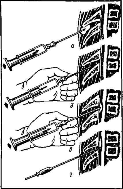
Fig. 20. Technique of epidural space puncture:
a — needle passing through the skin; b — advancing deeper; c — passing through the ligamentum flavum; d — needle in the epidural space
Anesthesia sets in within 15–30 minutes and lasts for 1.5–2 hours. To achieve adequate blockade of a single spinal segment in an adult patient, 1–2.5 ml of local anesthetic solution is required.
Complications during epidural anesthesia are relatively rare. The primary complication is collapse associated with the inhibition of A large number ofvasomotor fibers.
High epidural anesthesia may impair pulmonary ventilation, necessitating artificial ventilation. Less dangerous complications include headache, pain at the puncture site, and traumatic radiculitis. A hazardous complication of this type of anesthesia is infection of the epidural space.
Table 2. Site of anesthetic injection depending on the surgical area
|
Surgical zone |
Injection site |
|
Chest |
T3-T6 |
|
Upper abdomen |
Т7-Т10 |
|
Lower abdomen |
Т10— Т12 |
|
Rectum, perineal region |
L 2- 5 |
|
Prostate and urinary bladder |
L 2-4 |
|
Lower extremities |
L 3-5 |
Epidural anesthesia is indicated for major surgeries on the lower abdomen, urological and proctological procedures, and lower extremity surgeries (Table 2). It is the method of choice for elderly and senile patients, patients with cardiorespiratory pathology, Metabolic Disorders, hepatic or Renal Dysfunction, and in obstetrics.
Epidural anesthesia is widely used to relieve postoperative pain, rapidly restore bowel motility following abdominal surgery, and as part of combination therapy for various conditions (acute pancreatitis, Peritonitis, intestinal obstruction).
To maintain prolonged postoperative analgesia, the catheter is left in the epidural space (continuous/prolonged epidural anesthesia). Local anesthetic solutions are readministered as needed (with the trimecaine dose reaching 3–5 mg/kg).
Contraindications to this method include:
1) presence of inflammatory processes at the injection site;
2) hypovolemia, hypotension;
3) hypersensitivity to local anesthetics. Relative contraindications include spinal Column lesions, as well as disorders of the central and peripheral nervous systems.
CAUDAL ANESTHESIA
Caudal anesthesia, a variant of epidural anesthesia, can be used for surgeries in the perineal region and rectum. In this technique, the anesthetic solution is injected into the distal portion of the epidural space via the sacral hiatus or sacral canal. The solution spreads up to the first lumbar vertebra, anesthetizing all lumbosacral segments. The dose of the anesthetic is the same as in conventional epidural anesthesia.
SPINAL ANESTHESIA
In this type of regional anesthesia, a local anesthetic solution (lidocaine, trimecaine, novocaine) is injected into the subarachnoid space following a dural puncture. The anesthetic rapidly binds to the nerve roots, producing anesthesia in all PARTS OF THE body distal to the puncture site. If the specific gravity of the injected solution is lower than that of the CEREBROSPINAL FLUID (e.g., a 4–5% novocaine solution), it will ascend to higher segments. Typically, up to 3 ml of a 5% novocaine solution or 2–3 ml of a 5% trimecaine solution is used. The duration of anesthesia with trimecaine is 1.5–3 hours.
Currently, spinal anesthesia is primarily used for surgeries on organs located below the diaphragm and on the lower extremities. The needle should not be inserted higher than T12, as administering the anesthetic above this level may disrupt the cardiovascular and respiratory centers. The technique of spinal anesthesia is similar to that of epidural anesthesia. Most commonly, the puncture is performed in the interspinous space between L1 and L2, or between T12 and L1.
INTRAOSSEOUS ANESTHESIA
Intraosseous anesthesia can be performed for surgeries on the extremities. To do this, the limb is elevated and a tourniquet (or cuff) is applied
until the pulse in the peripheral Arteries disappears. After anesthetizing the skin, subcutaneous tissue, and periosteum near the joint, the soft tissues are punctured with a thick needle containing a stylet. Using rotary movements, the needle is advanced through the cortical bone into the spongy bone to a depth of 1–0.5 cm. A 0.5% solution of novocaine or trimecaine is then slowly injected through the needle. Anesthesia of the hand is achieved by injecting 35–40 ml of a 0.5% novocaine solution into the head of the metacarpal bone; for forearm surgeries, it is injected into the epiphysis of the radius or the olecranon.
With intraosseous anesthesia, the analgesic effect develops within 10–15 minutes and lasts until the tourniquet is released.
A variation of this pain management method is the anesthesia of a bone fracture site to relieve pain and reposition bone fragments. For this purpose, the fracture zone is punctured, and once blood appears in the needle, 5–20 ml of a 1–2% solution of novocaine, trimecaine, or xycaine is injected into the hematoma, depending on the fracture Location and the patient's age. Pain relief occurs within 5–10 minutes.
COMPLICATIONS OF LOCAL ANESTHESIA, THEIR PREVENTION, AND TREATMENT
The clinical picture of poisoning by various local anesthetics shares many common features. The effect of these substances on the central nervous system manifests as yawning, restlessness, disorientation, tremor, headache, nausea, vomiting, localized muscle twitching, and generalized clonic-tonic seizures. In severe cases, death ensues from respiratory paralysis.
The initial cardiovascular effect of the anesthetic manifests as tachycardia and arterial hypertension. Subsequently, electrical excitability and conductivity decrease, myocardial contraction is impaired with bradycardia and arterial hypotension, potentially progressing to cardiac arrest. These complications arise from anesthetic overdose or rapid intravenous administration.
A frequent complication of local anesthesia is anaphylactic reactions in patients hypersensitive to local anesthetics, manifested by allergic skin reactions (dermatitis, urticaria, subcutaneous edema), signs of cardiovascular collapse (pallor, cold extremities, cold clammy sweat, sharp drop in blood pressure, fainting), or anaphylactic shock.
To prevent allergic reactions, all patients with a history of hypersensitivity to local anesthetics should undergo sensitivity testing and receive antihistamines prior to surgery. The administration of diazepam or phenobarbital is effective in preventing central nervous system overexcitation in cases of local anesthetic overdose.
The treatment of toxicity depends on the clinical manifestations. In the presence of excitation and seizures, barbiturates (hexenal and sodium thiopental, 100–200 mg of a 2% solution) are administered intravenously and repeatedly until the desired effect is achieved. In case of respiratory depression, assisted or artificial pulmonary ventilation via an anesthesia machine mask is prescribed. Dithyline is administered at 20 mg every 2–3 minutes until seizures stop, accompanied by mandatory artificial ventilation. If hypotension develops, calcium chloride (10 ml of a 10% solution), ephedrine, or mesaton (15 mg every 30–60 seconds until effective) is administered intravenously; for hypertension, a 25% magnesium sulfate solution (10–20 ml) is given.
To reduce cerebral vasospasm, the patient should be placed in a horizontal position and allowed to inhale 2–3 drops of amyl nitrite. If an allergic reaction has developed and signs of cardiovascular collapse have appeared, adrenomimetic agents (adrenaline, 25 mg), glucocorticoids (dihydrocortisone), and antihistamines (promethazine, 50 mg, or suprastin, 25 mg) are prescribed.
CARDIOPULMONARY RESUSCITATION
Clinical death occurs As a result of circulatory and respiratory arrest. It typically lasts 3–5 minutes. In a hypoxic Organism, irreversible changes occur more rapidly before clinical death sets in, and the period of clinical death is shortened. Conversely, if circulation stops abruptly against the Background of a satisfactory or good general condition, the duration of clinical death increases.
Cardiac Activity can cease under The Influence of intracardiac and extracardiac factors. Circulatory arrest associated with cardiac factors is most commonly caused by myocardial infarction, myocardial trauma, cardiac tamponade, or coronary artery embolism.
The primary extracardiac factors causing circulatory arrest include intoxication (including from narcotic and analgesic substances), blood electrolyte imbalances, hypovolemia, and Acute Respiratory Failure.
The main pathogenic factors contributing to circulatory arrest are most frequently hypoxia, hypercapnia, and acidosis. Their impact on cardiac activity manifests as impaired metabolic processes in the myocardium, as well as reduced excitability and conductivity. Specifically, hypoxia disrupts energy production and utilization. Hypercapnia increases cardiac excitability and enhances the vagal effect on the heart. Acidosis impairs myocardial contractility and diminishes the hemodynamic efficacy of catecholamines.
An important role in the Pathogenesis of cardiac arrest is played by plasma electrolyte imbalances, most commonly manifested as hyperkalemia. The latter, along with hypoxia and acidosis, causes depression of the cardiac conduction system and contributes to myocardial atony. A decrease in extracellular potassium concentration triggers ventricular fibrillation.
Sudden cardiac arrest can result from sinus node inhibition caused by a sharp dominance of vagal tone. This may occur during direct stimulation of the heart or during procedures involving other organs and tissues innervated by the vagus nerve. Hypoxia and hypercapnia play a critical role in the pathogenesis of such asystole.
The following forms of circulatory arrest are distinguished:
1) asystole (or the complete cessation of mechanical and electrical cardiac activity);
2) ventricular fibrillation (uncoordinated contraction of individual muscle fibers);
3) "ineffective heart" (cardiac function is so severely impaired that it fails to sustain the body's vital activities).
Harbingers of circulatory arrest:
1) a drop in blood pressure exceeding 60 mmHg;
2) a sharp decrease in heart rate (below 40 bpm);
3) pronounced tachycardia with arrhythmia and ventricular extrasystoles;
4) a sharp impairment of atrioventricular conduction, particularly the onset of complete heart block.
Causes of respiratory arrest:
1) impairment of respiratory center function (central nervous system diseases and injuries, Acute Poisoning, drug overdose—especially general anesthetics and analgesics);
2) airway obstruction (aspiration, suffocation, drowning);
3) trauma to the external respiration apparatus (crushed chest, pneumothorax);
4) respiratory muscle dysfunction (effects of muscle relaxants, myasthenia gravis, etc.). Complete inability to inhale, leading to subsequent respiratory arrest, occurs during upper airway obstruction caused by the tongue falling back due to unconsciousness and decreased tone of the glossopharyngeal muscles.
This is exacerbated by attempts to take a gasping breath, which essentially causes the Base of the larynx to suction against the laryngeal inlet (post-anesthesia states, brain trauma, stroke, poisoning, etc.). Complete laryngeal occlusion may develop during laryngospasm triggered by the inhalation of irritating anesthetic agents, gases, smoke, hot steam, general anesthesia complications, drowning, or seizures.
It develops rapidly in the larynx during anaphylactic shock or bee stings to the laryngeal and chin areas. Respiratory disturbances—bordering on arrest—develop more slowly in botulism and severe myasthenia gravis. Circulatory arrest ensues immediately following respiratory arrest (within 30–40 s).
Signs of respiratory arrest: absence of movements in the chest, epigastric region, and larynx, as well as no evidence of airflow through the nose and mouth.
Signs of circulatory arrest:
a) unconsciousness;
b) absence of breathing;
c) pupil dilation;
d) absence of a pulse in major arteries (carotid and femoral). Checking the radial pulse should be avoided, as its absence does not definitively indicate circulatory arrest (arterial spasm may obscure the pulse even while the heart continues to function).
Additionally, the physician's own finger pulse may be misperceived as the patient's radial pulse;
d) pallor of the mucous membranes and skin (if respiration stops first, cyanosis is observed);
e) total atonia (which may be preceded by a brief convulsive seizure).
The presence of any three of the four specified signs (unconsciousness, pupil dilation, absence of pulse, respiratory arrest) in any combination provides grounds to diagnose "circulatory arrest" and requires cardiopulmonary resuscitation. Establishing this Diagnosis takes no more than 8–10 s.
The effectiveness of vital function stabilization is largely determined by the speed of medical intervention: if cardiopulmonary resuscitation is initiated within the first 2 min, success can be achieved in 92 % of cases; any delay reduces its efficacy.
Cardiopulmonary resuscitation is performed in the following order:
1) establishing the diagnosis of "clinical death";
2) ensuring patent airways and performing artificial pulmonary ventilation using the mouth-to-mouth (2–3 times) or mouth-to-nose method;
3) placing the patient in a supine position on a hard surface;
4) performing closed (external) chest compressions combined with artificial pulmonary ventilation.
METHODS FOR MAINTAINING RESPIRATION
Since unconscious patients in a state of clinical death lying in a supine position have the root of the tongue blocking the laryngeal part of the pharynx, artificial pulmonary ventilation requires ensuring upper airway patency. This is achieved by hyperextension of the head at the atlanto-occipital joint (Fig. 21). In doing so, the tongue is pulled forward, opening the entrance to the trachea.
To this end, one hand of the rescuer is placed under the patient's neck and the head is tilted back. Two fingers of the other hand pinch the nose, while the palm of that hand presses on the patient's forehead, thereby helping to extend the head. The first hand, previously placed under the occiput, grips the lower jaw from below, advances and lifts it, and the thumb opens the mouth. When performing ventilation through the nose, the hand supporting the lower jaw closes the mouth, and air is blown through the nose.
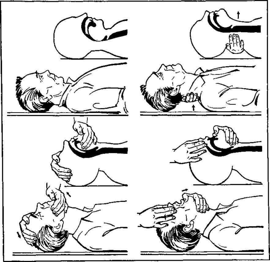
Fig. 21. Clearing the airways before performing artificial pulmonary ventilation by the mouth-to-nose and mouth-to-mouth methods
To perform artificial pulmonary ventilation, the rescuer takes a deep breath, opens the victim's mouth wide, presses their Lips firmly against the patient's mouth, and blows air into their lungs. Simultaneously ("out of the corner of the eye"), the rescuer monitors how the chest or epigastric region rises during inflation. Mouth-to-nose ventilation is performed when the patient's jaws are tightly clenched, or when it is difficult to achieve a seal during mouth-to-mouth breathing, as well as in cases of trauma to the lips, oral cavity, or lower jaw. Exhalation is passive due to the recoil of the elastic structures of the lungs.
If blowing air is ineffective, it is advisable to use a finger wrapped in a napkin or cloth to clear the oral cavity of mucus and vomit.
Inspirations should not be made too intensively, because in the case of partial airway obstruction, The Stomach is easily inflated, which may lead to regurgitation with aspiration of gastric contents into the lungs. If moderate gastric distension is observed, airways should be re-checked and secured—properly extend the head, push the lower jaw forward, and continue artificial pulmonary ventilation. Do not attempt to expel gas from the stomach, because trying to remove air from it by pressing on the epigastric region increases the risk of regurgitation. Should the latter occur, the patient is turned onto their side, the mouth and pharynx are cleared of gastric contents with a napkin-wrapped finger, and artificial pulmonary ventilation is resumed.
Resuscitation care for laryngeal foreign body occlusion and complete respiratory arrest has certain specific features. In these cases, the following maneuvers can be used:
1) back blows in the interscapular region;
2) chest compressions or abdominal thrusts. These two techniques are applied to remove the foreign body from the glottis;
3) attempting to remove the foreign body with a finger (carefully, so as not to displace it deeper). As soon as the foreign body is successfully removed, immediately intensify artificial pulmonary ventilation using the mouth-to-mouth (nose) method. If clearing the airways is impossible, a physician (feldsher) is authorized to perform a conicotomy on vital indications.
METHOD FOR MAINTAINING AND RESTORING CIRCULATION
The simplest and quite effective way to restore circulation is cardiac massage accompanied by mandatory artificial pulmonary ventilation, which ensures adequate circulation of oxygenated blood. Furthermore, the restoration of coronary blood flow and mechanical stimulation of the myocardium contribute to the recovery of cardiac activity. Massage is indicated not only in cardiac arrest but also during a sharp decline in cardiac function (in the absence of a pulse in the major vessels).
A favorable prognosis can be expected when massage is initiated no later than 4 minutes from the moment of sudden circulatory cessation.
There are two MAIN TYPES OF cardiac massage: closed (external, indirect) and open (internal, direct). The Essence of closed massage is that the heart, fixed in the Mediastinum between the chest and the spine, is compressed when pressure is applied to the chest in the anteroposterior direction, forcing blood from its chambers into the Vessels of the SYSTEMIC AND PULMONARY circulations.
When pressure is released, the chest expands due to its natural elasticity, allowing the heart to fill with blood once again. In most cases, this helps maintain a systolic blood pressure of 60—70 mm Hg for several hours. However, such circulation is insufficient to eliminate tissue hypoxia and its consequences.
Cardiac massage combined with artificial pulmonary ventilation (APV) and certain other therapeutic measures merely serves to sustain the body's viability for a few hours.
The technique for performing closed-chest cardiac massage is as follows. The patient is placed on their back on a firm surface (most often the floor), and buttons are undone or clothing is cut open. The rescuer kneels beside the victim, placing the palm of one hand on the lower-middle third of the Sternum, with the palm of the other hand placed on top crosswise (to increase pressure). Choosing the correct hand placement on the sternum prevents complications, as pressing directly on the middle of the sternum can cause a fracture. Pressing on the Ribs in the cardiac region may result in rib fractures, damage to the Pleura, lungs, heart, etc.
Using rhythmic thrusts and only the proximal part of the hand (the fingers should not participate), press on the sternum to displace it toward the spinal column by approximately 4—5 cm. When performing cardiac massage in adults, it is advisable to use not only arm strength but also body weight.
After pressing on the sternum, the hands should not be lifted from the chest, but the pressure must be completely released to allow the chest to return to its original position. The duration of the compression and relaxation phases should be equal. The rate of chest compressions should be within 60—80 per minute. For children under 10—12 years of age, closed-chest cardiac massage is performed using only one hand (70—80 thrusts per minute), and for newborns and infants, using the fingertips of two fingers.
There are two methods of performing cardiopulmonary resuscitation. In the first method, resuscitation is performed by a single person (Fig. 22, a). After every 2—3 breaths, 10—15 chest compressions are performed. In the second method, two rescuers assist: one performs APV while the other performs closed-chest cardiac massage. Following inflation, 4—5 compressions are applied (Fig. 22, b). The correctness of the massage is monitored by the rescuer performing APV (by checking for pulse waves synchronous with chest compressions in the carotid or femoral artery).
The effectiveness of cardiopulmonary resuscitation is confirmed by the following signs:
1) pupils constrict;
2) a pulse wave is detected in the carotid artery synchronously with chest compressions;
3) eyelid tone is restored (the palpebral fissure closes);
4) spontaneous laryngeal movements appear;
5) the color of the skin and mucous membranes improves.
Every 2 minutes, resuscitation is paused for a few seconds to check whether a pulse has appeared in the major vessels. Cardiac massage and APV should be continued until cardiac activity is restored or for as long as sufficient circulation can be maintained to support brain function.
If signs of clinical death persist for 30—40 minutes and an emergency medical team cannot be called (e.g., during expeditions, in remote residential areas, etc.), resuscitation is discontinued.
It should be noted that far from all cases allow even an experienced specialist to determine with a sufficient degree of certainty that resuscitation is hopeless; therefore, if there is even the slightest doubt, it is correct to continue full-scale assistance. Cardiopulmonary resuscitation is withheld only in the presence of signs of biological death.
During Stage II of cardiopulmonary resuscitation, a range of special diagnostic and therapeutic measures are employed, namely:
1) using suction devices (to remove mucus and vomit), airways to improve airway patency, and special devices and apparatus for APV;
2) ECG Diagnostics;
3) defibrillation in cases of fibrillation;
4) correction of metabolic acidosis.
After clearing mucus, vomit, etc., from the oral cavity, APV is performed using Guedel or Safar airways, manual resuscitators such as RDA-1,2, an AMBU bag, or portable APV devices such as "Pneumat", etc.
If there is no effect and resuscitation is prolonged, endotracheal intubation should be performed. After inflating the cuff of the intubation tube, the airway should be isolated from the esophagus to prevent aspiration, and APV should be continued. However, intubation is indicated only if performed by an experienced anesthesiologist within 20 seconds.
APV in this state should ideally be performed using pure oxygen.
During cardiopulmonary resuscitation, central analeptic drugs (caffeine, bemegride, corazole, cytiton, lobeline, cordiamine, etc.) must not be used to restore breathing, because under hypoxic conditions these drugs can stimulate the central nervous system for only a very brief period. This is followed by an even greater depression of the central nervous system, including the respiratory center.
Special suction devices are used to aspirate mucus and vomit from the airways. Meanwhile, closed-chest cardiac massage is continued according to general guidelines.
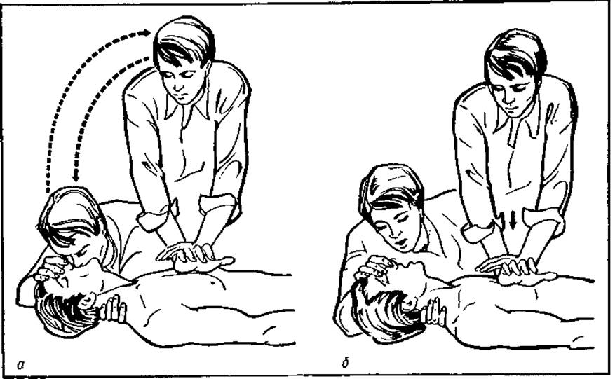
Fig. 22. Cardiopulmonary resuscitation:
a — performance of artificial lung ventilation and external chest compression by a single rescuer; b — performance of artificial lung ventilation and external chest compression by two rescuers
Electrocardiographic diagnosis of cardiac arrest-induced cardiac rhythm disorders is one of the priority emergency measures, as subsequent therapeutic decisions depend on it. Most frequently, ECG findings reveal:
a) ventricular fibrillation (irregularly shaped waves of varying amplitude, absence of cardiac complexes);
b) asystole (an isoelectric line indicating the absence of electrical cardiac activity);
c) "ineffective heart" (electrical activity of the heart is preserved, but cardiac contractions are sharply weakened or absent). All these ECG abnormalities are accompanied by signs of circulatory arrest.
In all cases of circulatory arrest (ECG signs of asystole, sluggish fibrillation, or an "ineffective heart"), 1 ml of 0.1% adrenaline solution and 1 ml of 0.1% atropine sulfate are administered intravenously (diluted in 10 ml of isotonic sodium chloride solution, with 5–7 ml of this mixture administered every 5 min). Closed chest compressions are performed during the injection.
For infants and newborns, adrenaline is administered at a dosage of 0.05 mg/kg, and atropine sulfate at a dosage of 0.03 mg/kg.
If establishing rapid venous access is impossible, adrenaline and atropine sulfate are administered via intracardiac injection. For this purpose, chest puncture is performed in the 3rd–4th intercostal space, 1–2 cm lateral to the left sternal border.
Atropine sulfate is prescribed to reduce vagal tone, as increased vagal activity can trigger reflexive cardiac arrest.
Adrenaline increases perfusion pressure during external chest compressions, stimulates spontaneous cardiac contractions, increases fibrillation amplitude (i.e., converts fine-wave fibrillation into coarse-wave fibrillation), which facilitates the restoration of normal heart rhythm.
If ventricular fibrillation is registered on the ECG, electrical defibrillation is performed. Its objective is the simultaneous depolarization of all myocardial fibers using an electric current to suppress ectopic foci of excitation, allowing rhythmic contractions driven by sinus node impulses to resume.
Since circulatory arrest is accompanied by metabolic acidosis—the manifestations of which are not alleviated by chest compressions and exert a detrimental effect on the myocardium—all patients urgently require the earliest possible intravenous infusion of an 8.4% sodium bicarbonate solution at a dose of 1 ml/kg, or a 4% solution at 2 ml/kg. Administration is repeated every 10 minutes until cardiac activity is restored.
In addition, clinical settings provide opportunities for direct (open) cardiac massage. Indications for its performance include:
1) cardiac tamponade;
2) stab wound of the heart;
3) myocardial rupture;
4) spinal column deformity (Scoliosis, lordosis, Kyphosis);
5) mediastinal shift (pneumothorax, hemothorax, Pulmonary Atelectasis following pneumonectomy);
6) flail chest;
7) during open-chest surgeries;
8) persistent ventricular fibrillation;
9) severe mitral stenosis (when commissurotomy is required);
10) in cases of profound hypothermia (during open-chest procedures), the heart can be rewarmed more rapidly using warm solutions.
Open cardiac massage is performed according to the following procedure. Without special preparation and anesthesia, an incision is made in the IV–V intercostal space. It begins 2 cm from the left sternal border and ends at the mid-axillary or posterior axillary line. After opening the chest cavity, a hand is inserted, and cardiac massage is initiated at a rate of 60–70 compressions per minute. If cardiac activity does not resume within 30–60 seconds, the Pericardium is incised, and direct massage of the exposed heart is performed.
Resuscitation is continued until cardiac activity is restored, after which the patient is transferred to the intensive care unit. If, despite all the aforementioned measures, the ECG shows a flat line (asystole) for 30 minutes, cardiopulmonary resuscitation should be considered ineffective and discontinued.
It should be remembered that following a cardiac arrest, close monitoring of major vital Organ Systems (central nervous system, Cardiovascular system, respiratory system, liver, kidneys, etc.) is essential.
CNS function is most frequently impaired following circulatory arrest. This is due to the high sensitivity of brain cells to hypoxia and the mismatch between the brain's high energy demands and its low energy reserves, which leads to the rapid disruption of all energy-dependent processes—specifically, the paralysis of ion pumps required to maintain transmembrane ion concentration gradients. Therefore, the outcomes of resuscitation are often determined by the reversibility of CNS changes. The prognosis is most favorable in patients with brief loss of consciousness, becomes questionable in prolonged comatose states, and is unfavorable if protective reflexes are absent for more than 48 hours.
In cases of successful resuscitation, a rapid transition from deep coma to stupor and, ultimately, to full consciousness is typically observed. In other words, the recovery of brain function proceeds in reverse order.
If the patient remains unconscious for a prolonged period, primary attention is directed toward measures aimed at restoring CNS function. For this purpose, a comprehensive set of therapeutic interventions is carried out. Mechanical ventilation with pure oxygen is continued in a hyperventilation mode to reduce PaCO2 to 30–35 mm Hg, as hypocapnia induces vasoconstriction and thereby helps reduce brain edema.
The administration of glucocorticoids (prednisolone at a dose of 300–500 mg per day) is mandatory in cases of cerebral edema.
To achieve dehydration, intravenous administration of a 20% mannitol solution (at 1–2 g/kg) is prescribed, and glycerin (1–2 g/kg) or Diuretics (furosemide 40–60 mg, ethacrynic acid 50–100 mg) are administered intravenously or via a gastric tube.
During dehydration therapy, a moderately negative fluid balance should be maintained. This is done to ensure that the cerebrospinal fluid pressure neither exceeds 100–120 mm H2O nor drops below 20–30 mm H2O, since CSF hypotension also has an adverse effect on CNS functional recovery.
For seizures and psychomotor agitation, the administration of medications such as dolantin, chlorpromazine, pipolfen, seduxen, diphenhydramine, or intravenous barbiturates is indicated.
Cerebral circulation is stimulated by administering vasodilators (aminophylline, cavinton), agents that improve blood rheology (rheopolyglucukin), and anticoagulants to lower blood clotting (heparin).
To replenish energy reserves and intensify metabolic processes in the CNS, glucose solutions (5–10%) with Insulin are prescribed, along with vitamin supplements, notably pyridoxine, cyanocobalamin, and ascorbic acid.
Nootropic medications (nootropic, piracetam) are effective.
Given the potential for secondary neurological deterioration, even with the most favorable and rapid recovery process, patients' physical activity should be restricted (for 10–12 days), and they should be discharged no earlier than 2 to 3 weeks after clinical death and resuscitation.
Complications Associated with Cardiopulmonary Resuscitation
Rib fractures frequently occur during closed-chest cardiac massage. They are typically transverse and located in the III–IV intercostal space along the mid-clavicular line or at the costochondral junctions.
In multiple rib fractures, lung volume decreases due to chest wall deformation. Additionally, small petechial hemorrhages appear on the anterior and posterior surfaces of the heart, coalescing in areas corresponding to the site of cardiac compression between the sternum and the spinal column.
Damage to the liver, Spleen, or Pancreas is less common and is invariably accompanied by clinically undiagnosed hemoperitoneum.
Complications associated with open-chest cardiac massage are somewhat different. Subendocardial hemorrhages on the anterior wall of the left and right ventricles are more frequently observed. A small amount of blood is occasionally found in the pericardial cavity. Thoracotomy performed for cardiac massage is sometimes complicated by lung collapse due to pneumothorax, hemothorax, or laceration of the lung tissue.
Pathomorphological changes related to cardiac puncture manifest as punctate and slit-like defects surrounded by a narrow zone of subepicardial Hemorrhage. In isolated cases, 50–60 ml of blood is found in the pericardium following this procedure. The development of a massive hematoma at the base of the interatrial septum is significantly less common. Closed defibrillation reveals no microscopic Changes in the myocardium. Open defibrillation is accompanied by scattered punctate subepicardial hemorrhages at the sites where the electrodes were applied.
One of the life-threatening complications of resuscitation (in the absence of a tracheal intubation tube) is the regurgitation of gastric contents into the respiratory tract. This occurs as a result of air entering the stomach during forced ventilation. As a rule, this happens when the head is insufficiently extended, causing the base of the tongue to partially block the entrance to the trachea, so that the majority of the air or oxygen enters the stomach instead of the lungs, causing acute gastric distension. Since the cardiac sphincter is relaxed in an unconscious state, gastric contents flow out and enter the lungs.
Last update: 08/08/2026
Editorial and Educational Adaptation: This material has been compiled based on the primary/original source text. The project team performed an editorial review, corrected technical inaccuracies, structured sections, and adapted the content for an educational format.
What was processed:
- elimination of formatting defects (OCR errors, structural breaks, corrupted characters);
- editorial organization of content;
- standardization of terminology in accordance with academic sources;
- verification of factual statements against the original source text.
All mentions of the author, publication year, and origin of the primary text have been preserved in accordance with the source.