Medical Radiology - Lazar A.P. 2008
Radiation Examination of the Genitourinary System
Inflammatory Diseases
Acute Pyelonephritis clinically manifests with lower back pain, dysuria, high fever, and a positive Pasternatsky's sign (costovertebral angle tenderness). Plain abdominal radiography often reveals an obscured contour of the psoas Muscle and enlargement of the affected Kidney, accompanied by a radiolucent halo around it due to edema of the perirenal adipose tissue. Excretory urography demonstrates signs of impaired renal function, specifically delayed and diminished contrast enhancement. The reduction in the kidney's concentrating capacity leads to delayed excretion of the contrast agent. Additionally, deformation of the renal pelvis and calyces is observed resulting from edema and infiltration of their walls (Fig. 246). Later, dilation of the deformed renal pelvis up to 2-3 cm develops, known as pyelectasis.
Class="center">
Fig. 246. Acute pyelonephritis on a radiograph (diagram).
1 - parenchymal atrophy with indentations located opposite the calyces;
2 - flat or convex bases of the calyces.
Ultrasonography (US) can reveal renal enlargement, calculi, Hydronephrosis, as well as intrarenal or perinephric abscesses resulting from the progression of pyelonephritis. Computed tomography (CT) in the Cytology/cytology/16.html">Early stages of pyelonephritis may detect patchy inflammatory areas within the renal parenchyma (Fig. 247), while later stages show kidney enlargement, blurred contours, thickening of the renal fascia, and edema of the perirenal fat pad.
Scintigraphy with 99mTc-gluceptate detects inflammatory foci in pyelonephritis earlier than US and CT. Renography records a decreased excretion rate of the radiopharmaceutical (99mTc-MAG-3, 131I-hippuran), followed by flattening of the peak of the renographic curve and elongation of its First and Second segments (Fig. 248).
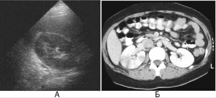
Fig. 247. Acute right-sided pyelonephritis.
A - Ultrasound examination reveals kidney enlargement with a loss of corticomedullary differentiation (edema).
B - computed tomography shows enlargement of the right kidney and areas of decreased densitometric density within the renal parenchyma.

Fig. 248. Right-sided pyelonephritis (renogram).
The curve of the left kidney is of normal type (Tmax = 3 min, T1/2 = 17 min), showing initial impairment of excretory function. The curve of the right kidney is of parenchymal type (Tmax = 6 min, T1/2 - not determinable).
Acute pyelonephritis may progress to Chronic Pyelonephritis or become complicated by The Development of an abscess, carbuncle, or perinephritic abscess (paranephritis), which are detected by US, CT, and MRI.
Glomerulonephritis is typically characterized by bilateral renal involvement and manifests as altered renal dimensions, along with widening and thickening of the cortical layer, as detected by US, CT, and MRI. Renography is preferably performed using 99mTc-DTPA, because this radiopharmaceutical is excreted by the renal glomeruli, which are primarily affected in this pathology. The renographic curve in glomerulonephritis is flattened.
Renal tuberculosis initially affects predominantly the tips of the renal pyramids; therefore, in the early Stages of the disease, papillitis develops, which is radiologically characterized by ulceration of the papillae and penetration of the contrast medium into the parenchyma. Furthermore, papillary Asymmetry, irregular contours of the calyces, and their deformation are observed (Fig. 249). In advanced cases, tuberculous cavities—irregularly shaped cavities with ragged contours, heterogeneous content, and surrounded by dense tissue—are detected within the renal parenchyma. Calcifications, severe deformations, and strictures can also be found in The Kidneys in cases of tuberculosis.

Fig. 249. Renal tuberculosis on a radiograph (diagram).
1 - additional image of a calyceal cavity (cavern);
2 - narrowing of the minor calyx;
3 - compression of the Base of the minor calyx and Atrophy of the cortical parenchyma.

Fig. 250. Renal tumor on a radiograph (diagram).
1 - calyceal margin stretched peripherally;
2 - rounded protrusion of the external renal contour;
3 - flattening of the calyx base.
Last update: 08/08/2026
Editorial and Educational Adaptation: This material has been compiled based on the primary/original source text. The project team performed an editorial review, corrected technical inaccuracies, structured sections, and adapted the content for an educational format.
What was processed:
- elimination of formatting defects (OCR errors, structural breaks, corrupted characters);
- editorial organization of content;
- standardization of terminology in accordance with academic sources;
- verification of factual statements against the original source text.
All mentions of the author, publication year, and origin of the primary text have been preserved in accordance with the source.