Medical Radiology - Lazar A.P. 2008
Radiation Examination of the Genitourinary System
Acquired Pathology of the Urinary System
Nephroptosis, or Kidney prolapse, is detected when the patient is in an upright position. This may reveal a tortuous course of the Ureter, its kinking, sometimes dilation above the site of the kink, and deformity of the renal pelvis and calyces. Prolapsed Kidneys can impair urine outflow and lead to Hydronephrosis—a persistent, progressive dilation of the pelvicalyceal system that results in Atrophy of the renal parenchyma. Hydronephrosis frequently develops due to an obstruction in the Urinary Tract caused by ureteral blockage by a calculus or compression of the ureter by a tumor. Morphological changes (dilation of the calyces and pelvis, atrophy of the renal parenchyma) in hydronephrosis are readily visualized via ultrasound, CT, MRI, and excretory urography (Fig. 243), whereas functional impairments are identified during radionuclide imaging by an obstructive-type renographic curve.
Class="center">
Fig. 242. Renal cyst on ultrasound.
1 - cyst; 2 - acoustic enhancement effect behind the echo-negative lesion.

Fig. 243. Right-sided hydronephrosis on excretory urogram (A), sonogram (B), and computed tomography scan (C).
1 - dilated calyces with rounded bases; 2 - dilated pelvis.
Urolithiasis. Most commonly, stones form in the renal pelvis and calyces. Calculi can vary greatly in size—from a few millimeters to several centimeters. Large stones replicate the shape of the pelvis and calyces, giving them a branched, staghorn appearance ("staghorn" calculi). Occasionally, stones begin to fragment and pass through the ureter, triggering sudden attacks of Renal Colic. During their passage through the ureter, stones most frequently become trapped in its abdominal or intramural segments (Fig. 244). Urinary Bladder stones can reach significant sizes. Sometimes, The chemical composition of stones can be determined by their shape and contours: smooth, rounded stones are typical of phosphates, whereas those with uneven, spiky projections are characteristic of oxalates.
Inorganic stones are often visible on standard plain abdominal radiographs, whereas organic ones are detected using intravenous urography and other Diagnostic Imaging Modalities (Fig. 245).

Fig. 244. Obstructive syndrome on an excretory urogram.
1 - right ureter without pathological changes;
2 - calculus in the abdominal segment of the left ureter.
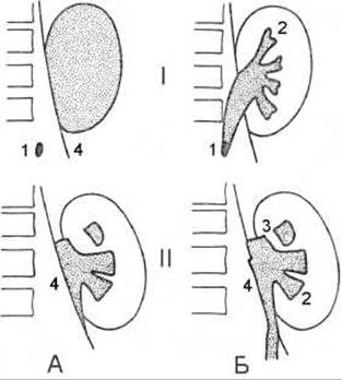
Fig. 245. Obstructive syndrome on
excretory urography (diagram). A - after 4 min;
B - after 20 min;
I - complete obstruction;
II - after removal of obstruction;
1 - calculus;
2 - flattened bases of the calyces;
3 - vascular trace;
4 - edge of the psoas major Muscle.
Arterial Hypertension is frequently of nephrogenic origin. The renovascular form of nephrogenic hypertension develops As a result of renal artery stenosis caused by atherosclerosis, thrombosis, kinking in nephroptosis, etc. The parenchymal form of nephrogenic hypertension occurs when intrarenal Blood flow is impaired due to Glomerulonephritis and Chronic Pyelonephritis.
A nephrogenic Etiology of arterial hypertension is highly probable in patients with high diastolic blood pressure (>110 mmHg) that is refractory to medical therapy, young individuals, those with a sudden onset of elevated blood pressure, and those with a positive captopril test.
When renal perfusion is reduced, Glomerular Filtration is maintained at a constant level due to constriction of the efferent arteriole induced by angiotensin II. Captopril blocks The formation of angiotensin II and eliminates the factors maintaining filtration, whereas the decrease in glomerular filtration can be detected using 99mTc-DMSA or 99mTc-DTPA scintigraphy, where the affected kidney is not visualized (the "pharmacological nephrectomy" sign).
Last update: 08/08/2026
Editorial and Educational Adaptation: This material has been compiled based on the primary/original source text. The project team performed an editorial review, corrected technical inaccuracies, structured sections, and adapted the content for an educational format.
What was processed:
- elimination of formatting defects (OCR errors, structural breaks, corrupted characters);
- editorial organization of content;
- standardization of terminology in accordance with academic sources;
- verification of factual statements against the original source text.
All mentions of the author, publication year, and origin of the primary text have been preserved in accordance with the source.