Medical Radiology - Lazar A.P. 2008
Diagnostic Imaging Modalities
Radionuclide Examination
Radionuclide imaging (RNI) is based on detecting gamma rays emitted by a radiopharmaceutical administered to a patient for diagnostic purposes. The radiopharmaceutical (RP) is selectively taken up by specific Organs and Tissues, containing a radionuclide that decays by releasing gamma photons (Fig. 18). For instance, the radiopharmaceutical Tc-99m-DTPA consists of diethylenetriaminepentaacetic acid molecules labeled with the radioactive isotope technetium-99m. Following intravenous injection, this agent is rapidly excreted by the Kidneys, making the Urinary Tract radioactive.
During scintigraphy, gamma radiation is captured by a gamma camera, which comprises a collimator, a detector, and a photomultiplier tube. The collimator, positioned at the front of the camera, filters out stray radiation. A scintillation crystal (typically a sodium iodide crystal up to 50 cm in size) serves as the detector, where gamma rays produce light flashes that are registered by photomultiplier tubes (PMTs). The summation of thousands of such scintillations forms an image, which is recorded on photographic film or paper. Areas of increased RP uptake ("hot spots") indicate inflammation, hyperplasia, certain tumors, and metastases. Conversely, decreased RP uptake is observed in regions of sclerosis, cysts, and specific types of tumors and metastases.
The underlying principle of radionuclide scanning is identical to that of scintigraphy, except that it employs a small (several centimeters wide) scintillation crystal that moves systematically, line by line, over the organ under investigation to build the image. This yields lower image resolution and requires a longer acquisition time compared to a gamma camera.
Class="center">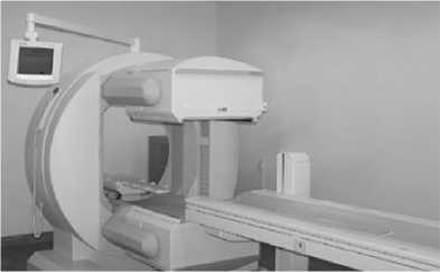
Fig. 19. Gamma camera with an open gantry frame capable of positron emission tomography.
In radiometry, a detector placed over The Organ of interest measures the intensity of gamma radiation emitted by the organ after the patient has received a radiopharmaceutical. The change in this intensity over time (expressed as counts per second or minute) can be recorded graphically (radiography). Both radiometry and radiography are used to assess the functional state of an organ.
Single-photon emission computed tomography (SPECT) utilizes radionuclides that emit a single gamma photon per radioactive decay, alongside mobile detectors that rotate around the object to generate cross-sectional images of RP distribution. SPECT enables clinicians to focus on changes within a specific body region, render a three-dimensional image of the organ on a computer monitor, and minimize Background radiation Interference from surrounding anatomical structures. Capable of detecting very subtle changes, SPECT allows for the early Diagnosis of pathological alterations at pre-structural stages with higher confidence. This technique is widely utilized in cardiology, neurology, and oncology.
Positron emission tomography (PET) employs radionuclides incorporated into Biomolecules as radiopharmaceuticals. These radionuclides are produced in cyclotrons and have half-lives ranging from a fraction of a minute to a couple of hours: 15O - 2.03 min, 13N - 10 min, 11C - 20.4 min, 18F - 110 min. Their decay is accompanied by positron emission; upon annihilation, these positrons generate pairs of gamma photons traveling at 1800 angles to one another, which are detected by opposing detectors (Fig. 19). PET makes it possible to trace metabolic processes involving molecules tagged with the radionuclide, helping to differentiate between benign and malignant tumors, metabolically active recurrent tumors and metabolically inactive post-Radiation therapy tissue, as well as diagnosing myocardial infarction. RP accumulation in metabolically active areas creates a "hot spot" sign (Fig. 20). To achieve precise anatomical and topographical localization of pathological processes, PET and CT scanners are frequently combined into a single hybrid unit (Fig. 21). Furthermore, PET facilitates not only qualitative but also Quantitative evaluation of radionuclide concentrations.
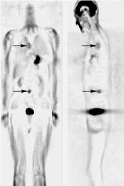
Fig. 20. Positron emission tomography in a patient with Central Lung Cancer and a metastasis in the fourth lumbar vertebra. Pathological "hot spots" (arrows) are visible in the right lung hilum and the fourth lumbar vertebra.
The main drawbacks of positron emission tomography include The Need for complex medical cyclotrons and radiochemistry laboratories to produce the radionuclides, which has slowed the adoption of PET into routine clinical practice in Ukraine. Consequently, PET Procedures are primarily performed in specialized research centers and come at a high cost.
In 1982, in vitro radionuclide diagnostic techniques were introduced into clinical practice. During in vitro radionuclide testing, specific human body components—such as Blood serum, tissue samples, or other excretions—are added to a radioactive tracer contained in a test tube. THE PRINCIPLE OF radioimmunoassay relies on the competitive binding between unlabeled target substances and their labeled analogs for a specific binding system (antigen-antibody interaction). If a labeled antibody is used, the assay is termed immunoradiometric; if tissue receptors serve as the binding system, it is referred to as a radioreceptor assay.
Standard reagent kits are commercially available for in vitro testing, each designed to measure the concentration of a specific target substance. The most common labels are the gamma emitter I-125 or the beta emitter H-3. Performing an in vitro assay requires a solution where the labeled antigen is always present in excess relative to the Antibodies. This sets up a competition between labeled and unlabeled Antigens for antibody binding sites. The antibodies must be highly specific, meaning they should react exclusively with the target antigen. Concurrently, under identical conditions and using the same kits, standard sera with pre-determined target antigen concentrations are tested. A calibration curve is then constructed by plotting radioactivity against known antigen concentrations. Comparing the radioactivity of the patient sample against this calibration curve allows for the precise Determination of the target substance concentration in the sample.
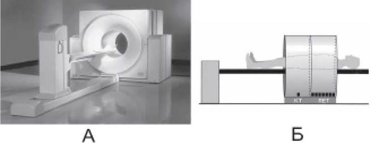
Fig. 21. Combined PET-CT scanner for computed and positron emission tomography.
In vitro radionuclide testing is applied in endocrinology (Insulin, T3, T4, TSH), oncology (carcinoembryonic antigen - CEA, alpha-fetoprotein, human chorionic gonadotropin - hCG), cardiology (Myoglobin), pediatrics (Growth Hormone - GH, thyroid-stimulating hormone - TSH), obstetrics and gynecology (luteinizing hormone - LH, follicle-stimulating hormone - FSH), allergology (IgE), and toxicology (pharmaceutical drugs).
Last update: 08/08/2026
Editorial and Educational Adaptation: This material has been compiled based on the primary/original source text. The project team performed an editorial review, corrected technical inaccuracies, structured sections, and adapted the content for an educational format.
What was processed:
- elimination of formatting defects (OCR errors, structural breaks, corrupted characters);
- editorial organization of content;
- standardization of terminology in accordance with academic sources;
- verification of factual statements against the original source text.
All mentions of the author, publication year, and origin of the primary text have been preserved in accordance with the source.