Medical Radiology - Lazar A.P. 2008
Radiation therapy
History of the development of therapeutic radiology
The Development of therapeutic radiology (frequently referred to simply as radiology) as a scientific discipline began in the late 19th century when Wilhelm Conrad Röntgen discovered X-rays. Less than a year later, the French physicist Antoine Henri Becquerel (1852–1908) discovered The phenomenon of radioactivity. In 1896, while conducting experiments with uranium sulfate, he found that the salt emitted radiation similar to X-rays. After testing other uranium salts, Becquerel concluded that the source of this 'invisible phosphorescence,' as he termed it at the time, was uranium itself. The following year, E. Rutherford established that uranium emits alpha and beta particles. In 1898, Pierre and Marie Curie investigated uranium and thorium ores and discovered that their radioactivity (a term they coined) was greater than that of pure uranium or thorium. From 8 tons of uranium ore, they managed to extract 1 g of a new chemical element, which was named radium, meaning 'radiant' in Ukrainian. Later, they discovered polonium, named in honor of Marie Curie’s homeland, Poland. Thus, natural radioactive elements were discovered.
In 1919, in experiments involving nitrogen and alpha-emitting radium, E. Rutherford achieved the first nuclear reaction, transforming one element into another. Subsequently, in 1934, the married couple Irene and Frédéric Joliot-Curie produced artificial radioactive elements in the laboratory by bombarding aluminum, boron, and magnesium with alpha particles.
Almost immediately following the discovery of X-rays and artificial radioactive elements, they began to be used for the Treatment of Skin diseases, Blood disorders, and malignant neoplasms. Alongside the Structure/182.html">Practical Application of ionizing radiation, research into the BIOLOGICAL EFFECTS OF radiation on various Tissues, Organs, and systems of living organisms was initiated.
Researchers working with radioactive substances were the first to experience The properties of these imperceptible emissions firsthand. A. Becquerel was not only the first to establish the existence of natural radioactivity, but also the first to suffer the damaging effects of radiation. In April 1902, at the request of P. Curie, Becquerel prepared a radium sample to demonstrate its properties at a meeting of the Physics and Technology Society. He placed a small Glass tube containing radium in his waistcoat pocket, where it remained for nearly 6 hours. Ten days later, an erythema appeared on the skin beneath the pocket, followed a few days later by an ulcer that persisted and refused to heal for a long time. Meeting with Pierre and Marie Curie later, Becquerel remarked, '...I love radium dearly, but I am rather angry with it.'
Class="center">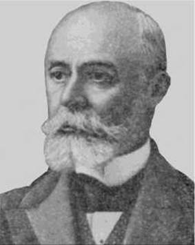
Henri Becquerel
The Russian researcher E.S. London (1868–1939) is considered the founding father of modern radiobiology. He studied the effects of radium radiation on Enzymes, toxins, and various tissues of living organisms, demonstrating the high radiosensitivity of the hematopoietic system and Gonads. He authored the world's first monograph on radiobiology, Radium in Biology and Medicine (1911). E.S.
London discovered the lethal effect of radium rays on mice. Largely influenced by E.S. London's experiments, the German scientist H. Heineke (1878–1922) incorporated X-rays into his own research.
Heineke was the first to describe radiation-induced anemia and leukopenia, and he drew attention to macroscopic damage to Hematopoietic organs in the form of splenic atrophy. He provided a detailed histological description of typical cellular Changes in the Bone Marrow and Lymph Nodes.
French physicians J.A. Bergonié (1857–1925) and L.M.F.A. Tribondeau (1872–1918) discovered that different Cells possess varying sensitivities to radiation. Based on these experiments, in 1906 they formulated a principle that entered radiobiology as the Bergonié-Tribondeau law: cellular radiosensitivity is directly proportional to mitotic activity and inversely proportional to the degree of Cell Differentiation. Certain adjustments to the Bergonié-Tribondeau law were made later.
X-rays were first used for Cancer treatment as early as 1896 by the American physicist Emil Grubbe (1875–1960).
Studies on the effects of ionizing radiation on Embryogenesis revealed the occurrence of various developmental abnormalities at specific stages of embryonic development. In 1925, in experiments on Yeast cells and Molds, G.N. Nadson and G.S. Philippov demonstrated that ionizing X-radiation affects the cellular genetic apparatus, leading to heritable phenotypic changes—Mutations. In 1927, the American scientist H.J. Muller successfully induced various Selection/21.html">Types of mutations in Drosophila using X-ray irradiation, increasing mutation frequency more than 150-fold compared to control groups.
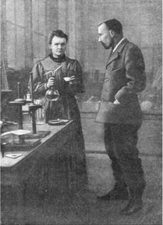
Marie and Pierre Curie
Radiobiological research experienced a particularly intense surge following the atomic bombings of Hiroshima and Nagasaki, which brought to the forefront the urgent task of developing radioprotective agents and Methods for treating radiation injuries. This necessitated a detailed Study of the mechanisms underlying the biological action of ionizing radiation and the Pathogenesis of radiation sickness. In 1957, the International Atomic Energy Agency (IAEA) was established as an autonomous intergovernmental Organization dedicated to the peaceful use of atomic energy. The International Commission on Radiological Protection formally recognized the linear no-threshold model regarding the health effects of low-level radiation.
Last update: 08/08/2026
Editorial and Educational Adaptation: This material has been compiled based on the primary/original source text. The project team performed an editorial review, corrected technical inaccuracies, structured sections, and adapted the content for an educational format.
What was processed:
- elimination of formatting defects (OCR errors, structural breaks, corrupted characters);
- editorial organization of content;
- standardization of terminology in accordance with academic sources;
- verification of factual statements against the original source text.
All mentions of the author, publication year, and origin of the primary text have been preserved in accordance with the source.