Medical Radiology - Lazar A.P. 2008
Diagnostic Imaging Methods
Medical Thermography
Medical thermography is a safe and non-invasive diagnostic imaging method that records infrared (thermal) radiation emitted from The Human Body surface. Most of human electromagnetic radiation has a wavelength of 10 µm, which lies in the infrared region of the spectrum.
Contact thermography is performed using liquid crystals embedded in a thin, flexible plastic base that change color depending on Temperature. Each thermal indicator has a specific color-temperature characteristic, which allows for studying the temperature distribution across the patient's body surface.
Non-contact (remote) thermography captures infrared rays from the human body surface using a specialized thermal imager that records them and displays a color image on a monitor screen or prints it on paper. The Main Components of a remote infrared thermography device include: a) a mirror scanning system; b) a lens system; c) a receiving sensor; d) an amplifier; e) a computer system for data acquisition, Processing, and presentation; f) a video monitor (Fig. 22). Areas of the body with different temperatures appear in different colors on the monitor screen.
Class="center">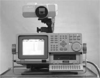
Fig. 22. Exterior view of a thermal imager.
Normally, each area of the human body has a specific temperature, which should be nearly identical in symmetrically positioned areas with a difference not exceeding fractions of a degree. Any change (increase or decrease) in the intensity of infrared radiation over a pathological focus is driven by a corresponding change (increase or decrease) in metabolic activity and Regional Blood Flow within it.
While standard medical thermography (in the micron range) examines the temperature at the body surface, the millimeter and decimeter ranges of the infrared spectrum make it possible to assess the condition of the human body at a certain depth.
In arterial Circulation disorders (angiospasm, constriction, complete vascular stenosis), thermography detects a hypothermic zone corresponding to the area of reduced blood flow. Conversely, pathological changes in venous vessels (thrombophlebitis, venous thrombosis) reveal a region of elevated temperature.
Thermography is used in the Diagnosis of Circulatory Disorders, inflammatory conditions, tumors, and certain occupational diseases. Thermograms help detect cerebrovascular accidents and occlusions of the Arteries and Veins OF THE extremities. Regarding oncological diseases, thermography has proven most useful in breast examination.
Patient preparation for a thermographic examination is quite complex and includes the following steps.
1. The examination is performed on an empty Stomach; follow-up examinations should be scheduled at the same time of day to account for circadian temperature fluctuations.
2. Examinations in women should be performed on the 10th–12th day after the onset of menstruation.
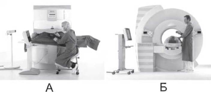
Fig. 23. MRI-guided invasive intervention: A — open type; B — closed type.
3. Two days prior to the examination, patients must avoid physiotherapy Procedures, medical cups, mustard plasters, irritating ointments, vasoconstrictor or vasodilator drugs, and cosmetics.
4. Twenty-four hours before the examination, oral or parenteral administration of vasoconstrictor or vasodilator medications is prohibited.
5. When examining the extremities, the patient should take a warm bath the evening before to remove the stratum corneum, Skin oils, and exfoliated epidermis, trim the fingernails/toenails short, and remove any nail polish.
6. The body surface must be free of any ointments and cosmetics.
7. Patients are forbidden to smoke for 4 hours prior to the examination.
6. When undergoing HEAD examination, men should shave the evening before to avoid fresh cuts.
Furthermore, a significant limitation of thermography is its low Specificity in disease diagnosis.
Last update: 08/08/2026
Editorial and Educational Adaptation: This material has been compiled based on the primary/original source text. The project team performed an editorial review, corrected technical inaccuracies, structured sections, and adapted the content for an educational format.
What was processed:
- elimination of formatting defects (OCR errors, structural breaks, corrupted characters);
- editorial organization of content;
- standardization of terminology in accordance with academic sources;
- verification of factual statements against the original source text.
All mentions of the author, publication year, and origin of the primary text have been preserved in accordance with the source.