Medical Radiology - Lazar A.P. 2008
Diagnostic Imaging Methods
Interventional Radiology
Interventional radiology is a branch of medical radiology based on The Use of diagnostic imaging techniques to perform minimally invasive Procedures for diagnostic and therapeutic purposes.
The Essence of interventional radiology lies in combining diagnostic and therapeutic manipulations within a single Procedure (Fig. 23). At The First stage of the study, The Nature and extent of the lesion are determined; at the Second Stage, typically without interrupting the procedure, the required therapeutic maneuvers are performed using specialized instruments.
Class="center">
Fig. 24. CT-guided needle biopsy. A – the horizontal CT scan reveals an infiltrative mass in the pelvic cavity, with the puncture needle entering a bowel loop; B – the needle passes past the bowel loop; C – the puncture needle is embedded within the infiltrative mass, with the pathomorphological report indicating rectal carcinoma.
Currently, the main areas of interventional radiology include endovascular interventions, endobiliary interventions, endoesophageal interventions, endoureteral interventions, endobronchial interventions, aspiration biopsy (Fig. 24), percutaneous drainage of cysts and abscesses, and percutaneous surgeries on various Organs and systems.
Most manipulations are performed percutaneously using specialized instruments. In terms of efficacy, these interventions are generally comparable to conventional surgery; however, they eliminate The Need for open surgical access and shorten the patient's hospital stay. Interventional procedures are performed under real-time imaging guidance. These procedures can be carried out using X-ray, ultrasound, computed tomography (CT), or MRI scanners. Aseptic and antiseptic rules are strictly observed during such procedures. Patient preparation includes premedication and anesthesia.
Fluoroscopically guided vascular dilatation was first proposed by Americans Ch. Dotter and M. Judkins in 1964. It became especially promising with the Introduction of a fundamentally new balloon catheter design in 1979 by Swiss physician A. Gruentzig.
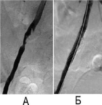
Fig. 25. Endovascular intervention (stenting) shown on a series of radiographs.
A – the angiogram reveals a greater than 50% stenosis of the external iliac artery;
B – after stent deployment, the stenosis has completely disappeared.
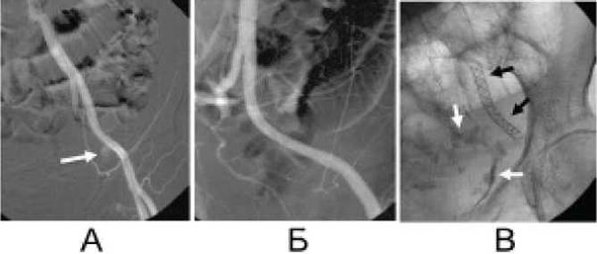
Fig. 26. Stenting on a series of radiographs. A – the external iliac artery angiogram in a female patient with a large pelvic tumor reveals Hemorrhage (white arrow); B – deployment of the stent in the damaged vessel halts the bleeding; C – the final radiograph shows the stent (black arrow) and residual uncontrasted Blood in the pelvic cavity (white arrows).
Nowadays, balloon angioplasty is one of the primary Methods for treating arterial stenoses. However, in 30–40% of patients, restenosis occurs relatively quickly after this procedure. Although balloon angioplasty can be repeated, stenting is generally preferred. A stent is an endovascular prosthesis consisting of a tubular metallic Structure used to reinforce the vessel wall (Figs. 25, 26).
One of the subtypes of endovascular interventions is vascular embolization. Embolization is used to stop bleeding (Fig. 27), treat angiomas, aneurysms, vascular malformations, and fistulas, as well as to occlude tumor-feeding vessels. Modern synthetic Materials are employed for embolization.
In many cases, the use of interventional radiology methods helps avoid major open surgeries. Consequently, interventional radiology is currently one of the most dynamic and rapidly evolving fields in medical radiology.
Last update: 08/08/2026
Editorial and Educational Adaptation: This material has been compiled based on the primary/original source text. The project team performed an editorial review, corrected technical inaccuracies, structured sections, and adapted the content for an educational format.
What was processed:
- elimination of formatting defects (OCR errors, structural breaks, corrupted characters);
- editorial organization of content;
- standardization of terminology in accordance with academic sources;
- verification of factual statements against the original source text.
All mentions of the author, publication year, and origin of the primary text have been preserved in accordance with the source.