Review of Medical Physiology - William F. Ganong 2002
Circulation
Regional Blood Flow
Placental and Fetal Circulation
Uterine Circulation
Uterine Blood flow runs parallel to the METABOLIC ACTIVITY OF the myometrium and endometrium, undergoing cyclical changes that closely correlate with the Menstrual cycle in non-pregnant women. The Functions of the spiral and basal Arteries of the endometrium during menstruation are described in Chapter 23. During Pregnancy, blood flow increases rapidly in tandem with uterine enlargement (Fig. 32-18). The production of vasodilatory metabolites undoubtedly occurs in the Uterus just as it does in other active Tissues. In early pregnancy, the uterine arteriovenous O2 difference is small, leading to the hypothesis that estrogens induce an increase in uterine BLOOD FLOW IN response to heightened tissue O2 demand. However, even with a 20-fold increase in uterine blood flow during pregnancy, the embryo grows vastly larger, developing from a single Cell into a fetus and Placenta that together weigh 4–5 kg at term. As a result, more O2 is extracted from the uterine blood in late pregnancy, leading to a decrease in uterine venous O2 saturation. Immediately prior to labor, There is a marked reduction in uterine blood flow, though the precise physiological significance of this phenomenon remains unclear.
Class="center">
Fig. 32-18. Changes in uterine blood flow and venous blood oxygen content during pregnancy (from Barcroft H. Modified with permission from Keele CA, Neil E: Samson Wright’s Applied Physiology, 12th ed. Oxford Univ Press, 1971).
Placenta
The placenta serves as the "Lungs of the fetus." Its maternal portion functions as a large blood sinus, into which project the villi of the fetal portion, containing small Branches of the fetal umbilical arteries and Veins (Fig. 32-19). Through this vascular interface, the fetus takes up O2 and discharges CO2 into the maternal circulation across the villous walls, much like O2 and CO2 exchange in the lungs. However, because The Cell layer covering the villi is thicker and less permeable than the alveolar membranes in the lungs, this exchange is considerably less efficient. The placenta also acts as the conduit through which nutrients reach the fetus and Metabolic waste products pass into the maternal blood.
The pathways of fetal circulation are schematically illustrated in Fig. 32-20. Approximately 55% of the fetal Cardiac Output passes through the placenta. Umbilical vein blood in humans is thought to be about 80% saturated with O2, compared with 98% saturation in the adult Arterial System. The ductus venosus (Fig. 32-21) channels a portion of this blood directly into the INFERIOR VENA CAVA, while the remainder mixes with the fetal portal blood. Portal and systemic venous blood in the fetus is only about 26% saturated with oxygen, and the saturation of mixed blood in the inferior vena cava is approximately 67%. Most of the blood entering The Heart via the inferior vena cava passes directly into the left atrium through the foramen ovale. The bulk of the blood from the SUPERIOR VENA CAVA enters the right ventricle and then flows into the pulmonary artery. Because resistance in the unexpanded lungs is high, pulmonary arterial pressure is somewhat higher than aortic pressure; consequently, most of the blood in the pulmonary artery flows through the ductus arteriosus into the aorta. Through this pathway, relatively deoxygenated blood from the right ventricle is delivered to the trunk and lower body of the fetus, whereas the HEAD receives more well-oxygenated blood from the left ventricle. From the aorta, some blood passes into the umbilical arteries and back to the placenta. The O2 saturation of blood in the lower aorta and umbilical arteries of the fetus is approximately 60%.

Fig. 32-19. Diagram of a cross-section through the human placenta, showing the penetration of fetal villi into maternal sinuses (reproduced with permission from Benson RC: Handbook of Obstetrics and Gynecology, 8th ed. McGraw-Hill, 1983).
Fetal Gas Exchange
The tissues of mammalian fetuses and newborns exhibit a remarkable, though not fully understood, resistance to Hypoxia. However, the O2 saturation of maternal blood in the placenta is so low that hypoxic injury could develop in the fetus unless fetal erythrocytes possessed a higher oxygen affinity than adult erythrocytes (Fig. 32-22). Fetal erythrocytes contain fetal Hemoglobin (hemoglobin F), whereas adults contain adult hemoglobin (hemoglobin A). The difference in affinity between these two forms is due to the fact that hemoglobin F binds 2,3-DPG less effectively than hemoglobin A. The reduction in oxygen affinity caused by binding to 2,3-DPG is described in Chapter 35. Quantitative Aspects of placental gas exchange, based on experiments performed in cattle, are presented in Table 32-5.

Fig. 32-20. Schematic diagram of the circulation in the fetus, newborn, and adult; DA = ductus arteriosus; FO = foramen ovale (modified and reproduced with permission from Born GVR et al: Changes in the heart and lungs at birth. Cold Spring Harbor Symp Quant Biol 1954; 19:102).
small amount of hemoglobin A is detectable during the fetal period (see Fig. 27-19). After birth, hemoglobin F is normally no longer produced, and by four months of age, 90% of the circulating hemoglobin is hemoglobin A.
Table 32-5. Gas exchange across the bovine placenta1
Vessel |
Hemoglobin saturation, % |
Partial pressure, mm Hg |
|
O2 |
СО2 |
||
Maternal artery |
90 |
70 |
41 |
Uterine vein |
70 |
41.5 |
46.5 |
Umbilical vein |
11.5 |
48 |
|
Umbilical artery |
5.5 |
50 |
|
1 Data from Roos J, Romijn C: J Physiol (Lond) 1938;92:261.
Circulatory and Respiratory Changes at Birth
Because the ductus arteriosus and foramen ovale remain patent (see Fig. 32-21), the left and right sides of the fetal heart work in parallel rather than in series, as they do in the adult. Following birth, placental circulation ceases, and peripheral vascular resistance suddenly increases. Aortic pressure rises until it matches or exceeds pulmonary arterial pressure. Simultaneously, due to the cessation of placental blood flow, the infant experiences a state of profound asphyxia. This triggers several spontaneous breaths, causing the lungs to expand. The negative intrapleural pressure (-30 to -50 mm Hg) generated during these breaths plays a key role in lung expansion, though other less understood factors are also involved. The suction effect of the first breath, combined with umbilical vein spasm, forces about 100 mL of blood from the placenta into the infant (placental transfusion).
Once the lungs have expanded, pulmonary vascular resistance drops to about 20% of its intra-uterine level, leading to a substantial increase in pulmonary blood flow. The return of blood from the lungs raises left atrial pressure, closing the foramen ovale by pushing the valve flap over it in the interatrial septum. The ductus arteriosus constricts within minutes of birth, though in sheep, for example, it does not close completely for 24–48 hours. Ultimately, both the foramen ovale and the ductus arteriosus close in normal infants, establishing an adult-type circulatory pattern by the end of the first few days of life. The mechanisms responsible for the obliteration of the ductus arteriosus, as well as the initial expansion of the lungs, are not fully understood, although evidence suggests that the postnatal rise in arterial PO2 plays a critical role. Bradykinin has been shown to constrict the umbilical vessels and ductus arteriosus while dilating the pulmonary vascular bed. Prostacyclin helps maintain the patency of the ductus arteriosus before birth. Rectal administration of one or two small doses of indomethacin—a drug that inhibits the synthesis of prostacyclin and Prostaglandins (see Chapter 17)—induces ductal closure in many infants, thereby avoiding surgical correction. Conversely, premature closure of the ductus before birth causes pulmonary Hypertension. A mild predisposition to pulmonary hypertension is observed in infants whose mothers took prostaglandins to delay the onset of labor. Continuous intravenous infusions of prostacyclin are valuable in the Treatment of pulmonary hypertension in adults.

Fig. 32-21. Fetal circulation. Most of the oxygenated blood reaching the heart via the umbilical and inferior venae cavae passes through the foramen ovale and from the aorta to the head, whereas deoxygenated blood returning via the superior vena cava is preferentially pumped through the pulmonary artery and ductus arteriosus to the lower limbs and umbilical arteries.
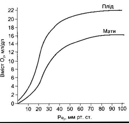
Fig. 32-22. Oxygen hemoglobin dissociation curves for maternal and fetal (human) blood.
Last update: 10/08/2026
Editorial and Educational Adaptation: This material has been compiled based on the primary/original source text. The project team performed an editorial review, corrected technical inaccuracies, structured sections, and adapted the content for an educational format.
What was processed:
- elimination of formatting defects (OCR errors, structural breaks, corrupted characters);
- editorial organization of content;
- standardization of terminology in accordance with academic sources;
- verification of factual statements against the original source text.
All mentions of the author, publication year, and origin of the primary text have been preserved in accordance with the source.