Medical Radiology - Lazar A.P. 2008
Radiation Examination of the Thoracic Organs
Lung Cancer
Lung Cancer develops insidiously and almost asymptomatically, which is why 60–70% of patients seek medical attention only at advanced Stages of the disease. Clinical symptoms of lung cancer include rapid fatigue, weight loss, chest pain, cough, and sometimes Blood-tinged sputum. Laboratory blood tests typically reveal a slightly elevated ESR and anemia.
Lung cancer almost always originates from the bronchial epithelium. The large Bronchi—main and lobar—are most frequently affected, followed less commonly by segmental bronchi and, even more rarely, small peripheral ones. Among lobar bronchi, those in the upper lobe of the right lung are most commonly involved. Out of numerous classifications proposed for this disease, the most widely accepted is that of Yu. M. Sokolov (1956), who distinguishes the following forms of lung cancer: 1) central; 2) peripheral; 3) mediastinal; 4) Pancoast (superior sulcus); and 5) miliary pulmonary carcinosis (carcinomatosis).
Central Lung Cancer develops from the epithelium of the main, lobar, or segmental bronchi. In some cases, the tumor grows primarily into the bronchial lumen endobronchially; in others, it grows exobronchially and peribronchially around the bronchus, invading the adjacent lung parenchyma. The clinical symptoms of central cancer appear at the stage of tumor development when external Respiration, the drainage function of the bronchus, and inflammatory reactions in surrounding Tissues become impaired.
The radiologic presentation of central cancer comprises manifestations of the tumor itself (visualized as a shadow on radiography and as a nodule or mass on computed tomography), signs of impaired bronchial patency, and complications arising from progressive tumor growth and metastasis. Due to the narrowing of the bronchial lumen, the following signs are observed: a) signs of hypoventilation (subatelectasis) manifested by decreased transparency of a lobe or the entire lung, against which an enhanced pulmonary pattern is visible; b) narrowing of the intercostal spaces; c) a slightly elevated position of the Diaphragm dome on the affected side; d) a slight mediastinal shift toward the hypoventilation, primarily involving the Esophagus (upon contrast examination) and the Trachea.
Class="center">
Fig. 121. Central Cancer of the right lung on chest radiographs in the direct (A) and lateral (B) projections, and on a computed tomogram (C).
1 - tumor; 2 - upper lobe Atelectasis; 3 - right main bronchus.
In endobronchial cancer, the tumor obturates the bronchial lumen, leading to atelectasis or hypoventilation and The Development of secondary inflammation. In the early Selection/3.html">Stages of development, the tumor nodule may be well-demarcated from the surrounding tissue. However, as the tumor process progresses, it loses clear boundaries and begins to spread beyond the bronchus into the lung tissue. Computed tomography and standard radiography reveal a tumor obturating the bronchial lumen (Fig. 121).
In the exobronchial growth pattern, the tumor appears on the radiograph as a rounded shadow only when its dimensions exceed the diameter of the large pulmonary vessels. In the Cytology/cytology/16.html">Early stages of tumor development, there are no impairments to bronchial patency. Only later, upon infiltration of the bronchial wall followed by tumor enlargement to several centimeters, do ventilation disorders occur (hypoventilation, check-valve emphysema, atelectasis). The lung ROOT is widened, featureless, and stringy.
In the peribronchial growth pattern, thick cuffs form around the bronchi and vessels. An enhanced pulmonary pattern is detected, which subsequently transforms into coarse strands extending fanwise from the root into the lung tissue. The bronchial lumens remain patent, and signs of impaired lung ventilation are absent. Computed and standard tomography reveal thickening of the bronchial walls and visualize the tumor mass itself, while bronchography demonstrates a concentric, uniform narrowing of the bronchial lumen over a significant extent associated with the thickening of its wall.
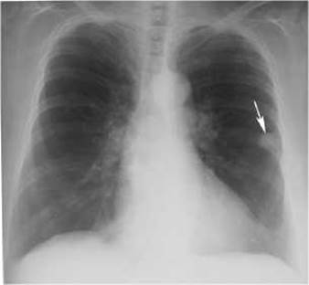
Fig. 122. Peripheral cancer of the left lung (arrow) on a plain chest radiograph.
Peripheral cancer originates from the mucosal epithelium of subsegmental bronchi and bronchioles. The clinical symptoms of this form of cancer depend on the Location OF THE tumor nodule relative to the chest wall and major bronchi. Invasion of the Pleura by the tumor causes chest pain. When the tumor infiltrates a large bronchus, cough, sputum production, and hemoptysis appear.
X-ray and CT examinations detect peripheral Cancer as a nodule or mass with a diameter of 2–4 cm or more, typically located in the upper lobes of the Lungs, more frequently on the right. Important signs of peripheral lung cancer include: 1) an irregular spherical shape revealed on CT and multi-axis X-ray Examination of the patient (Figs. 122, 123); 2) an inhomogeneous Structure OF THE tumor shadow consisting of separate nodules, as determined on radiographs, standard tomograms, and computed tomograms; 3) uneven, lobulated, and in places indistinct, blurred contours of the shadow; 4) unaltered lung tissue surrounding the shadow of the cancerous nodule; 5) the presence in some cases of a "trail" leading to the lung root, caused by small metastatic disseminations or tumor growth along the Lymphatic vessels (carcinomatous lymphangitis); 6) pleural thickening, adhesions, synechiae, or pleural effusions observed when the tumor is located close to the pleura; 7) an irregular-shaped cavity with uneven edges, without a fluid level, which appears during tumor necrosis and is better visualized on computed or linear tomograms (Figs. 124, 125).
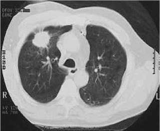
Fig. 123. Peripheral cancer of the right lung on a computed tomogram.

Fig. 124. Peripheral cancer of the right lung (arrow) with breakdown on a radiograph.
Peripheral cancer presenting as a nodule is often difficult to differentiate from non-malignant focal diseases, such as benign tumors, Tuberculoma, and others. In such cases, depending on the availability of imaging modalities, short-term dynamic X-ray monitoring, contrast-enhanced CT, or PET can be utilized (Fig. 126).
Mediastinal lung cancer is a manifestation of metastasis from a primary malignant tumor, most commonly located at the level of the tracheal bifurcation or the medial wall of the main or lobar bronchi. Metastasis occurs to the tracheobronchial and paratracheal Lymph Nodes. Clinically, the mediastinal form of lung cancer is characterized by compression syndrome of the Mediastinum (SUPERIOR VENA CAVA syndrome, compression of the esophagus, recurrent laryngeal and phrenic nerves), notably presenting with facial and neck edema. The radiologic picture is characterized by a rounded shadow on one side of the mediastinum, obscuring the shadow of the lung root. CT scans reveal hyperplasia of the affected lymph nodes.

Fig. 125. Peripheral cancer of the VI segment of the left lung with breakdown on a computed tomogram.
A - a peripheral cavitary mass in the left lung and solitary small foci in both lungs of the patient;
B - the same patient 4 months later: peripheral cancer with breakdown in the left lung, right-sided pneumothorax, and metastatic dissemination in both lungs.

Fig. 126. Peripheral cancer of the left lung on CT (mediastinal window) and 18F-fluorodeoxyglucose PET.
Pancoast tumor (superior sulcus tumor) is a carcinoma of the upper lobe of the lung originating from branchial cleft remnants. The tumor grows as a thin layer along the subpleural surface of the lung. Its early invasion into the Ribs and involvement of nerve plexuses determine the Clinical presentation: pain in the shoulder joint and chest, as well as Horner's syndrome (ptosis of the upper eyelid, enophthalmos, miosis). However, these are symptoms of an advanced process that has spread beyond the lung. The radiological picture of an apical tumor is characterized by a homogeneous shadow in the apical region of the lung with a lower polycyclic convex contour. Tomography reveals a rounded homogeneous mass in the lung apex, located close to the posterior chest wall. A characteristic feature is the destruction of the posterior segments of one or two ribs, as well as the transverse processes of several vertebrae.
Miliary carcinomatosis (carcinosis) is a form of metastatic spread of lung cancer. The primary tumor node is small and cannot be detected by X-ray and computed tomography examinations. Chest radiographs and CT scans reveal multiple shadows up to 3 mm in size. Over time, these shadows increase, predominantly in the lower lobes. The pulmonary pattern is obliterated.
Last update: 08/08/2026
Editorial and Educational Adaptation: This material has been compiled based on the primary/original source text. The project team performed an editorial review, corrected technical inaccuracies, structured sections, and adapted the content for an educational format.
What was processed:
- elimination of formatting defects (OCR errors, structural breaks, corrupted characters);
- editorial organization of content;
- standardization of terminology in accordance with academic sources;
- verification of factual statements against the original source text.
All mentions of the author, publication year, and origin of the primary text have been preserved in accordance with the source.