Orthopedics - Oleksa A.P. 2006
Tumor-like bone lesions and bone tumors
Clinical symptoms of tumors
Benign tumors (tumores benigni) are typically clinically silent and do not affect the patient's general condition. An exception is osteoid osteoma (Fig. 402), which develops within the Bone Marrow cavity and causes severe pain, Sleep disturbances, and loss of appetite, ultimately exhausting the patient. Occasionally, large benign tumors may compress adjacent Blood Vessels or nerves, causing pain and Circulatory Disorders. When located near joints, they may restrict the range of motion. In most cases, small benign tumors are incidental findings, while osteoblastoclastoma is sometimes diagnosed following a pathological fracture of the affected bone. Benign tumors are predominantly located in the metaepiphyseal regions; they feature clear margins, are painless, and do not metastasize. Figure 398 illustrates the most common anatomical sites of various tumors in long tubular bones.
Palpation of osteomas and osteochondromas reveals a hard, fixed, painless, smooth, or nodular bone neoplasm with normal surrounding Tissues. The rate of tumor growth is a crucial diagnostic indicator: benign tumors grow slowly, whereas accelerated growth (following trauma) over a short period may indicate malignant transformation.
Malignant tumors (tumores maligni) with a primary localization in the bones account for 1.2% of all tumors in men and 0.9% in women in Ukraine. They manifest clinically through both local and systemic Changes in the patient's body. Unfortunately, in most cases, patients seek medical attention due to pain only after the tumor process has spread beyond the bone into the surrounding soft tissues. Malignant tumors are characterized by infiltrative growth and the absence of a distinct boundary between the tumor and healthy tissue.
As the tumor progresses, the pain intensifies—especially at night—and becomes constant. The condition progresses extremely rapidly in osteogenic Sarcoma (Figs. 403, 404). This leads to antalgic contracture, impaired limb function, local Swelling, and a hallmark sign of malignancy: the dilation of subcutaneous Veins resembling caput medusae. Palpation of the tumor site reveals local hyperthermia and a "plus tissue" mass that is typically fixed, poorly defined, and occasionally exquisitely tender. A palpable mass indicates that the process has extended from the bone into adjacent structures. If tumor growth and bone destruction are aggressive and the mass enlarges rapidly, a pathological fracture may occur. The tumor may also invade the soft tissues and eventually break through the Skin surface.
Class="center">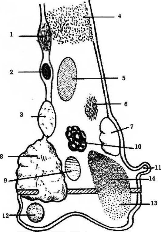
Fig. 402. Anatomical localization of tumor-like lesions, benign, and malignant tumors in long bones (Medewell J.E., Ragsdale B.D., Sweet D.E., 1981). In the diaphyses: 1 — adamantinoma, 2 — osteoid osteoma, 3 — chondromyxoid fibroma, 4 — Ewing's sarcoma, histiocytic lymphoma, myeloma, 5 — Fibrous Dysplasia, 6 — Fibrosarcoma.
In the metaphyses: 7 — fibrous cortical defect, 8 — osteogenic sarcoma, 9 — enchondroma. In the diaphyses and metaphyses: 10 — Bone Cysts and Cancer metastases, 11 — osteochondroma, 12 — chondroblastoma, 13 — giant Cell tumor, 14 — aneurysmal bone cysts.
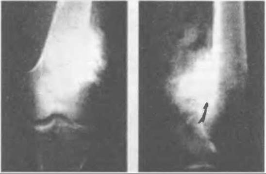
Fig. 403. Periosteal osteosarcoma of the distal Femur (anteroposterior and lateral radiographs).
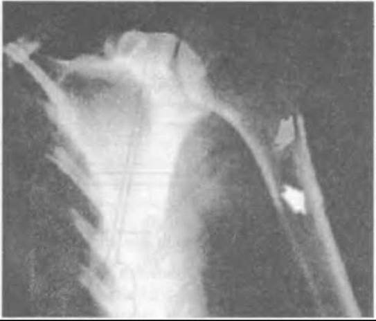
Fig. 404. Radiograph of periosteal osteosarcoma of the humerus.
Malignant tumors cause systemic changes, pain, insomnia, and loss of appetite, leading to severe cachexia. The patient loses weight, develops anemia and generalized weakness. Regional Lymph Nodes become affected, and metastases develop in the Lungs and other Organs, ultimately resulting in the patient's death.
Last update: 10/08/2026
Editorial and Educational Adaptation: This material has been compiled based on the primary/original source text. The project team performed an editorial review, corrected technical inaccuracies, structured sections, and adapted the content for an educational format.
What was processed:
- elimination of formatting defects (OCR errors, structural breaks, corrupted characters);
- editorial organization of content;
- standardization of terminology in accordance with academic sources;
- verification of factual statements against the original source text.
All mentions of the author, publication year, and origin of the primary text have been preserved in accordance with the source.