Medical Radiology - Lazar A.P. 2008
Radiation examination of the chest organs
Metastatic lung tumors
Lung Metastases can originate from any primary malignant tumor. However, they are most frequently observed in breast Cancer, Gastric Cancer, Adrenal Cancer, osteogenic Sarcoma, and Choriocarcinoma. A characteristic feature of these metastases is their multiplicity.
Class="center">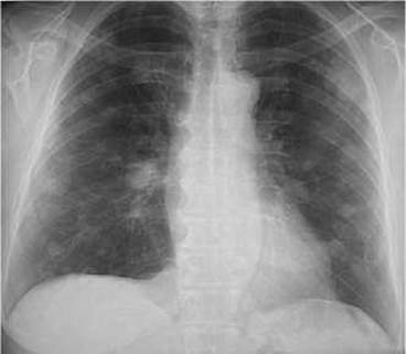
Fig. 127. Multiple lung metastases on a radiograph.
Tumor masses invade the lung tissue via hematogenous, lymphogenous, and bronchogenic pathways. In hematogenous spread, tumor emboli become lodged in the smallest pulmonary vessels (arterioles, capillaries, venules) and initiate The Development of metastases, which are most commonly localized in the superficial Regions of the lung parenchyma (often subpleurally). On radiographs, metastases appear as rounded opacities (round nodules on CT scans) with clear, smooth, or lobulated margins (Fig. 127). They can vary in number and may be present in one or both Lungs.
In lymphogenous dissemination of tumor masses, the Lymph Nodes surrounding the lung roots and Mediastinum are affected, leading to tumor transformation and enlargement (hyperplasia) of the lymph nodes.
Last update: 08/08/2026
Editorial and Educational Adaptation: This material has been compiled based on the primary/original source text. The project team performed an editorial review, corrected technical inaccuracies, structured sections, and adapted the content for an educational format.
What was processed:
- elimination of formatting defects (OCR errors, structural breaks, corrupted characters);
- editorial organization of content;
- standardization of terminology in accordance with academic sources;
- verification of factual statements against the original source text.
All mentions of the author, publication year, and origin of the primary text have been preserved in accordance with the source.