Nephrology for the Family Physician - O.I. Bakaliuk 2003
Primary Semiotics of Kidney Diseases
Renal Colic
In most cases, renal colic is a hallmark clinical symptom of urolithiasis. It develops As a result of the sudden cessation of urine outflow from the Kidney, triggered by a calculus obstructing the lumen of the upper Urinary Tract. Complete obstruction can also be driven by a functional factor: a local spasm of the ureteral wall Muscles occurs at the site of even a small calculus. Ureteral dyskinesia can likewise trigger renal colic—for instance, in Herpes progenitalis, when acute ureteral occlusion develops in response to the virus fixing onto the ureteral mucosa.
Particular significance is attributed to Nephroptosis, where colic is caused less by acute impairment of urine passage than by acute disorders of renal Blood Circulation.
Acute impairment of ureteral patency may also occur when a fragment of a disintegrating tumor, a blood clot, caseous masses, or bits of mucus and pus pass through in patients with Renal tuberculosis or Chronic Pyelonephritis. This leads to an increase in intrapelvic pressure; because the Innervation of the pelvicalyceal system is closely intertwined with that of the renal Vascular System, any urodynamic disturbances in the upper urinary tract immediately provoke renal hemodynamic disorders.
These disorders manifest as a pronounced decrease in renal vein tone and a spasm of intrarenal Arteries, resulting in ischemia of the nephrons and upper urinary tract (as the pelvicalyceal system and the upper part of the Ureter are supplied primarily by the renal arteries). Initially, hyperkinesia and dyskinesia of the upper urinary tract occur, which give way to hypokinesia as Hypoxia intensifies.
Thus, upper urinary tract urodynamic disorders set the stage for impaired renal blood flow, while the hypoxia of renal structures potentiates ureteral dyskinesia, creating a vicious circle.
Venous congestion triggers interstitial edema, which cannot be resolved even by the enhanced transport of fluid through The Lymphatic system of the kidney. Overloading the lymphatic system exacerbates the interstitial edema; the kidney enlarges and is additionally compressed by its fibrous capsule, which has little capacity for significant stretching.
Severe interstitial edema and the compression of the kidney by the fibrous capsule cause edema in the adipose tissue of the renal sinus and perirenal fat, giving rise to a second vicious circle.
It should be noted that perirenal Adipose tissue is viewed not merely as a Structure that fixes The Kidneys in place. It also serves as a backup pathway that facilitates fluid Transport from the kidneys and Functions as a unique "Shock absorber." This prevents injury to the renal parenchyma during intense physical exertion or sharp increases in intrapelvic pressure. The renal sinus fat persists under any conditions (such as starvation or other processes leading to severe bodily exhaustion). Prolonged and intense contractions of the calyces and renal pelvis, which alter their volume through stretching and contraction, never normally traumatize the renal parenchyma, as this is counteracted by the inherent shock-absorbing capacity of this exact structure.
Consequently, any disruption to the state of the perirenal fat is accompanied by hemodynamic Disorders of the Renal Circulation that are far too severe to be ignored.
During a renal colic attack, we must also highlight the negative role played by various emerging refluxes. In the Cytology/cytology/16.html">Early stages of urinary tract occlusion, pelvicalyceal refluxes are fornix-related, resulting from the rupture of the caliceal fornices. Later on, In addition to pyelovenous refluxes, pyelolymphatic and pyelosinus refluxes develop.
The occurrence of pyelosinus refluxes underlies The Development of pedunculitis—inflammation of the fatty tissue in the region of the renal hilum, which is encountered in 80–100% of urolithiasis cases (Yu.I. Udovytskyi, 1998).
The stretching of the caliceal fornices due to elevated intrapelvic pressure is also accompanied by The formation of additional clefts between epithelial Cells, through which urine penetrates (along an osmotic gradient) into the renal medulla, and—under a high hydraulic gradient—into the renal sinus adipose tissue. This process culminates in the fibrosing and sclerosis of the sinus.
Repeated attacks of renal colic create conditions wherein the caliceal fornices become obliterated, making their rupture impossible. In such a scenario, during a subsequent renal colic attack, tubular refluxes occur instead, with urine seeping into the Tissues surrounding the tubular System of the kidneys. All of this combined creates extremely unfavorable conditions for renal function during an attack of renal colic.
In other words, renal colic—previously perceived as a rather banal condition—is a formidable symptom that demands prompt medical intervention by a physician to prevent severe, and sometimes life-threatening, complications.
Elevated intrapelvic pressure and venous congestion stimulate the sensory nerve receptors of the renal hilum and its fibrous capsule, which clinically presents as acute pain. Because the nerve plexuses of the kidney are closely linked (both anatomically and functionally) with the mesenteric and celiac plexuses, renal colic features a characteristic pattern of referred pain that AIDS in accurate Diagnosis. When a calculus is localized in the kidney or the upper third of the ureter, the pain radiates to the umbilical region; when localized in the middle or lower segments of the ureter, it radiates to the iliac region, genitalia, and the inner thigh (Fig. 18).
Sometimes, patients experience an urgent need to urinate accompanied by the release of only a few drops of urine. Due to the periodic relaxation of the spastic ureteral wall, urine slowly trickles between the calculus and the wall, causing the pain to temporarily subside before intensifying once again.
Renal colic typically strikes suddenly, during or after physical exertion, brisk walking, bumpy rides, or the consumption of large amounts of fluid. The acute pain spreads across the entire corresponding half of the abdomen; changing body position does not affect (!) the intensity of the pain, which causes patients to behave extremely restlessly.
Intense pain can last anywhere from a few minutes to several hours or even days, subsiding periodically.
A renal colic attack is frequently accompanied by nausea, vomiting, dysuria, intestinal paresis, reflex tension of the anterior abdominal wall muscles, and more rarely, anuria (of reflex origin).
Intoxication syndrome manifests as weakness, dry Mouth, headache, elevated body Temperature, and chills.
Clinical examination helps confirm the diagnosis of renal colic. Bimanual Palpation of the kidney intensifies the pain, and Percussion tenderness (tapping sign) in the lumbar region is observed. In some cases, tenderness is elicited upon pressing the abdominal wall along the course of the affected ureter at the so-called ureteral points. The upper ureteral point is located 7–8 cm lateral to the umbilicus; the middle point is at the intersection of the horizontal line connecting the anterior superior iliac spines and the vertical line passing through the junction of the middle and outer thirds of the inguinal ligament; and the lower point is at the level where the ureter enters the Urinary Bladder.
Urinary sediment is characterized by gross Hematuria occurring after the pain attack, along with the presence of salts or small calculi.
The long-held view that gross hematuria during renal colic is primarily caused by trauma to the ureteral wall from the stone should be reconsidered.
The onset of gross hematuria is more frequently attributed to the rupture of the fornix Zones of the renal calyces and the venules located in these regions as a result of elevated intrapelvic pressure.
It must be emphasized that at the peak of renal colic caused by complete ureteral obstruction, the urinary sediment is normal (!), because in this case, the urine being examined is that which enters the bladder from the contralateral, healthy kidney.
Class="center">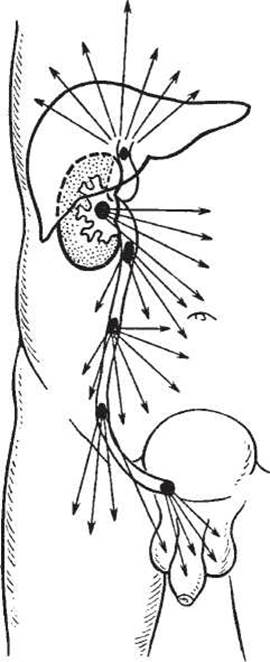
Fig. 18. Diagram of pain irradiation during biliary, renal, and ureteral colic attacks (adapted from Yu.A. Pytel et al., 1985).
In such situations, Instrumental Diagnostic Methods, particularly chromocystoscopy, are highly valuable. Timely excretion of the dye from both Ureters (within 3–5 minutes following intravenous administration of 5 ml of a 0.4% indigo carmine solution) rules out ureteral occlusion as the cause of renal colic-type pain.
Renal colic is also diagnosed using plain radiography (which detects radiopaque calculi), ultrasound scanning, excretory urography, and computed tomography.
An indirect sign of a urinary tract calculus on a plain radiograph is excessive gas accumulation in the Abdominal cavity, most frequently on the side of the renal colic, caused by paresis of certain loops of the Small Intestine (P. Bayuk et al., 1967).
Renal ultrasound (US) detects calculi of any composition measuring greater than 0.15–0.20 mm.
Excretory urograms during renal colic reveal imbibition of the renal parenchyma with the contrast agent—the so-called "white kidney"—whereas the absence of a contrast shadow on the affected side is attributed to enhanced reabsorption of the pelvicalyceal system contents, including the contrast medium, by the fornical apparatus.
Excretory urography is performed when it is necessary to assess the presence and functional status of the contralateral kidney, such as when planning Surgical Treatment for colic. In all other cases, excretory urography should be avoided during an episode of renal colic.
Establishing a diagnosis of renal colic requires ruling out appendicitis, cholelithiasis, perforated gastric and duodenal ulcers, acute pancreatitis, mesenteric vessel thrombosis, salpingo-oophoritis, the gastralgic variant of acute myocardial infarction, and radicular syndrome associated with Spinal osteochondrosis.
Last update: 08/08/2026
Editorial and Educational Adaptation: This material has been compiled based on the primary/original source text. The project team performed an editorial review, corrected technical inaccuracies, structured sections, and adapted the content for an educational format.
What was processed:
- elimination of formatting defects (OCR errors, structural breaks, corrupted characters);
- editorial organization of content;
- standardization of terminology in accordance with academic sources;
- verification of factual statements against the original source text.
All mentions of the author, publication year, and origin of the primary text have been preserved in accordance with the source.