Tuberculosis Study Guide - M.M. Savula 2002
Extrapulmonary tuberculosis
Tuberculosis of the urinary and genital organs
Renal tuberculosis
Genitourinary tuberculosis accounts for about 30% of all extrapulmonary sites of this disease.
Pathogenesis. Mycobacteria enter the Kidneys hematogenously, less commonly via the lymphatic route. The specific process begins with The formation of small tubercles in the renal cortex (renal parenchymal tuberculosis). At this stage, healing may occur, and the process halts. If the disease progresses, the lesions spread to the renal papillae, and the tubercles coalesce into circumscribed foci, which subsequently undergo necrosis. This gives rise to an acute destructive form of Renal Tuberculosis—Tuberculosis of the renal papilla (papillitis). This condition is characterized by destructive Changes in the apex of one or more pyramids. Subsequent liquefaction of necrotic masses, followed by rupture and drainage into the renal calyces and pelvis, initiates cavernous renal tuberculosis. Concurrently, sclerotic changes occur within the Kidney. Complete destruction and purulent liquefaction of the kidney lead to tuberculous Pyonephrosis. The kidney becomes resembling a sac filled with pus and caseous material. The process spreads to the Ureter and Urinary Bladder. Significant anatomical changes in the kidney lead to a decline in its function, resulting in The Development of chronic renal failure.
Clinical Features and Diagnosis. Most patients (60%) with renal parenchymal tuberculosis present with no subjective symptoms. Occasionally, they may complain of malaise, intermittent low-grade fever, and, rarely, a dull ache in the lumbar region. Their routine urinalysis results are normal. Sometimes, urine culture can detect MTB.
More pronounced symptoms appear with the development of destructive forms of renal tuberculosis. General symptoms include malaise, fever, and sometimes arterial Hypertension. Patients complain of a dull, aching pain in the lumbar region and lower abdomen, and occasionally frequent, painful urination.
During physical examination, it is sometimes possible to palpate a tender kidney and detect a positive Pasternatsky's sign.
Urinalysis shows an increased number of leukocytes (more than 8 per high-power field); erythrocytes are detected in the urine of 75% of patients, and protein in 90%. If Hematuria is accompanied by ureteral obstruction with Blood clots, paroxysmal pain (Renal Colic) occurs. The urine pH is typically acidic.
With the development of pyonephrosis, the urine becomes turbid, and A large number of leukocytes (pyuria) are detected. In patients with renal tuberculosis, biochemical blood tests (creatinine, urea) must be performed to diagnose chronic renal failure.
Renal tuberculosis is frequently detected in individuals with asymptomatic pyuria or in patients undergoing long-term Treatment for Chronic Pyelonephritis or cystitis. Therefore, at the slightest suspicion of tuberculosis of the kidneys or Urinary Tract, the patient must be referred to a specialized facility for further evaluation. Definitive confirmation of the tuberculous Nature of the disease is the detection of mycobacteria in the urine, which is examined using sediment Cell/15.html">Microscopy, flotation, and culture Methods.
Before collecting urine for mycobacterial testing, proper hygiene of the external genitalia is performed. A morning urine sample is collected in a sterile container and delivered to the bacteriological laboratory.
The Mantoux test with 2 TU is positive in patients with renal tuberculosis. Subcutaneous administration of tuberculin (Koch's test) is more informative. The occurrence of a systemic reaction (fever, changes in blood count) and a focal reaction (increased leukocyturia, detection of MTB in urine) confirms the diagnosis.
X-ray Examination is of great importance. A plain radiograph sometimes reveals calcification foci in the renal area. Retrograde pyelography or excretory urography can show dilation of the renal cavities, destructive changes in the papillae, and cavities (Fig. 30). Structural changes in the kidney are determined by ultrasound and radioisotope studies. Concurrently, patients must undergo chest photofluorography. Detecting tuberculous changes in the Lungs significantly facilitates diagnosis.
Differential diagnosis is carried out with pyelonephritis, cystitis, and Nephrolithiasis.
Class="center">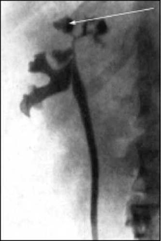
Fig. 30. Retrograde urography. Cavernous renal tuberculosis (a cavity in the renal parenchyma filled with contrast medium, communicating with the renal pelvis).
Treatment of patients with renal tuberculosis is carried out using a combination of antituberculosis drugs according to General Principles. This therapy is highly effective in the Cytology/cytology/16.html">Early stages of renal tuberculosis. In patients with manifestations of chronic renal failure, the doses of antituberculosis drugs are reduced. In destructive forms of renal tuberculosis, following preliminary antibacterial treatment and depending on indications, conservative renal resection or cavernectomy (removal of the cavity) is performed; in cases of a completely destroyed kidney (pyonephrosis), nephrectomy is indicated. Postoperatively, treatment with antituberculosis drugs is continued.
Last update: 10/08/2026
Editorial and Educational Adaptation: This material has been compiled based on the primary/original source text. The project team performed an editorial review, corrected technical inaccuracies, structured sections, and adapted the content for an educational format.
What was processed:
- elimination of formatting defects (OCR errors, structural breaks, corrupted characters);
- editorial organization of content;
- standardization of terminology in accordance with academic sources;
- verification of factual statements against the original source text.
All mentions of the author, publication year, and origin of the primary text have been preserved in accordance with the source.