Nephrology for the Family Physician - O.I. Bakaliuk 2003
Pyelonephritis
Among Kidney diseases, acute and Chronic Pyelonephritis (PN) rightfully occupy one of the leading positions in terms of incidence. Among all hospitalized patients, those suffering from PN account for 0.6%, general therapeutic department patients for 6%, and nephrological patients for 30% (A.Ya. Pytel, 1997). Overall, the prevalence of renal infections in Ukraine has increased by 46.7% (1.53-fold, L.P. Pavlova et al., 1995) over recent years and continues to grow (L.A. Pifig, 2001), with chronic PN being the cause of uremia in 1/3 of cases. PN is more frequently diagnosed in women (2:1), with 75% of female patients developing PN under the age of 40 (O.L. Tiktinsky et al., 1996). Over the past 15–20 years, an increasing incidence of PN has been observed among young people, which is attributed to Impaired Blood supply to the lower Urinary Tract resulting from wearing tight jeans and synthetic underwear.
The term PN refers to a nonspecific inflammatory process localized primarily in the pelvicalyceal System of the Kidneys and its tubulointerstitial zone. Bacterial invasion of the renal pelvis, calyces, and parenchyma, which lies at the ROOT of this disease, is primarily associated with the local impact of Bacteria followed by an inflammatory reaction characterized by focal-destructive and focal-granulomatous changes, scarring of all renal tissue structures, and the subsequent acquisition of "nephritis" features, i.e., glomerular involvement and progressive renal shrinkage. Due to this specific Pathogenesis, PN constitutes an independent pathological entity with a morphogenesis unique to itself. Furthermore, even the involvement of immune and autoimmune mechanisms, microcirculatory disorders, alterations in the lipid layer Structure of Introduction/36.html">Biological Membranes, and changes in their permeability—to which considerable attention has been paid recently (O.F. Vozianov et al., 1995, 1997; Ye.M. Neiko et al., 1995)—do not alter the infectious and inflammatory essence of the disease.
Currently, The Theory of so-called "bacterial modulins"—a new Class of microbial virulence factors—has been proposed. This class of factors includes bacterial forms possessing cytokine-inducing properties, such as The ability to stimulate the synthesis and release of tumor necrosis factor-alpha by macrophages, promote incomplete phagocytosis, and so on (N.V. Beloborodova et al., 2002; W. Liano et al., 1993).
The most frequent causative agents of PN are members of the intestinal microflora—Saprophytes and opportunistic flora—including Escherichia coli, enterococci, hemolytic variants of E. coli, Proteus, Klebsiella, Staphylococcus aureus, and Pseudomonas aeruginosa. Isolation frequencies for various flora are as follows: Staphylococcus aureus and Staphylococcus epidermidis — 50–70%, Escherichia coli — 60–66%, Proteus vulgaris and Proteus mirabilis — 30–56%, enterococci — 13–30%, Pseudomonas aeruginosa — 17–23%. Microbial associations are detected in 20–54% of cases. Thus, the leading role in the onset of PN belongs to the intestinal flora group (most commonly E. coli types 025, 026, 044, 0128), and less frequently to pyogenic flora originating from existing inflammatory foci in the body. Mention should also be made of nosocomial infections, which account for 10–20% of PN cases.
The identification of bacterial pathogens is based on detecting their aggressive factors using specialized tests (hemolytic, fibrinolytic, and coagulase activity, dermonecrotic properties, production of DNase, lecithinase, urease, hyaluronidase, etc.). However, in 15% of cases, the pathogen cannot be found by any method, including culture of renal Tissues obtained during surgical interventions, which historically led to the hypothesis of non-bacterial forms of PN. This situation has a clear explanation: recent studies have proven the potential for bacteria to transform into unique forms characterized by the absence of typical bacterial Cell walls while retaining pathogenic properties and Antibiotic Resistance.
Three such forms are distinguished: protoplasts, spheroplasts, and L-forms (A-stable and B-unstable). In protoplasts and A-stable L-forms, The Cell wall is completely absent, whereas in spheroplasts and B-stable L-forms, it is partially preserved. The pathway of Bacterial Transformation into one of these forms depends on the Osmotic Pressure of the surrounding environment. For instance, at an osmotic pressure lower than the intracellular pressure of the bacterium (noting, for example, that the latter reaches 15–20 atmospheres in Staphylococcus aureus), bacteria transform into protoplasts, whereas at higher pressures, they convert into L-forms.
Thus, in The Human Body, altered bacterial forms persist temporarily as protoplasts, while only L-forms survive and preserve their antigenic structure.
These forms are localized predominantly in the renal medulla, where conditions are favorable for their vegetation (slow blood flow, hyperosmolar environment). Bacterial L-forms act as a specific reservoir of infection, particularly in elderly individuals, and their ability to revert to parent bacterial forms upon even minor disruptions in urine outflow drives their active Participation in the pathogenesis of chronic PN. The isolation rate of bacterial L-forms in inflammatory urinary tract disorders ranges from 11 to 17%. These facts indicate that achieving clinical and laboratory remission in PN is not always a sign of patient recovery, and disease exacerbations may be linked to the reverse reversion of the microflora.
In recent years, a certain role in the pathogenesis of PN has been attributed to Mycoplasmas—Mycoplasma hominis, Mycoplasma fermentans, and T-Mycoplasma. The activation of mycoplasmal infection is promoted by various endogenous and exogenous factors capable of reducing body resistance, as well as by bacteria and Viruses. Conversely, the persistence of mycoplasmas can itself diminish systemic resistance.
Viruses (ECHO, Coxsackie, Adenoviruses) and Fungi (most commonly Candida albicans) also play a significant role in The Development of urinary tract pathologies, particularly PN, especially in elderly patients. All these facts clearly demonstrate that the onset and exacerbation of PN are 100% associated with infection—PN does not occur without an infectious trigger.
Factors that to some extent limit the proliferation of Microorganisms in the urinary tract include (C.R.V. Edwards et al., 1994): high urinary flow rate, regular and complete bladder emptying, and an adequate level of Tamm-Horsfall protein and local secretion of classes A and G IMMUNOGLOBULINS.
Recent years have yielded data that allow for the evaluation of another component absolutely essential for the development of PN: impaired urine outflow. The latter includes not only mechanical but also (and to a greater extent) functional disorders of urinary tract activity (hyper- and hypokinesia, refluxes, dystonia). These findings have convincingly proven that without prior impairment of urine outflow or renal hemodynamics, the pyelonephritic process does not develop. Consequently, the distinction between Primary and secondary PN is called into question (M. Kolesnyk, 1997), although its Classification into acute and chronic, unilateral or bilateral, obstructive or non-obstructive, as well as phases of exacerbation and clinical-laboratory remission for chronic PN, remains valid.
The urinogenous (ascending) pathway is recognized as the primary route of infection entry into renal structures (Yu.I. Udovytskyi, 1998), driven by the structural characteristics of certain microorganisms. Specifically, it has been established that most Gram-negative bacteria possess so-called fimbriae (pili)—glycoprotein-based Organelles whose receptors are Glycolipids in the membrane of the urinary tract epithelium. The presence of fimbriae enables bacteria to successfully attach to the urinary epithelium, a phenomenon known as adhesion (J.A. Roberts, 1991). This phenomenon facilitates the expression of bacterial virulence associated with the presence of so-called K and O Antigens. The K antigen inhibits bacterial phagocytosis, while the O antigen exerts systemic and local toxic effects. WHAT IS A local toxic bacterial effect? It is The impact of endotoxin on the smooth musculature of the urinary tract via the prostaglandin system, leading to suppressed peristalsis and potentially complete blockage. This results in so-called physiological urinary tract obstruction, accompanied by increased pressure within the tract and subsequent reflux, i.e., retrograde urine flow. Therefore, urodynamic disorders can be both a cause and a consequence of PN. Bacterial attachment to the urinary epithelium allows them to resist mechanical washout from the urinary tract, while the local toxic effect accompanied by reflux facilitates the upward migration of bacteria along the Ureters. In this regard, pyelovenous and pyelolymphatic refluxes are of great importance, whereby microbes—due to increased pelvic pressure—enter adjacent Veins and Lymphatic vessels from the fornix, and subsequently spread via the blood or Lymph stream to other renal structures, extending all the way to the cortical layer along medullary rays. Thus, a combination of ascending and hematogenous-lymphogenous (already within renal structures) pathways of infection spread is most commonly observed in PN.
Furthermore, it has been demonstrated that individuals susceptible to PN exhibit a significantly higher density of specific glycoprotein adhesion receptors for pathogenic microorganisms on their urothelium (I.A. Borisov, 1997; E. Donald et al., 2000; C. Svanborg et al., 1997).
A "purely" hematogenous route of bacterial entry into the kidney is likely less common than previously believed and typically occurs in patients with bacteremia or Sepsis. It is well established that The structure of the kidneys and Urinary Bladder hinders the spread of infection—bacteria cannot penetrate intact glomerular and tubular basement membranes. For instance, according to E. Kass (in studies conducted as early as 1972), even when 1 million microbial Cells of E. coli are placed on the bladder mucosa in the absence of urine, only a few hundred bacteria can be cultured from the mucosa after 1 hour. This phenomenon is not observed when residual urine is present in the bladder, as it impairs local phagocytic activity. The Role of bladder dysfunction (neurogenic bladder) in the genesis of PN has been confirmed by other researchers (V.I. Zaitsev, 1997). Conversely, when microorganisms are administered intravenously, only 1/100,000 of the injected bacterial load is detected within the renal parenchyma. This implies that infecting the kidney is virtually impossible unless fewer than 1 billion microorganisms are administered intravenously. Kass also noted that injecting 10,000 bacteria into the renal cortex is insufficient to trigger an inflammatory process, whereas injecting 10–100 bacteria directly into the medulla induces PN. This is because BLOOD FLOW IN the renal medulla is extremely sluggish compared to the cortex, creating favorable conditions for intensive bacterial proliferation. Animal experiments have also demonstrated that bacteria pass from the blood into the kidney via peritubular capillaries and from there into the interstitium, initially infecting the cortex and subsequently other renal zones.
The macroorganism's response depends on the state of nonspecific immune reactivity—specifically T-cell Immunity Functions (decreased both antibody-dependent cell-mediated and spontaneous cytotoxicity, depletion of reserve capacities of this system to mount a normal response to antigenic stimuli) (V.Ye. Drianska, 1998), the synthesis levels of serum Antibodies against microbial O and K antigens, and The production of interleukins-1 and 2 by macrophages and monocytes (G.N. Drannik et al., 1998). This condition may be genetically determined; it has been proven that the development of PN is associated with the presence of HLA antigens A1 and B17 as well as antigenic combinations A1 + B5, A1 + A2, A1 + B7, and A1 + B17 (V.Ye. Drianska, 1998; V.A. Zhmurov et al., 2000).
In recent years, reports have emerged regarding the immunopathogenetic role of Tamm-Horsfall protein in the development of PN (A.R. Maurer et al., 1995). It is possible that under certain circumstances, this protein may act as an autoantigen when it penetrates renal tissue As a result of reflux.
Acute pyelonephritis. Acute PN ranks second among all infectious diseases (following respiratory tract inflammations) and accounts for 14–15% of all renal diseases. Acute PN is quite frequently triggered by acute urinary obstruction, urinary tract manipulations, and hypothermia. The onset of the disease is also promoted by Diabetes Mellitus, adnexitis, congenital renal malformations, prolonged use of analgesics, glucocorticoids, immunosuppressants, and oral contraceptives. Essentially, acute pyelonephritis is an acute infectious interstitial nephritis characterized by the appearance of neutrophilic infiltrates in the kidneys. Infection can occur via ascending or hematogenous routes. In ascending infection, the medulla is affected first, whereas in hematogenous infection, the cortex is primarily involved, often with The formation of microabscesses. The ascending pathway of infection spread is most frequently caused by the following flora: Escherichia coli, Klebsiella, Proteus, Pseudomonas, Streptococcus faecalis, Serratia, Alcaligenes, and Staphylococcus albus; the hematogenous pathway by Staphylococcus aureus, Actinomyces, Brucella, Filamentous fungi, and Mycobacterium tuberculosis (G.S. Hill et al., 1989).
According to the morphological classification, three forms of acute PN are distinguished: serous, purulent, and purulent with pronounced mesenchymal reaction (V.V. Serov et al., 1973). All these forms are considered separate, Sequential Stages of the inflammatory process. The most frequent form of purulent PN is apostematous (pustular) nephritis, and less commonly, Renal abscess and carbuncle. Emphysematous PN, xanthogranulomatous PN, and renal papillary necrosis are regarded as distinct forms.
The onset of acute PN resembles an infectious disease. Clinically, it is characterized by hyperthermia, chills, general Intoxication syndrome, myalgia, arthralgia, signs of dehydration, dull lower back pain of varying severity, pollakiuria, and dysuria.
Intoxication syndrome features a phased clinical course—ranging from initial endotoxemia (resulting from the accumulation of toxic products in the blood, manifesting as weakness, sweating, and decreased appetite) to systemic endotoxicosis (a systemic inflammatory response characterized by fever, decreased blood pressure, and tachycardia—Systemic Inflammatory Response Syndrome (SIRS), A.L. Kharkiv, 1997; M.I. Kuzin, 2000; R.C. Bone, 1992).
Occasionally, the severity of dysuric disorders and pain mimics a Renal Colic attack. Chills are extremely pronounced, with body Temperature reaching 41 °C. Within 1–2 hours, profuse sweating occurs, and body temperature drops for a short period. Such episodes happen daily, up to 4–6 times a day. Physical examination reveals a severe patient condition, hyperthermia, dry Skin, occasional herpetic eruptions, a dry coated Tongue, tachycardia, hypotension, tenderness upon Palpation of the affected kidney, a positive costovertebral angle tenderness (pudge) sign, and dysuric disorders. A characteristic feature (in 99% of patients) is the so-called psoas sign—forced flexion of the legs toward the torso, which alleviates pain.
In some cases, patients with a renal carbuncle, renal abscess, or acute obstructive PN develop a picture of severe bacteremic Shock accompanied by tachycardia, a drop in blood pressure, oliguria, acidosis, and potentially ACUTE RENAL FAILURE. It is believed that the etiological factor of bacteremic shock is most frequently Gram-negative flora (E. coli, Proteus, Pseudomonas aeruginosa), while an important pathogenetic factor is marked impairment of urine outflow. The latter occurs in cases of obstructive renal and Ureteral calculi or following prostatectomy. Signs of collapse and decreased urine output typically appear after intense chills lasting 2–3 hours.
An important factor for the timely Diagnosis of circulatory collapse is information regarding baseline blood pressure levels. Thus, for a patient suffering from Hypertension, signs of shock may include a systolic blood pressure drop to 100–105 mmHg rather than the universally accepted thresholds (80 mmHg and below).
The second leading clinical symptom of bacteremic shock is a decrease in diuresis. Even in the Cytology/cytology/16.html">Early stages of shock, urine output may drop to 25–30 mL/h, and in its fully developed stage, oliguria progresses to anuria.
The Clinical symptoms of shock are compounded by metabolic acidosis, respiratory disorders, confusion, and loss of consciousness. Anuria, Cheyne-Stokes Respiration, and cerebral coma are considered three prognostically unfavorable signs of bacteremic shock.
Examination of a patient with acute pyelonephritis reveals signs of dehydration, moderate abdominal distension, local tenderness in the renal area, and a positive costovertebral angle tenderness (Pasternack's sign). Blood tests show leukocytosis with a pronounced left shift, aneoosinophilia, accelerated ESR, and occasionally Hyperbilirubinemia. In uncomplicated acute pyelonephritis, azotemia does not develop; only pyelonephritis complicated by papillary necrosis causes significant Impairment of the nitrogen-excreting renal function.
The Toxic Effect of the infectious agent primarily disrupts the function of the nephron tubular apparatus, manifested by impaired urine concentration, decreased or lost individual energy-dependent transport processes, and reduced sodium reabsorption. Visually, the urine becomes turbid, with a purulent flaky sediment and an unpleasant odor.
Urinary changes are characterized by moderate proteinuria, significant leukocyturia (pyuria), bacteriuria, and cylindruria (hyaline and leukocyte casts). Notably, the absence of bacteriuria does not rule out pyelonephritis. In complete renal obstruction, urinalysis may show no abnormalities at all (!), creating certain diagnostic challenges. In such cases, chromocystoscopy is absolutely indicated.
Data on the frequency of Hematuria in acute pyelonephritis vary from 30% to 80%, and it is more common in secondary pyelonephritis (50% of cases). Along with proteinuria, leukocyturia, hematuria, and bacteriuria, the diagnosis of pyelonephritis takes into account the urine reaction, which is persistently alkaline due to stasis, infection, and/or impaired renal acid-excreting function (Zh.D. Semidotska, 2001).
Indicators of local antibody production can also be used to diagnose pyelonephritis. Elevated titers of antibacterial antibodies in the urine are such an important diagnostic criterion for pyelonephritis that negative results of such a study allow ruling out the diagnosis of pyelonephritis with a high degree of certainty and attributing bacteriuria to urine contamination (B.I. Shulutko et al., 1993; V.N. Zhuravlev, 1999).
Diagnosis also utilizes data from plain radiography (kidney size, outer contour configuration) and excretory urography (infiltration of the renal medulla), ultrasound (signs of inflammatory infiltrative changes – altered contours and enlarged kidneys, calyceal-pelvic system deformation), isotope renography (impaired secretory function of renal tubules, curve Asymmetry), thermography (presence of a zone of intense infrared skin radiation over the respective area), and computed tomography (signs of obstruction, involvement of the perirenal adipose tissue).
Emphysematous pyelonephritis is a type of acute Purulent Pyelonephritis characterized by gas formation within renal structures. In 60–80% of cases, the CAUSATIVE AGENT OF emphysematous pyelonephritis is Escherichia coli. Gas initially infiltrates the Connective Tissue structures of the kidney and then, as a result of breakthrough through the renal sinus, spreads into the perirenal tissue.
The clinical picture is characterized by a severe patient condition (intense lower back pain, fever), a pronounced endogenous intoxication syndrome (nausea, persistent vomiting, tachycardia, dehydration, hepatorenal syndrome, anemia), and impaired renal function (acidosis, hypercreatininemia, hyperazotemia). Palpation reveals an enlarged, severely tender kidney.
Pathognomonic symptoms of the disease include the absence of the psoas Muscle shadow on the affected side and the presence of gas in the perirenal tissue on plain urograms. Urinalysis reveals proteinuria, leukocyturia, microhematuria, and bacteriuria. Due to glucose decomposition, the urine acquires a sharply acidic (!) reaction.
A specific variant of the course is acute pyelonephritis caused by Proteus mirabilis – xanthogranulomatous pyelonephritis. Its morphological manifestation is granulomatosis with A large number of lipid-laden macrophages (M.W. Griffing et al., 1996), strongly resembling chronic granulomatous disease in children due to a congenital phagocytosis defect. In this pathology, microorganisms are engulfed by macrophages, but phagocytosis proves incomplete, and macrophages act as a protective "capsule" (A. Cohen et al., 1998). Thus, the infection persists in the body for a long time. Granulomas comprising histiocytes, plasma cells, foamy macrophages, and Cholesterol crystals are formed in the kidneys, Liver, Lymph Nodes, and Lungs. The urinary syndrome is non-specific, and clinical manifestations depend on the Location OF THE granulomas. Diagnosis is based on the Analysis of the clinical picture and histological findings. A frequent cause of death, besides renal failure, is a progressive pulmonary process.
Renal papillary necrosis (necrotizing papillitis, papillary necrosis, renal medullary necrosis, necrotizing renal papillitis) is a form of pyelonephritis whose pathogenesis involves three components, the severity of which varies in each specific case:
- infectious (a prerequisite for the development of this form of necrosis is the presence of prior pyelonephritis);
- vascular (papillary necrosis is associated with impaired Blood supply to the renal medulla);
- ischemic (papillary necrosis occurs as a result of interstitial edema and sclerosis accompanied by compression of the vessels supplying the papilla).
Clinically, renal papillary necrosis is characterized by symptoms of an acute inflammatory process and urinary tract obstruction – high fever, chills, endogenous intoxication syndrome, and pain of varying intensity, from dull aching to sharp and unbearable. However, the most characteristic clinical symptom is total macrohematuria caused by the sloughing of the necrotic papilla. The passage of a necrotic papilla or its fragment through the urinary tract can cause occlusion with Typical symptoms of renal colic. A specific symptom is the presence of necrotic elements of the renal medulla in the urinary sediment. Dynamic X-ray Examination AIDS in diagnosis – plain radiography (signs of impregnation of the necrotic papilla), excretory urography, or retrograde pyelography (small calcification shadows in the papillary-forniceal zone of the calyx, a triangular calculus with a radiolucent core, indistinct, seemingly "gnawed" contours of the papilla and calyceal fornix, presence of a canal (fistula) in the papilla, formation of a niche with irregular contours in the distal part of the pyramid at the site of the sloughed papilla, multiple filling defects of the renal pelvis and calyces (Fig. 72, 73, 74).
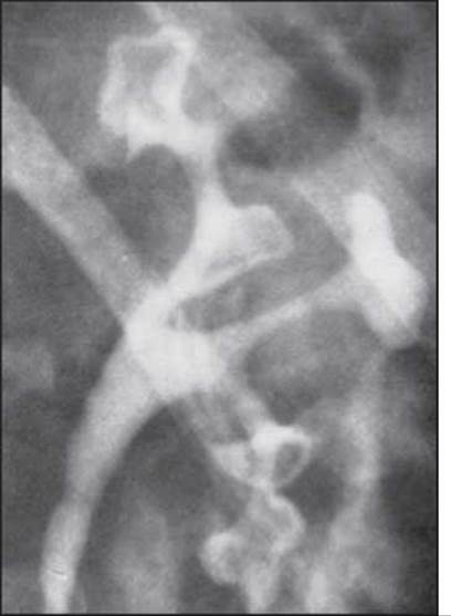
Fig. 72. Papillary necrosis (excretory urogram): papillae are sloughed (sequestered), typical rounded shadows are present.
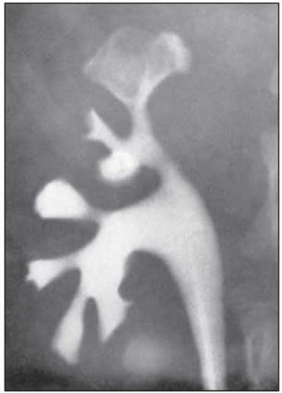
Fig. 73. Papillary necrosis (right-sided excretory urogram): contrast medium surrounds the sloughed papilla from the inside in the lower calyx area.
Verification of the diagnosis of acute pyelonephritis requires the exclusion of sepsis, Influenza, Pneumonia, malaria, typhoid and typhus fevers, food-borne toxicoinfections, salmonellosis, appendicitis, adnexitis, cholecystopancreatitis, perforated gastric and duodenal ulcers, renal anomalies, tuberculosis, as well as papillary tumors in renal papillary necrosis.
The management algorithm for patients with acute pyelonephritis and the scheme of specialized care for it were developed by O.F. Vozianov et al. (1998).
Chronic pyelonephritis is diagnosed when complete clinical and laboratory recovery does not occur during three months of Treatment. However, this premise does not exclude the possibility of primary chronic latent pyelonephritis developing, the exacerbation of symptoms of which is often interpreted as acute pyelonephritis. For instance, according to A.N. Shpygel (1985), 74.8% of patients with a preliminary diagnosis of "acute pyelonephritis" were diagnosed with the exacerbation phase of chronic pyelonephritis as a result of comprehensive examinations. This issue is quite fundamental, since 40.4% of the examined patients already showed varying degrees of impaired renal function.
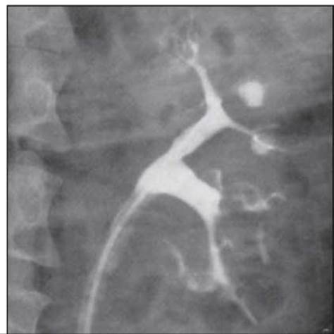
Fig. 74. Papillary necrosis (left-sided retrograde pyelogram): the renal edge is not visualized, arbitrary-shaped distortion and destruction of all calyces are determined, and contrast medium extravasates are located parallel to the papillae and extended toward the renal cortex.
The relationship between acute and chronic pyelonephritis (PN) is another fundamental issue. For instance, B.I. Shulutko (1996) views acute and chronic PN as essentially distinct diseases, denying the possibility of acute PN progressing into the chronic form. The author proposes an alternative theory of the pathogenesis of these pathological forms, attributing the development of acute PN to bacterial factors, while chronic PN is considered the result of immune and autoimmune damage to renal structures followed by an inflammatory response.
The clinical and laboratory manifestations of chronic PN depend on the phase of the pathological process and its extent (unilateral or bilateral). They are minimally pronounced during remission, presenting as low-grade fever, endogenous intoxication syndrome, nocturia, dysuria, and pollakiuria. Occasionally, the initial symptom of chronic PN may be an isolated urinary syndrome, iron-deficiency anemia, arterial hypertension (AH), or even Chronic Kidney Disease (CKD).
Urinary syndrome is characterized by nocturia and dysuria, whereas the urinary sediment reveals leukocyturia and bacteriuria (exceeding 100,000 microbial bodies per 1 ml of urine). Proteinuria (PU) is moderate (up to 1 g/day), and hematuria (HU) is rare.
Söderlund syndrome (G. Söderlund), a variant of interstitial nephritis (pyelonephritis), is classified among the forms of chronic PN without bacteriuria against the Background of typical clinical and radiological findings. It predominantly affects young males. Clinically, patients present with lower back pain, sometimes resembling renal colic, along with nocturia, dysuria, and stranguria. Body temperature remains normal; urinalysis reveals leukocyturia, proteinuria, and microhematuria in the absence of bacteriuria. Radiological examination demonstrates impaired tone of the renal pelvis, calyces, and ureters, as well as a spastic urinary bladder, while cystoscopy indicates purulent-fibrinous cystitis.
Renal function impairments in chronic PN primarily involve tubular mechanisms, manifesting as hyposthenuria, nocturia, decreased excretion of ammonium and hydrogen ions, and impaired sodium reabsorption (Thorn syndrome). Inadequate ammonia synthesis and hydrogen ion secretion also exacerbate sodium loss due to impaired bicarbonate reabsorption and the onset of acidosis. Aside from worsening acidosis, inadequate hydrogen ion secretion is accompanied by a compensatory increase in potassium excretion (which may lead to muscle weakness, flaccid paralysis, Cardiac Arrhythmias, and impaired myocardial inotropic function). Uncompensated sodium loss may eventually result in salt-wasting nephropathy (chloropenic kidney syndrome), which is rooted in the redistribution of body fluid spaces toward a reduced extracellular fluid volume. This syndrome is characterized by a drastic deterioration in general condition, stupor, gastric and intestinal atonia, vomiting, tachycardia, a drop in blood pressure, and muffled first Heart sounds.
Diagnosing chronic PN is an extremely challenging task, and none of the numerous instrumental Methods can be considered definitive. This challenge can only be resolved through a comprehensive and detailed evaluation of multiple criteria: medical history, dynamic clinical assessments, biochemical tests (including the evaluation of enzymuria patterns), bacteriological urine analysis, as well as radiological, radioisotope, and ultrasound imaging. Naturally, the structural Changes in the kidneys associated with PN, as revealed by these instrumental techniques, will depend on the phase and stage of the pathological process.
Below are the most specific alterations observed.
On plain radiography, pyelonephritic kidneys assume a vertical orientation, their contours become irregular, and physiological mobility is significantly reduced. A reduced renal shadow indicates cicatricial and sclerotic changes, provided that congenital Renal Hypoplasia has been previously ruled out.
Excretory urography offers greater diagnostic value. In the early stage of chronic PN or a protracted course of acute PN, radiological changes manifest as hypertonus and hyperkinesia of the calyces, renal pelvis, and Ureter resulting from inflammatory infiltration of the renal structures. The necks of the calyces become narrowed, and the calyces themselves acquire a triangular shape. With pronounced infiltration, the calyces appear elongated and displaced by the infiltrate (Fig. 75).
As the disease progresses, hypotonia and hypokinesia of the pelvicalyceal system and ureter develop, accompanied by the rounding of fornices and deformation of the calyces, as well as the blurring and gradual disappearance of the papillary contours. Fibrous remodeling is characterized by flattening of the renal papillae, changes in the shape of the calyceal necks, and the appearance of numerous secondary calyces with rigid necks (Fig. 76). In the terminal stage of chronic PN, radiography reveals a reduction in kidney size, increased density of the renal shadow, cicatricial retractions along the outer contour, delayed excretion of the contrast agent, and a renocortical index exceeding 0.38.
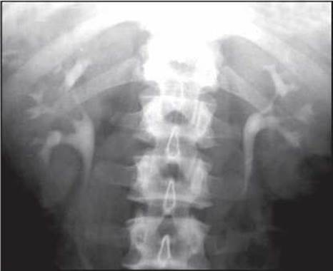
Fig. 75. Chronic PN, early stage (excretory urogram).
Overall, the typical radiological features of advanced chronic PN stages include:
- deformed, "mushroom-shaped" calyces combined with the narrowing of their necks;
- flask-shaped, hypotonic calyces;
- vertical configuration of a moderately dilated renal pelvis with small, triangular calyces featuring thin, elongated necks;
- moderate pyelectasis combined with deformed, atonic calyces;
- calyces with flattened and thinned fornices;
- tubular, forniceal, and venous refluxes;
- shortening and narrowing of the necks of individual calyces;
- papillary necrosis and sloughing. A Secondary contracted kidney is considered an almost absolute sign of chronic PN on Ultrasound examination, characterized by reduced
kidney size, deformation, and high echogenicity of the renal structures due to ongoing sclerotic processes (Fig. 77).
In the absence of renal shrinkage, there are no absolute pathognomonic echographic signs of chronic PN. For instance, during an exacerbation phase, renal infiltration may simulate completely normal kidney dimensions.
Radioisotope studies are used in combination with radiological methods; proper Interpretation of Results is possible only through this combined approach. Specifically, a normal renographic curve does not rule out PN, whereas its alterations (prolongation of the maximum peak time and isotope half-excretion time) are identical to those in Chronic Glomerulonephritis (GN). Nevertheless, curve asymmetry remains a more characteristic feature of PN.
Computed tomography in chronic PN reveals cysts of varying sizes, unilateral or increased density of the pyramid apices, their calcification, thickening of the renal capsule, irregularity of its outer contours (Fig. 78), and diffuse fibrous strands in the pararenal adipose tissue (O.I. Dhyadyk et al., 1997).
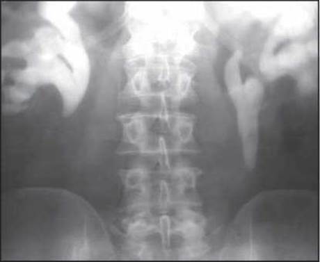
Fig. 76. Chronic pyelonephritis, stage of pronounced morphological changes (excretory urogram).
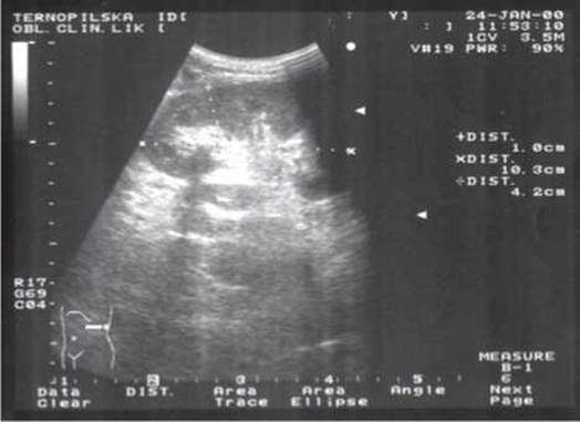
Fig. 77. Chronic pyelonephritis (ultrasound examination).
However, according to the same authors, computed tomography revealed no renal or urinary tract abnormalities in 90 out of 178 examined patients with primary pyelonephritis.
Indications for renal biopsy in patients with pyelonephritis are strictly regulated, and the Procedure is rarely used, primarily in diagnostically challenging cases.
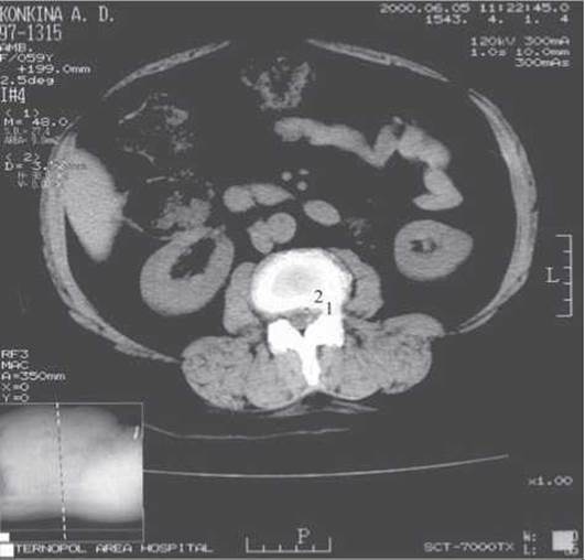
Fig. 78. Chronic pyelonephritis (computed tomography showing cysts in the renal parenchyma).
Distinct adult population groups in whom pyelonephritis develops most frequently are clearly identified: pregnant women and the elderly. The Specific features of pyelonephritis in these age groups are discussed by us separately, while below we outline the clinical characteristics of pyelonephritis in specific physiological states of the human body.
Pyelonephritis in patients with diabetes mellitus. Its incidence is 5–7% with a tendency to increase as obstruction arises or progresses.
The onset of the disease is predominantly primary-chronic; the clinical picture is dominated by endogenous intoxication and anemia syndromes (V.I. Sovalkin, 1999). Renoparenchymal hypertension syndrome is often masked by the presence of diabetic nephropathy. Among laboratory criteria, the predominant impairment of the renal concentrating function should be noted; bacteriuria is moderate.
The likelihood of pyelonephritis activation largely depends on unstable glycemic control; conversely, an exacerbation of pyelonephritis can serve as a factor causing decompensation of diabetes mellitus. Hyperglycemia creates favorable conditions for the spread of the inflammatory process to the perirenal adipose tissue, leading to the rapid development of severe septic complications. This justifies a more active medical approach in acute obstructive pyelonephritis among patients with diabetes mellitus—open surgical interventions within 24 hours in the absence of positive clinical dynamics (L.P. Sarychev, 2000).
Iatrogenic pyelonephritis. It typically occurs as a consequence of diagnostic and therapeutic instrumental Procedures (cystoscopy, retrograde pyelography, cystolithotripsy).
The clinical course is characterized by a "lightning-fast" onset—fever, chills, diaphoresis, dysuria, and endogenous intoxication. Such pyelonephritis requires aggressive therapy using reserve Antibiotics, glucocorticoids, relief of urinary outflow disorders, and improvement of central and renal hemodynamics (similar to bacteremic shock).
Post-radiation pyelonephritis most commonly occurs in women who have received Radiation therapy for uterine and adnexal neoplasms. The Development of the disease is facilitated by post-radiation strictures of the lower ureters or reflux.
The course of the disease is usually latent, with exacerbations developing after a radiation session or an intercurrent infection. The clinical picture is dominated by syndromes of endogenous intoxication, anemia, and hypertension. The prognosis depends on The rate of progression of ureteral stenosis; in some cases, surgical correction must be considered.
Pyelonephritis in Spinal Cord injury patients. Bladder atonia resulting from impaired neural regulation of its function leads to the development of ureteropyelectasis. In addition to urinary stasis in the bladder, frequent catheterization contributes to urinary tract infection. The ascending route of infection spread in this situation is compounded by a hematogenous route due to venous stasis in the kidneys. Electrolyte disturbances, particularly calcium imbalance resulting from prolonged immobilization and hypokinesia, are accompanied by urolithiasis. The clinical course of such pyelonephritis is progressive, treatment efficacy is minimal, and remissions are short-lived. The condition typically rapidly culminates in end-stage chronic renal failure.
Pyelonephritis following prostatectomy. Its incidence is determined by the severity of preoperative urinary outflow disorders, the invasiveness of the surgery, the proper management of the postoperative period, and the state of the patient's major homeostatic systems (cardiovascular, respiratory, and immune systems).
Features of its development include acute hyperthermia, intense chills, a collapse-like state, as well as the rapid transformation of the initial serous form into an apostematous one. Due to the similarity in symptoms, this transition from one form to the other is extremely difficult to diagnose.
Post-hemodialysis pyelonephritis. Renal infection is caused by frequent medical manipulations, regular contact of blood with dialysis membranes, the presence of chronic renal failure with profound Mineral METABOLISM disorders (secondary hyperparathyroidism, hypocalcemia, hyperphosphatemia, hyperkalemia), acid-base balance disorders (acidosis), and anemia.
For obvious reasons, timely diagnosis of such pyelonephritis is difficult. Treatment must be comprehensive, including antibiotic therapy that takes into account the antibiotic clearance pathways and its nephrotoxicity, alongside effective correction of homeostatic parameters against the background of an appropriate diet.
Post-transplantation pyelonephritis. The incidence of infection following kidney transplantation reaches 71% (J. Virka et al., 1997). Infection occurs primarily from the patient's own microflora (Escherichia coli, Klebsiella, Enterococcus, Enterococcus faecalis, Candida), although cytomegalovirus and herpes infections are also frequent causes of pyelonephritis. The pathogenetic basis of such pyelonephritis is continuous, high-dose immunosuppressive therapy (sandimmune, glucocorticoids) and impaired normal urine drainage from the transplanted kidney, as well as vesicoureteral reflux.
The efficacy of treatment for such pyelonephritis is minimal, and the clinical course resembles the picture of chronic graft rejection, which complicates the Selection of optimal medical management.
Differential diagnosis OF chronic pyelonephritis is most frequently performed with chronic glomerulonephritis, interstitial nephritis, Hydronephrosis, Pyonephrosis, Polycystic Kidney Disease, hypertensive disease, tuberculosis, Renal Tumors, cystitis, and urethritis.
Treatment of pyelonephritis. The rationale for etiotropic and Pathogenetic Treatment of pyelonephritis is obvious—without eliminating the infection in the kidneys and urinary tract, patient recovery is impossible. At the same time, one must take into account the ability of pathogenic flora to transform under The Influence of antibiotics into protoplasts and L-forms (J. Sanford et al., 1996), the presence of already resistant strains, their temporary insensitivity during periods when they predominantly reside in the renal medulla, the nephrotoxicity of the drug, urodynamics, and the patient's immune reactivity. All this dictates The Need for an individualized approach to treating such patients not only during the acute phase of the process but also during remission.
The primary treatment for renal diseases should be considered active therapy for acute exacerbations using antibiotics and urinary antiseptics, followed by long-term secondary prophylaxis after the infection is resolved (I.A. Borisov, 1997).
Among the existing classifications of antibiotics, the most clinically convenient one is based on their chemical structure and antibacterial spectrum: Penicillins, Cephalosporins, Aminoglycosides, polymyxins, Tetracyclines, chloramphenicol, macrolides, and antifungals. In addition, There is a group of chemotherapeutic agents with antibiotic-like properties (sulfonamides, nitrofurans, quinoxaline derivatives, and quinolones).
Let us briefly characterize the main groups of these agents.
Penicillins. Among penicillins, semi-synthetic forms have gained widespread use: ampicillin (synonyms: ampicillin trihydrate, pentrexyl, roscillin, penbritin, pentarcin, semicillin; daily dose: 2-10 g), carbenicillin (synonyms: piopen, unipen, geopen, carindacillin; daily dose: 4-10 g), amoxicillin trihydrate (gramox-A, flemoxin; daily dose: 1-1.5 g), amoxicillin (hincocil, daily dose: 1.5-3 g), flemoxin solutab (daily dose: 1.0 g), vancocin (daily dose: 2 g), amoxiclav (a combination of amoxicillin and clavulanic acid, daily dose: 2-4 g), and unasyn (a combination of ampicillin and sulbactam, daily dose: 3-12 g). These antibiotics exhibit a bactericidal effect against gram-negative and gram-positive flora, including certain Proteus strains, have low renal toxicity, and can be used for extended periods. Among the penicillin derivatives active against Pseudomonas aeruginosa, carficillin is noteworthy (daily dose: 1.5-3 g). Reserve antibiotics in this group include azlocillin (securopen, 8-20 g/day), mezlocillin (baypen, 80 mg/kg body weight/day), and piperacillin (icipen, piprax; 100-200 mg/kg body weight/day).
The cephalosporin group of antibiotics is used in the presence of gram-negative and gram-positive flora, as well as Proteus.
First-generation representatives of this group include cephalexin (daily dose: 4-5 g), cephalothin (ceporin, keflin; daily dose: 4-6 g), and kefzol (cefazolin, zolfin, cephamezin, orizolin, totacef; daily dose: 4-6 g).
Second-generation agents include cefaclor (ceclor, alfacet, vercef; daily dose: 0.75-1.5 g), cefuroxime (zinacef, ketocef, cefogen; daily dose: 0.75-1.5 g), zinat (daily dose: 1 g), claforan (cefamandole, cefotaxime; daily dose: 2-6 g), and tamycin (cefpiramide; daily dose: 1-2 g). Cephalosporins with a prolonged effect that are resistant to most bacterial beta-lactamases include ceftazidime (mirocef, fortum) and cefobid (cefoperazone). These agents are administered intramuscularly or intravenously at a dose of 2 g twice daily.
Third-generation cephalosporins, which are effective against virtually all pathogenic flora, are represented by cefixime (suprax, cefspan; administered orally at a dose of 400 mg once daily), cefotaxime (daily dose: 2-4 g), cedax (ceftibuten; daily dose: 400 mg once daily orally), and oframax (ceftriaxone sodium salt, administered intravenously as an infusion over 15-30 minutes at a daily dose of 1-2 g).
Fourth-generation cephalosporins are represented by maxipime (cefepime).
Among antibiotics, mention should be made of tienam (a mixture of imipenem and cilastatin sodium), a novel broad-spectrum beta-lactam antibiotic. Tienam is administered by intravenous drip for polymicrobial aerobic-anaerobic infections of any localization, including renal, prior to pathogen identification, at 1-2 g 3-4 times daily. Also worthy of note is tazocin (a mixture of piperacillin and tazobactam), which is likewise an inhibitor of most microbial beta-lactamases. This agent is particularly effective in renal Infections caused by Streptococcus faecalis. Monotherapy with tazocin is not inferior in efficacy to combination therapy with cephalosporins, macrolides combined with aminoglycosides, and metronidazole.
The fluoroquinolone group of antibiotics is particularly effective against gram-negative microorganisms (Pseudomonas aeruginosa, Escherichia coli), Proteus infections, chlamydia, mycoplasmas, and ureasplasmas. Widely used antibiotics include quintor (ciprofloxacin, cyflox, cyprinol, ciprobay, ciprobid, ciprolet, cyprocinal, cifran 400-500 mg orally or intravenously), tarivid (ofloxacin; daily dose: 0.4 g), abactal (pefloxacin, daily dose: 0.8 g), and nolicin (norfloxacin, norbaktin, norilent, daily dose: 0.4-0.8 g). Newer generations of fluoroquinolones are also being developed, such as rufloxacin, sparfloxacin, pazufloxacin, and trovafloxacin (Zh.D. Semydotska, 2001).
From this group, the most cost-effective and safe agents are norbaktin (B.M. Vornik, 1997; Y. Sakata et al., 1995) and zanocin (E.N. Padeyskaya et al., 1998; J. Gilbert et al., 1995). Norbaktin is distinguished by its bactericidal action on bacteria in both resting and multiplying phases, as well as high efficacy (87–97%) against uropathogen strains (gram-negative and gram-positive) resistant to penicillins, cephalosporins, aminoglycosides, tetracyclines, sulfonamides, and nalidixic acid. The drug is typically prescribed at 200 mg twice daily for 7–10 days, or 800 mg/day for 6 days followed by 400 mg/day for 4 days. Treatment may be extended up to 21 days in complicated urinary tract infections.
There are isolated reports in the literature regarding the advisability of using norbaktin in immunoinflammatory kidney diseases, even in the presence of stage I–II chronic renal failure (I.G. Pali et al., 1999).
Zanocin possesses a broad antimicrobial spectrum that includes gram-negative and gram-positive strains, as well as intracellular microorganisms. In addition to not causing immunosuppression, zanocin is capable of increasing interferon production and enhancing the phagocytic activity of polymorphonuclear leukocytes, which is crucial in the treatment of chronic conditions. Zanocin is typically prescribed orally or intravenously at 200 to 400 mg/day.
Aminoglycosides. This group of antibiotics, which acts against Escherichia coli, Pseudomonas aeruginosa, and Proteus infections, is characterized by varying degrees of neuro-, oto-, and nephrotoxic effects: kanamycin (cantrex, kamycin; daily dose: 0.5-1 g), gentamicin (garamycin, cidomycin, septopal; daily dose: 80-160 mg), tobramycin (nebcin, obracin; daily dose: 2-5 mg/kg body weight), sisomicin sulfate (schizomicin, sisocin; daily dose: 25-100 mg); and semi-synthetic agents—amikacin (10 mg/kg body weight/day), nipocin (dibekacin, 2 mg/kg body weight/day), and netromicin (netilmicin, 200 mg/day).
Polymyxins are classified as antibiotics with a high tropism for Pseudomonas aeruginosa, Proteus infections, chlamydia, and mycoplasmas, while certain agents (rifampicin) are effective against the tubercle bacillus. Widely used agents include polymyxin (polymyxin M sulfate, daily dose: 2-3 million IU orally), lincomycin (neloren, lincocin, cylimin; daily dose: 4-8 g), clindamycin (dalacin C, climicin; daily dose: 2.4-4.8 g), and rifampicin (rifadin, benemycin, tibicin, rimactane, tubocin; daily dose: 900-1200 mg).
Tetracyclines—natural and synthetic broad-spectrum bacteriostatic antibiotics—are effective against gram-negative and gram-positive flora. Renal excretion of 50% of the antibiotic necessitates reducing the daily dose in chronic renal failure. Tetracycline hydrochloride is used at a dose of 1-2 g/day, rondomycin at 0.6 g/day, and doxycycline (vibramycin) at 0.4 g/day on the first day, followed by 0.1 g/day thereafter. Tetracyclines are particularly effective in non-specific Inflammatory Diseases of the Urethra, prostate, and Seminal Vesicles.
Recently, the latest generation of semi-synthetic tetracyclines has been used to treat renal infections, other urogenital infections (Gonorrhea, Syphilis, chlamydia, mycoplasmosis, ureaplasmosis), and specific infections (brucellosis, tularemia, plague, anthrax, gas gangrene): doxybene (100-200 mg/day) and unidox solutab (ranging from 100-200 mg/day for uncomplicated urogenital infections to 400 mg/day for syphilis treatment).
Chloramphenicol (chlorocid C) is more frequently used in urological practice in the presence of gram-positive flora and Escherichia coli; the daily dose is 2-4 g.
Macrolides are classified as broad-spectrum antibiotics. They exert a bacteriostatic effect against gram-positive flora, as well as Escherichia coli and chlamydial infections, while lacking nephrotoxic and cumulative properties. The most common antibiotics in this group include oleandomycin (1-1.5 g/day), erythromycin (erythran, eracin, eric; daily dose: 2 g), azithromycin (sumamed, daily dose: 250-500 mg), josamycin (vilprafen, daily dose: 0.8-2 g), kitasamycin (leucomycin, daily dose: 150-300 mg), spiramycin (rovamycin, daily dose: 6-9 million IU), midecamycin (macropen, 800-1200 mg/day), roxithromycin (rulid, 150-300 mg/day), and clarithromycin (fromilid, klacid, 250-500 mg/day). Combination drugs in this group include oletetrin (1 g/day orally) and tetraolean (300-500 mg/day parenterally).
Antifungal (polyene) antibiotics include nystatin (500,000 IU three times daily) and levorin (ipetrophan, 1 tablet 2-4 times/day buccally).
Among sulfonamides, long-acting agents most frequently used in nephrology and urology should be noted. All of them affect gram-negative and gram-positive flora, including chlamydia and Proteus species. Sulfatene is prescribed on the first day at a daily dose of 0.8-1 g, and on subsequent days at 0.2 g/day for 7-14 days; trimethoprim (trimopan, triprim) is given at 0.02-0.1 g/day for 5-7 days. Bactrim (co-trimoxazole, berlocid-240, berlocid-480, biseptol, orybact, contribena, septrim, septrim forte, sumetrolin, trimosul, sulphatrim, oriprim) and primotren contain trimethoprim and sulfamethoxazole, which provides a dual blocking effect on bacterial metabolism. Bactrim and primotren are prescribed at a dose of 0.48-1.44 g/day after meals for 5-14 days, while sulfatone (0.25 g sulfamonomethoxine and 0.1 g trimethoprim) is given as 1 tablet twice daily for 7-14 days.
Nitrofurans exert a bactericidal effect on gram-negative and gram-positive flora as well as trichomonads. Widely used agents include furagin (0.3-0.4 g/day orally for 7-10 days), furazolidone (0.8 g/day orally for 7-10 days), levantin (nifurtoinol, 640 mg/day for 5-8 days), soluphar (furagin potassium salt, 40-200 mg/day by intravenous drip), and nitrofurantoin, nifureten, nitrofuranthon (5 mg/kg body weight/day orally for 7-10 days).
The quinoxaline group includes quinoxydine and dioxidine. Despite their high toxicity, these drugs are used for severe purulent inflammatory processes caused by gram-negative flora (Proteus, Pseudomonas aeruginosa) and strep-Staphylococcal infections. Quinoxydine is prescribed only to adults at 0.75 g/day orally for 7-14 days, and dioxidine is administered parenterally at 600-800 mg/day.
Quinolone group preparations are effective only against gram-negative flora. Quinolones containing nalidixic acid as their active substance (negramon, negram) are prescribed orally at 2 capsules 4 times daily for 7 days; pipemidic acid (palin, pipegal, pipemidic acid, pipem, pimydel) is given at 2 capsules twice daily for 10 days; and cinobac is administered at 500 mg twice daily intramuscularly for 7 days.
The quinolone derivatives, whose active substance is oxyquinoline, exhibit antibacterial, antiparasitic, and antifungal activity. These include 5-OK (nitroxoline) and Gramurin (oxolinic acid). 5-OK is prescribed at a daily dose of 400-800 mg; notably, this agent is effective against Candida fungi. Gramurin is prescribed at a maximum daily dose of 250 mg for children and 1.5 g for adults.
Table 4 lists the most common antibacterial agents recommended for the treatment of urinary tract infections, depending on the type of pathogen.
Antibiotic therapy should be combined with nonsteroidal anti-inflammatory drugs (NSAIDs), agents capable of normalizing impaired urine flow (Urolesan, AVISAN, Phytolysin, myolytics) and renal hemodynamics (Curantil, Agapurin, Ticlid), immunomodulators (methyluracil, pentoxyl, dibazol, Immunal, Splenin, Thymalin, T-Activin, Prodigiosan, Solcourovac, Intron A, Neovir, Echinacea purpurea), antioxidants (vitamin E, ascorbic acid, nicotinic acid), adaptogens (Eleutherococcus, Schisandra chinensis, ginseng, Saparal, Manchurian aralia, spiny thistle, garlic, soybeans, camphor basil, astragalus), sorbents (activated charcoal, SKN-P1 and SKN-P2 granular charcoal, Vaulen, Gastrosorb, white clay, dry pectin, Fibrabет, Enterosgel, Syllard-P), and medicinal herbs.
Application of antibacterial agents in urinary tract infections according to the pathogen type
|
Pathogen type |
First-line drug |
Reserve drug |
|
Escherichia coli |
Ampicillin, cephalosporins, gentamicin |
Biseptol, Negram, Furadantin, Gramurin, Tarivid, Claforan, Norbactin, Palin |
|
Proteus |
Ampicillin, cephalosporins |
Carbenicillin, levomycetin, neomycin, gentamicin, Furadantin, Neggram, Biseptol |
|
Enterococci |
Ampicillin, tetracycline, gentamicin |
Erythromycin, Rulid, ristomycin, Palin, Furadantin, 5-OK, Tarivid, Norbactin |
|
Pseudomonas aeruginosa |
Carbenicillin, gentamicin |
Polymyxin B |
|
Staphylococci |
Ampicillin, methicillin, oxacillin |
Cephalosporins, erythromycin, Rulid, oleandomycin, levomycetin, Furadantin, 5-OK, Tarivid |
|
Klebsiella |
Neomycin, gentamicin |
Polymyxin, nitrofurans, Palin |
|
Hemolytic streptococcus |
Ampicillin, oxacillin, methicillin |
Amoxicillin, cephalosporin, tetracycline, Rulid |
Let us characterize these drug groups.
NSAIDs are a class of medications with pronounced anti-inflammatory activity that are extremely widely used in clinical practice.
The majority of NSAIDs are derivatives of various acids: salicylic (aspirin, sodium salicylate), anthranilic (flufenamic acid, mefenamic acid), arylacetic (diclofenac, alclofenac), arylpropionic (ibuprofen, ketoprofen, naproxen, pirprofen, oxaprozin), heteroarylacetic (ketorolac, tolmetin), and indole/indenacetic (indomethacin, sulindac, etodolac). Non-acidic derivatives include proquazone, flufizone, and tinoridine; pyrazolidinediones (phenylbutazone, azapropazone, fenprazone) and oxicams (piroxicam, isoxicam, meloxicam (Movalis)) also form distinct groups (E.L. Nasonov et al., 1996).
The anti-inflammatory effects of NSAIDs are primarily associated with the inhibition of cyclooxygenase (COX, PH-endoperoxide synthase), the key enzyme in arachidonic acid metabolism (J.G. Hardin et al., 1992; T.G. Bushell et al., 1993). Some NSAIDs (such as ketorolac) can additionally block lipoxygenase, which is responsible for leukotriene synthesis.
It is well known that arachidonic acid metabolites—Prostaglandins and Leukotrienes—are potent inflammatory mediators. For instance, PGE2, whose concentration rises most markedly in the inflammatory focus, not only causes local vasodilation but also sensitizes receptors to pain mediators (histamine), lowering the pain threshold while simultaneously increasing the sensitivity of hypothalamic centers to pyrogens.
A major breakthrough in the 1990s was the discovery of two COX isoforms (COX-1 and COX-2) encoded by different genes (W. Xie et al., 1992; I.R. Vane, 1995). The first isoform, COX-1, is constitutively present in tissues and regulates the synthesis of prostaglandins that maintain normal cellular functions, including gastrointestinal mucosal integrity, platelet function, and renal blood flow. The second isoform, COX-2, is normally undetectable; it is induced by mitogens, endotoxins, and cytokines that trigger an inflammatory response. Consequently, the anti-inflammatory action of NSAIDs is attributed to COX-2 inhibition, whereas adverse effects stem from COX-1 inhibition (K. Amosova, 1997). From this perspective, preference is given to NSAIDs that selectively inhibit COX-2 activity with minimal impact on COX-1 (E.L. Nasonov, 1999; I.E. Tareyeva et al., 1999). In order of increasing selectivity for COX-2, NSAIDs are ranked as follows: tolmetin, aspirin, piroxicam, indomethacin, ibuprofen, flurbiprofen, naproxen, meloxicam (I.R. Vane, 1995); in terms of anti-inflammatory efficacy: aspirin, ibuprofen, naproxen, ketoprofen, piroxicam, diclofenac sodium, flurbiprofen, indomethacin, meloxicam; and for analgesic efficacy: ketoprofen, aspirin, ibuprofen, naproxen, pirprofen, piroxicam, meloxicam, flurbiprofen, indomethacin, diclofenac sodium (G.V. Dzyak, 1998).
Recent studies have slightly modified the notion that COX-2 is expressed exclusively under pathological conditions; it has been established that COX-2 is constitutively produced by the kidneys (Cells of the thick ascending limb of the Loop of Henle and macula densa cells), with its production increasing from the fourth decade of life onward (P. Harding et al., 1997; C.P. Vio et al., 1997).
This isoform plays a critically important role in regulating normal renal function. In 1998, A. Ichihara et al. demonstrated that COX-2-generated prostaglandins counteract afferent arteriolar vasoconstriction, inhibit prostacyclin synthesis (thereby preventing its pathological effects, such as vasoconstriction and enhanced platelet adhesion), and promote natriuresis and diuresis. COX-2 and the prostaglandins synthesized via its pathway are vital for the survival of renal medullary interstitial cells, likely through an anti-apoptotic mechanism (C.-M. Hao et al., 1999). Simultaneously, COX-2 induces renin secretion.
At the same time, this isoform is responsible for the pathophysiological effects of various renal dysfunctions, justifying its therapeutic inhibition (R. Komers et al., 2001). For instance, experiments (J.-L. Wang et al., 1998) proved that cortical COX-2 expression accelerates the development of glomerulosclerosis, hypertension, and renal failure, whereas COX-2 inhibitors slow this process by blocking pro-sclerotic cytokines, transforming growth factor-beta, and thromboxane. In contrast to normally functioning kidneys, which serve as a target for the Adverse effects of COX inhibitors, the latter exert a renoprotective effect in renal dysfunctions, particularly in non-inflammatory and immunoinflammatory models (glomerulonephritis, Tubulointerstitial Nephropathies, papillary necrosis, Renal Arterial Hypertension, diabetic nephropathy) (P.L. Sanchez et al., 1999; M.W. Brands et al., 2001). Notably, the adverse renal effects of selective COX-2 inhibitors (nimesulide, meloxicam), especially newer-generation agents such as celecoxib at 800 mg/day (marketed in Ukraine by Pfizer as Celebrex) and rofecoxib (25 mg/day), are minimal compared with other NSAID classes (ibuprofen, diclofenac, aspirin) (CLAAS study, F.S. Silverstein et al., 2000; SUCCESS study, A. Whelton et al., 2001).
At the same time, it should be emphasized that this group of drugs may play an important role in the Prevention of Colorectal Cancer, cancers of other sites, and mastopathy, given the recently proven crucial role of COX-2 in tumorigenesis (B. Levin et al., 2002).
The most frequent adverse effects of NSAIDs include gastrointestinal lesions (dyspepsia, erosions, peptic ulcers, bleeding), impaired autoregulation of renal blood flow and renal function (fluid and sodium retention, elevated blood pressure, hypercreatininemia), and nephrotoxicity (potential development of interstitial nephritis). Risk factors for renal complications include advanced age (over 65 years), prolonged use of NSAIDs or Diuretics, pre-existing kidney disease, and liver cirrhosis. NSAIDs are contraindicated in erosive and ulcerative gastrointestinal lesions, impaired hepatic and renal function, and cytopenias.
Restoring normal urine flow (whether due to functional or organic causes) as the foundation of urinary tract infection treatment is an axiom that requires no debate.
Local hemodynamic disturbances also significantly contribute to the chronic progression of renal inflammation. Currently, these changes are managed by prescribing various groups of medications that improve renal Blood supply and microcirculation (aminophylline, pentoxifylline, Agapurin, pentylin, flexital, dipyridamole, Ticlid, low-molecular-weight heparins), enhance venous outflow from the kidney (Venoruton, Troxevasin), increase the resistance of renal structures to inflammation (aspirin, 125 mg/day; heparin, 2,500–5,000 IU/day), or target specific pathways of inflammation (proteolysis inhibitors such as Contrykal, Trasylol, epsilon-aminocaproic and para-aminobenzoic acids).
Secondary immune deficiency requires correction at all stages of urinary tract infection treatment (G.M. Drannik et al., 1998). It is safe to say that all substances produced within the body or administered externally, as well as physical therapy modalities (ultraviolet irradiation, laser therapy, magnetotherapy, Microwave Resonance therapy, etc.), can directly or indirectly influence immune reactivity.
Several classifications of immunotropic agents exist, of which the following is most frequently used (G.N. Drannik et al., 1994; G.N. Drannik, 1996):
1. Products of physiological origin:
- Thymus-derived preparations: thymoptin, vilozen, T-Activin, thymalin, thymogen;
- Bone Marrow-derived preparations: myelopid;
- Spleen-derived preparations: Splenin;
- interferon preparations: Laferon, Realferon, Roferon-A, Leukinferon, Berofor, beta-interferon, Leukine (ukin);
- interleukins and tumor necrosis factor;
- immunoglobulins (pentaglobin, sandoglobulin).
2. Substances of microbial origin:
- bacteria, Vaccines;
- bacterial products: lysates (bronchovaxom, diribiotin, rhynovax), extracts (biostim, ribomunyl, bronchomunal), lipopolysaccharides (pyrogenal, prodigiosan), Yeast Polysaccharides (zymosan, sodium nucleinate), fungal polysaccharides (krestin, bestatin, lentinan).
3. Synthetic drugs.
4. Vitamins and vitamin preparations.
Let us specify the doses of the most common immunomodulators (immunostimulants): methyluracil - 0.5 g 3 times a day for 10-15 days; pentoxyl - 0.2 g 3 times a day for 15-20 days; dibazole - 0.02 g 2 times a day for 15-20 days; immunal - 30 drops 3 times a day for 15-20 days; thymalin - 10-20 mg/day for 5-7 days; T-activin - 100 mcg/day for 5 days; myelopid - 3 mg/day intramuscularly for 3 days; prodigiosan - 0.005% solution from 0.5 to 1 ml intramuscularly, for a course of 4-6 injections with an interval of 4-7 days; solco-urovac - 0.5 ml intramuscularly once every 1-2 weeks, 3 injections in total; intron A - 210 IU subcutaneously 3 times a week for 2 months; neovir - 250 mg/day intramuscularly every 48 hours, 5-7 injections; purple coneflower (Echinacea purpurea) tincture - 25-30 drops 3 times a day for 15-20 days.
Currently, in the treatment of many internal organ diseases, particularly those of the Urogenital System (chronic pyelonephritis, prostate adenoma, chronic prostatitis, urolithiasis), considerable attention is paid to systemic enzyme therapy (wobenzym, wobemugos, phlogenzym, V.I. Mazurov et al., 1995; V.N. Kovalenko, 1997; O.V. Sinyachenko et al., 2000; K. Nouza, 1994; K. Ransberger, 1995; H. Wrba, 1995, G. Stander, 1996), the approaches to which are outlined in the methodological recommendations by V.M. Kovalenko et al. (1996).
The MECHANISM OF ACTION and efficacy of wobenzym in chronic pyelonephritis have been studied in the works of N.I. Shved et al. (1999). According to their findings, enzyme therapy in chronic pyelonephritis promotes an increase in the blood concentration of all antibiotics (by 17-40%), while the selective accumulation of Enzymes enhances phagocytosis and prevents the formation of fibrous tissue in areas of inflammation, exerting simultaneous anti-inflammatory and immunomodulatory effects. Clinical effects are also observed when using other natural adaptogens (E.M. Neyko et al., 1999), quantum therapy (T.D. Nykula et al., 1997), and microwave resonance therapy (L.P. Dyachan, 1994).
With reference to G.N. Drannik et al. (1994), we provide a list of frequently used adaptogens that possess immunomodulatory properties. Adaptogens with immunomodulatory properties include certain plants and preparations derived from them. In terms of immunoadaptation, the most widely recognized are: eleutherococcus - 2 ml of alcohol extract 30 minutes before meals 2-3 times a day for 3-4 weeks; for children - 1 drop per year of life 1-3 times a day (stimulation of leukocyte phagocytic activity, production of serum antihemagglutinins, antineuraminidase antibodies, induction of endogenous interferon, anti-stress action (A.N. Naykhin et al., 1989); ginseng - 2 ml of alcohol extract 30 minutes before meals twice a day for 3-4 weeks (elimination of the immunosuppressive effect of cyclophosphamide, stimulation of natural T-killer cell activity (K.J. Joung et al., 1990); roseroot (Rhodiola rosea) - 5 drops of a 20% alcohol solution with the daily addition of 1 drop per dose (up to 30 drops) 3 times a day during meals for 2-3 months (normalization of the functional activity of neutrophilic granulocytes, positive effect on the proliferative activity of thymic cells (S.A. Ogreba et al., 1989); garlic - 5 g of extract per day for 6 weeks, then 10 g also for 6 weeks (increase in natural T-killer cell activity); bean pods and seeds - stimulation of interferon synthesis (M. Tsoneva et al., 1987); soybeans - intralipid preparation, 5% or 10% soybean oil emulsion (increase in natural T-killer cell activity (M. Kurzer et al., 1989).
To reduce the manifestations of endogenous intoxication that accompany the inflammatory process, various types of sorbents are used, among which activated charcoal has become widely distributed. Activated charcoal (carbolene) is a sorbent with a large adsorption surface - 1 g of carbolene has a total adsorption surface of about 100 m2. Carbolene is prescribed in doses of 10 g to 100 g, followed by the administration of a laxative 2-2.5 hours later to remove toxic substances adsorbed on the carbolene from the intestine. SKN-P1 and SKN-P2 granulated charcoals are produced in the form of fine granules and are prescribed at 1 tablespoon 3 times a day 1.5 hours before meals for 10-14 days. Vaulen (an activated carbon fiber sorbent) is prescribed in a similar manner. Gastrosorbent possesses significantly greater adsorption efficiency compared to granulated sorbents and is used at 1-2 g three times a day between meals. White clay (aluminum silicate combined with calcium and magnesium silicate) is prescribed at 15-25 g in half a Glass of Water 3-4 times a day. Dry pectin is used at a dose of 3-4 g/day for adults and 1-2 g/day for children; the daily dose can be divided in two and added to the first or third course dish at the end of cooking, avoiding boiling. Cereal decoctions, especially oat-based ones, also possess sorption properties.
Data on the efficacy of a dietary Supplement with enterosorbent properties - fibrabet - are presented by us in the chapter "Treatment of Chronic Renal Failure".
Following the elimination of the active inflammatory process, the patient should receive anti-relapse therapy. This includes the sequential course use of antibiotics, urinary antiseptics, herbal antiseptics, and diuretics. Below is an approximate course of such therapy: 1st week of the month - cranberry mors, rosehip decoction, vitamins, antioxidants; 2nd-3rd weeks - phytodiuretics, phytoseptics; 4th week - one of the antibacterial drugs, rotating to another for the following month. Against this background, courses of immunomodulators are recommended. Such treatment is carried out from 3 months to 2 years until complete normalization of clinical and laboratory parameters.
Table 5 lists the most common medicinal plants that are advisable to use for the treatment of chronic pyelonephritis (Ye.S. Tovstukha, 1974; F.I. Mamchur, 1984; Asp. Boychinov, 1972).
Medicinal plants used for the treatment of chronic pyelonephritis
|
Plant Name |
Diuretic action |
Bactericidal action |
Demulcent, astringent action |
Hemostatic action |
Analgesic action |
|
Marshmallow (root) |
- |
++ |
+ |
- |
+ |
|
Lingonberry (leaves, berries) |
++ |
++ |
- |
- |
- |
|
Silver birch |
+++ |
- |
- |
- |
- |
|
Black elderberry (fruits) |
+++ |
+ |
+ |
- |
- |
|
Cornflower (flowers) |
++ |
+ |
- |
- |
- |
|
Bistort |
+ |
++ |
+++ |
+ |
+ |
|
Rowan (fruits) |
++ |
- |
+ |
++ |
- |
|
Common yarrow |
- |
++ |
- |
++ |
++ |
|
Common oak (bark) |
- |
- |
++ |
- |
- |
|
Angelica (root) |
++ |
- |
- |
- |
++ |
|
St. John's wort (leaves, flowers) |
+ |
+++ |
++ |
+ |
+ |
|
Guelder rose |
- |
+++ |
- |
++ |
+ |
|
Cranberry (fruits) |
+ |
+ |
- |
- |
+ |
|
Caraway |
++ |
- |
- |
- |
++ |
|
Stinging nettle (leaves) |
++ |
- |
++ |
+++ |
+ |
|
Corn silk |
++ |
++ |
- |
- |
- |
|
Flax |
- |
+ |
+++ |
- |
+ |
|
Raspberry |
- |
++ |
- |
- |
+ |
|
Lemon balm |
++ |
- |
- |
- |
++ |
|
Bearberry |
+++ |
+++ |
- |
- |
- |
|
Calendula |
- |
+++ |
- |
- |
++ |
|
Orthosiphon / Kidney tea (leaves) |
+++ |
- |
- |
- |
- |
|
Elecampane (root) |
+ |
+++ |
++ |
- |
- |
|
Garden parsley |
+++ |
- |
- |
- |
+ |
|
Couch grass (rhizomes) |
++ |
- |
- |
- |
- |
|
Greater plantain |
- |
++ |
- |
+++ |
++ |
|
Chamomile (flowers) |
- |
++ |
- |
- |
++ |
|
Licorice |
+ |
+ |
+ |
- |
- |
|
Knotweed |
++ |
+ |
++ |
++ |
- |
|
Wild strawberry |
- |
++ |
++ |
- |
+ |
|
Fennel |
++ |
- |
- |
- |
++ |
|
Wild pansy |
+++ |
- |
- |
- |
+ |
|
Field horsetail |
+++ |
++ |
- |
++ |
- |
|
Sage |
- |
++ |
- |
- |
+ |
|
Horse sorrel |
- |
++ |
++ |
++ |
- |
|
Common juniper |
++ |
+ |
- |
- |
- |
In chronic pyelonephritis, the following herb combination is considered appropriate: one herb with diuretic properties and two with bactericidal properties (for 10 days), followed by one herb with bactericidal properties and two with diuretic properties (long-term - months, years).
The treatment of pyelonephritis also includes The Use of physiotherapeutic procedures. In acute pyelonephritis, UHF therapy to the renal area is most commonly prescribed (for 10-15 minutes daily, without a sensation of warmth), whereas in chronic pyelonephritis, electric light baths, Sollux lamps, microwave therapy to the renal area, FOOT baths at 39-40 °C for 10-15 minutes daily, general fresh-water baths at 37-38 °C for 10-12 minutes daily, and ultrasound therapy to the renal area are used.
Last update: 08/08/2026
Editorial and Educational Adaptation: This material has been compiled based on the primary/original source text. The project team performed an editorial review, corrected technical inaccuracies, structured sections, and adapted the content for an educational format.
What was processed:
- elimination of formatting defects (OCR errors, structural breaks, corrupted characters);
- editorial organization of content;
- standardization of terminology in accordance with academic sources;
- verification of factual statements against the original source text.
All mentions of the author, publication year, and origin of the primary text have been preserved in accordance with the source.