Nephrology for the Family Physician - O.I. Bakaliuk 2003
Dysmetabolic and Toxic Nephropathies
The Kidneys in Diabetes Mellitus
The extreme interest of internists in Renal Involvement in DM is entirely understandable, as Kidney pathology practically determines the life prognosis for patients with type 1 DM. The incidence of renal involvement in these cases reaches 30-50%, and death from uremia occurs in 48% of such patients under the age of 20.
Proteinuria in DM was identified 200 years ago. In 1936, G. Kimmelstiel and C. Wilson first described a distinctive clinical syndrome (prolonged diabetes, edema, massive proteinuria, Hypertension), which they termed diabetic glomerulosclerosis. Currently, the broader term "diabetic nephropathy" (DN) is used to describe various pathological Changes in the Kidneys associated with DM. This encompasses degenerative changes in the renal glomerular Arteries and arterioles, the tubular apparatus, as well as acute and Chronic Pyelonephritis against the Background of DM. Thus, the term "diabetic glomerulosclerosis" reflects significant, virtually irreversible morphological alterations of the nephrons in this patient group.
DN develops in 31% of cases with a DM duration of up to 10 years, and in 100% of cases lasting over 20 years. From the onset of the first clinical signs of DN and renal failure, the average life expectancy of patients with DM is 4.8 years, ranging from 2 to 7 years. It should also be noted that vascular changes in the kidneys occur earlier than those in the fundus oculi or Skin (A.S. Efimov, 1989; V.V. Ostapova, 1994). For this reason, the issue of renal involvement in DM has been, is, and will remain a subject of special focus for endocrinologists and nephrologists.
Morphological changes in The Kidneys in DM are diverse; the degree of their severity depends on the duration of DM and the localization of these changes.
Glomerular lesions involve basement membrane thickening, nodular and diffuse glomerulosclerosis, and the appearance of so-called "capsular drops" and "fibrinous caps" (F.K. Mostofi, D.E. Smith, 1972).
Among the forms mentioned, nodular glomerulosclerosis is considered the most pathognomonic for DM, resulting from the reaction of mesangial Cells in the glomerular capillary segment, whose characteristic properties include the capacity for phagocytosis and proliferation (Fig. 79).
Class="center">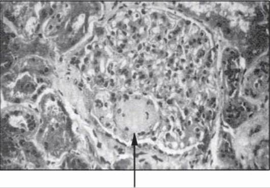
Fig. 79. Nodular glomerulosclerosis in DM (after C.R.W. Edwards and A.D. Boucher, 1994).
The earliest (one might say pre-diabetic) lesions involve changes in the basement membrane—the enlargement of existing partitions (branches) and The formation of new ones that depart from the thickened basement membrane and extend into the intracapillary space and the mesangial matrix. With the progression of DM, the thickness of these partitions increases, and mesangial cells lose their Organelles and Cytoplasm, giving rise to nodules. These structures are centrally located, which is considered the most specific feature of glomerular involvement in DM. At the same time, the capillary lumen may be locally narrowed or aneurysmally dilated.
Diffuse glomerulosclerosis is characterized by a uniform thickening of the Capillary Wall due to the deposition of material in the basement membrane and/or diffuse thickening and deposition within the mesangium.
Unlike these forms of glomerulosclerosis, "capsular drops" (homogenates located between the basement membrane and the capsule endothelium) and "fibrinoid caps" (eosinophilic lipid-containing formations in the lumen of the glomerular capillaries), which occur in DM, are not its specific hallmark.
Immunofluorescence studies have revealed that such hyaline-like deposits contain IMMUNOGLOBULINS of classes A, M, and G, fibrinogen, albumin, ceruloplasmin, and Complement. According to most researchers, the deposition of these substances and individual plasma components into the mesangium and the basement membrane of the glomerular capillaries is not of an immune nature (H.H. Marks, 1995).
Vascular lesions in DM are characterized by The Development of arteriolosclerosis and/or hyalinosis, particularly in the afferent glomerular arterioles, resulting in glomerular ischemia (Fig. 80).
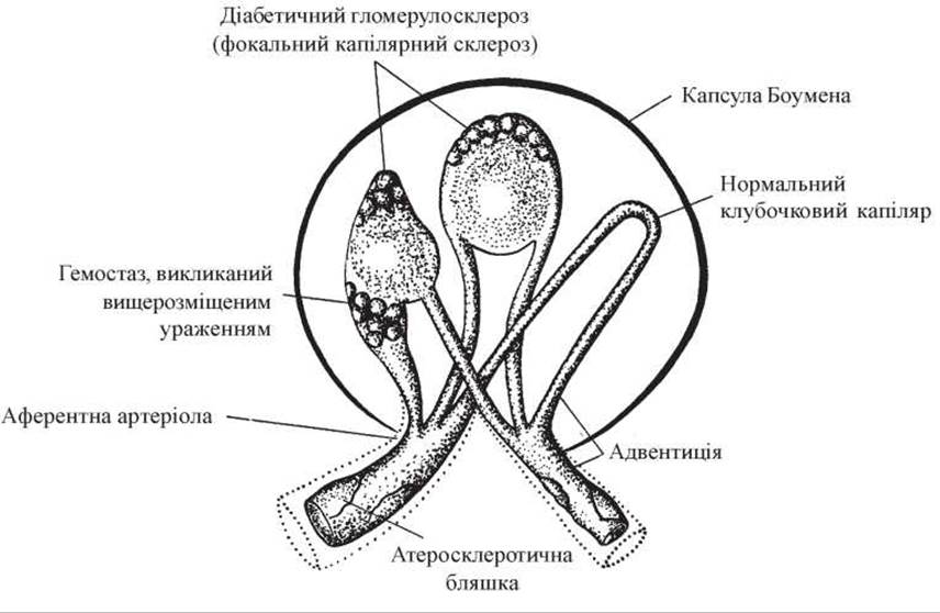
Fig. 80. Diabetic glomerulosclerosis: "capsular drops" and "fibrinoid caps", arteriolosclerosis (diagram after F.K. Mostofi et al., 1972).
Tubular lesions in DM consist of vacuolization of epithelial cells (Armanni-Ebstein cells) due to a reduction in their Glycogen content. A second pathognomonic sign of DM is the localization of these changes in the cortico-medullary junction area (in the terminal part of the straight portion of the proximal tubule). This may also be accompanied by thickening of the tubular epithelial basement membrane.
There are several hypotheses explaining The Mechanism of renal injury in DM.
The most widespread is the dysmetabolic hypothesis, which links all variants of renal pathology to disorders of carbohydrate, protein, and Lipid METABOLISM resulting from absolute or relative Insulin deficiency. This creates conditions for the prolonged Circulation of impaired glucose metabolism products in the Blood and their deposition in the vascular walls (E. Sim, 1985).
This process activates the glycosylation of various Proteins, including Hemoglobin. Under conditions of hyperglycemia, glucose is capable of forming bonds not only with hemoglobin but also with the Membrane Proteins of erythrocytes, blood serum, Collagen, neuronal myelin, Muscle Proteins, the proteins of renal glomerular basement membranes, and lens crystallin. It is also possible that insulin itself may undergo glycosylation, thereby losing some of its biological properties (A.S. Efimov et al., 1998). Glycosylated proteins, by interacting with leukocytes, trigger the release of cytokines, proliferation factors (M. Kirsten et al., 1990), and free radicals. The latter (which are essentially highly active oxidizers of biological substrates) act cytotoxically, causing Protein Denaturation and activating Lipid Peroxidation processes. Hyperglycemia, by also participating in the non-enzymatic glycosylation of serum Lipids, significantly potentiates the atherogenicity of Cholesterol and triglyceride fractions (M.V. Shestakova, 1999).
Alongside this, we should note the changes in blood levels characteristic of DM, namely an increase in total cholesterol, low-density lipoprotein cholesterol, and triglycerides, accompanied by a simultaneous decrease in lipolytic activity within the vascular wall (V.A. Galenok et al., 1990), which is similarly accompanied by impaired tissue oxygenation and the development of tissue Hypoxia.
However, it is also known that DN still develops in 30-40% of patients with DM despite meticulous and adequate Metabolic control. It is hypothesized that in these cases, the tendency to develop DN, as well as arterial hypertension, is associated with a genetic defect—an impairment of transmembrane cation transport. This defect manifests, in particular, as an increased rate of transmembrane sodium/lithium countertransport.
Other researchers link the development of DN to the hypothesis of a congenital defect in renal structures (A. Sahato et al., 1999). It is believed that 35-40% of the population is born with a reduced number of nephrons (approximately 800,000 in each kidney instead of 1.0-1.2 million). Under unfavorable conditions—such as DM—such a defect leads to hyperfiltration followed by Hypertrophy of the remaining nephrons, a reduced renal functional reserve, and the rapid development of arterial hypertension and progressive renal failure.
It should be noted that a reduced nephron number is most frequently found in children born with signs of intrauterine growth restriction and low birth weight. In adults, one should analyze The ratio of body weight in grams to body length in centimeters at birth; a value of 60 or less indicates a probable risk of intrauterine growth restriction and a reduced nephron count (K.M. Sergeeva et al., 2000).
According to another hypothesis, DM and its complications are viewed as a primary genetic defect of somatic cells that does not always depend on insulin deficiency (I.I. Dedov et al., 1998).
Active research continues into the genetic, immune, and autoimmune aspects of DM and the associated disturbances in microcirculation, hemostasis, and Fibrinolysis.
Recently, researchers have increasingly focused on the actual immune and autoimmune mechanisms of diabetic nephropathy (DN)—specifically, dysregulation of the Immune Response associated with certain HLA system loci, elevated levels of circulating immune complexes (CIC) and cytotoxic Antibodies in the blood, an inverted ratio of T-lymphocyte subpopulations, and the appearance of activated T-lymphocytes (S.F. Karagan et al., 1994; N.B. Lebedev et al., 1995). For instance, according to K.V. Salozhin et al. (1991), the majority of patients with Diabetes Mellitus (DM) exhibit a close association between DN and the overexpression of the interleukin-2 CD25 receptor as well as the class II DR antigen of the Major Histocompatibility Complex. These same patients showed an increased serum concentration of neopterin—an indicator that reflects the activation and proliferation of T-lymphocytes, macrophages, and monocytes and correlates directly with the severity of proteinuria (PU).
Activated T-lymphocytes produce a vascular permeability-enhancing factor and synthesize endoglycosidase (heparanase). The latter is an enzyme capable of degrading heparan sulfate Proteoglycans in the subendothelial matrix and the glomerular basement membrane (GBM) (K.V. Salozhin et al., 1991). Heparan sulfate largely determines the negative charge of the GBM and acts as a physiological anticoagulant. Thus, a decrease in the negative charge of the Glomerular Filtration GBM, a reduction in local anticoagulant potential, and an increase in vascular permeability resulting from immune alterations can significantly potentiate PU in DM.
When studying individual aspects of DN development, researchers also focus on systemic angiotensin II (AT-II). There is a well-substantiated hypothesis that AT-II is a pivotal pathogenetic factor in the onset and progression of DN. The reason is that a close relationship exists between insulin and the systemic renin-angiotensin-aldosterone system (RAAS), with potassium acting as the "connector" in this system. Under The Influence of insulin, potassium enters cells, thereby creating extracellular hypokalemia. The latter activates the RAAS, leading to an increased concentration of AT-II alongside a reduced production of aldosterone. Conversely, if the RAAS is activated primarily, the direction of these processes changes. In such cases, due to the enhanced synthesis of both AT-II and aldosterone, hypokalemia occurs, which stimulates the release of glucose-induced insulin, resulting in hyperinsulinemia. Prolonged hyperinsulinemia reduces the number of insulin receptors, subsequently leading to insulin resistance (Cs. Farsang, 1997).
Data from recent studies indicate that an important role in the onset and progression of DN also belongs to the local renal (glomerular and tubular) RAAS (K.D. Burns et al., 1993). The central mechanism of DN development is AT-II-mediated vasoconstriction of the efferent glomerular arteriole. This is accompanied by an increase in intraglomerular pressure (and consequently, in the glomerular filtration rate [GFR], as a response reaction to elevated blood osmolality under insufficient correction of hyperglycemia), the opening of additional Pores in the GBM, and The Emergence of microalbuminuria (MA).
The AT-II-induced onset and progression of PU are considered the primary pathogenetic mechanism and marker of renal injury in DM.
PU itself, both directly and through The stimulation of hyperlipidemia, is a leading cause of focal segmental glomerulosclerosis/hyalinosis and, consequently, the onset and progression of renal failure (RF).
The increase in intraglomerular pressure induced by AT-II and hyperglycemia stimulates mesangial matrix expansion, activates cytokines and potential mediators of sclerosis/hyalinosis—endothelium, transforming growth factor-beta (TGF-beta), and platelet-derived growth factor (PDGF) (M.S. Komadenko et al., 2000). These same factors are synthesized in enhanced amounts in the presence of hypertension (HTN). The combination of hyperglycemia and activated local AT-II synthesis acts as a mediator of enhanced sodium reabsorption in the tubules, which contributes to the onset or stabilization of HTN. This combination also inhibits The activity of intracellular proteinases responsible for the so-called small protein turnover, as well as the expression of nitric oxide synthase. All of this leads to the dysregulation of enzyme systems involved in cellular metabolism, excretion, and secretion processes within the tubules.
From this, at least two Conclusions follow: first, changes in local AT-II activity may not be reflected at the systemic level, making the determination of plasma renin activity uninformative for assessing the state of the renal RAAS; second, the adverse hemodynamic and metabolic effects of AT-II on the kidneys in DM are a direct indication for their therapeutic inhibition.
Furthermore, it was established that the activity of local AT-II synthesis in each individual patient with DM depends on the polymorphism of the ACE Gene, which plays a major role in regulating renal hemodynamics.
An insertion/deletion (I/D) polymorphism in intron 16 has been described for the ACE gene, containing either an extended (I) or a normal (D) DNA fragment (I.M. Kutyrina et al., 1999). The ACE gene containing two insertions (II genotype) is associated with a low concentration of the enzyme itself and blood AT-II; the gene lacking the insertion (DD genotype) encodes very high AT-II synthesis. Numerous studies have also proven that DN develops much less frequently or not at all in DM patients who carry the II genotype of the ACE gene. In other words, unlike the DD genotype, the II genotype of the ACE gene possesses nephroprotective properties; however, the DD genotype realizes its nephrotoxic effect only under conditions of inadequate hyperglycemia correction (M.V. Shestakova, 1998).
The earliest sign of DN is an increase in GFR (exceeding 150 mL/min), followed by MA. MA, as an early pathological sign of DN, is the urinary excretion of albumin ranging from 30 to 200 mg/day. M.V. Shestakova et al. (1991) established that a GFR exceeding 150 mL/min is combined with a complete absence of filtration reserve in 67% of DM patients. This means that all functioning nephrons are operating at maximal filtration load, and there are no non-working (silent) nephrons. Such a chronically heavy load leads to rapid "wear and tear" of the renal glomerular apparatus, followed by a progressive decline in GFR (V.A. Zhuk, 1998). MA is detected in 83% of patients with hyperfiltration, which further Supports the hypothesis of W. Brenner et al. (1996) regarding the damaging effect of high intraglomerular hydrostatic pressure gradients on the glomerular filter.
The Diagnostic significance of identifying MA rather than PU in DM lies in the fact that renal function impairment at this stage remains reversible. The mechanism of MA at this stage of DN differs fundamentally from the mechanism of PU, which develops later.
In the Cytology/cytology/16.html">Early stages of DM development, tubular reabsorption can fully compensate for the increased filtration of albumins. However, over time, high blood glucose concentrations, the presence of Ketone Bodies, and an increased urine flow rate through the tubular portion of the nephron (high diuresis) reduce the efficiency of reabsorption due to the impairment of tubular energy-dependent transport systems, leading to the appearance of PU, which is detected by standard clinical Methods. Such PU indicates the development of irreversible renal changes, even if blood creatinine levels remain within the normal range. According to V.Ya. Plotkin (1988), PU as a sign of irreversible renal damage subsequently activates the lysosomal-vacuolar apparatus of proximal tubular nephrocytes on its own, which is manifested by an increased production of lysosomal Enzymes. These enzymes are deposited beneath the apical and basal Plasma Membranes, followed by enzymuria, damage to the GBM, and the interstitium. In addition, intracellular structures degrade via autophagy, and the process culminates in interstitial fibrosis with the compression of peritubular vessels.
It must be emphasized that from the moment PU appears, the pathological process in the kidneys takes on a course independent of the metabolic disturbances that triggered it (M.V. Shestakova et al., 1999). In such cases, even prolonged correction of hyperglycemia is no longer able to stop the progression of renal failure, and an increase in blood creatinine levels to 0.2 mmol/L is a sign that 50% of renal glomeruli are functioning insufficiently. From the onset of stable PU, the GFR progressively declines (on average by 1 mL/min/month), and uremia develops within 6–7 years. The above holds true for both types of DM.
Thus, urinary albumin excretion is "...a powerful predictor of both DN and cardiovascular morbidity and mortality in patients with insulin-dependent and non-insulin-dependent types of DM..." (Proceedings of the International Congress of Nephrology, Sydney, Australia, 1997).
An early predictor of diabetic nephropathy, alongside MA, is an increased urinary excretion of enzymes—N-acetyl-beta-D-glucosaminidase, beta-galactosidase, and Alanine aminopeptidase (E. Kohler et al., 1996).
The onset of hypertension, sometimes accompanied by Nephrotic Syndrome (NS) in some patients, is a sign of the advanced stage of DN. It should be noted that hypertension can exist as a pre-existing condition or develop during the course of DN formation. According to I.I. Dedov et al. (2001), Glomerulonephritis is histologically detected in every third patient with type 2 DM and every tenth patient with type 1 DM, while in 10% of cases, hypertension against the background of DN is associated with other diseases. In both cases, this is an unfavorable factor (deterioration of effective renal blood flow due to hyalinosis and sclerosis of renal and other regional vessels) and requires correction. The relationship between hypertension and PU and their role in the progression of renal failure are illustrated in Figure 81.
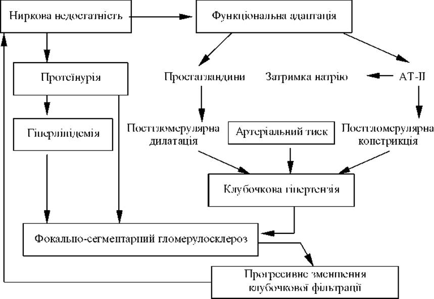
Fig. 81. The Role of hypertension and PU in the mechanism of renal failure progression.
The presence of MA, PU, hypertension, and edema serves as an indicator for determining the stage of DN.
Various classifications of DN stages exist.
The Classification of DN by H. Keen et al. (1981) involves distinguishing functional, structural, and clinical stages.
The classification of DN stages by V.R. Klyachko (1974) is based primarily on clinical criteria: Stage I (prenephrotic)—transient PU, normal blood pressure, unimpaired renal function, diabetic retinopathy is detected; Stage II (nephrotic)—edema, elevated blood pressure, persistent PU, hypercholesterolemia, hypo- and isosthenuria; Stage III (nephrosclerotic)—signs of stage II chronic renal failure + "improvement" in the course of DM—decreased hyperglycemia and glucosuria.
A more modern classification of DN is that by C.E. Mogensen (Table 6).
DN is a risk factor for soft tissue and Urinary Tract infections, tuberculosis, and candidomycosis. When a urinary tract infection is joined, pyelonephritis, apostematous nephritis, and papillary necrosis may develop. In 10–15% of cases, DN is accompanied by the development of NS with all the Characteristic Features of this condition and the rapid onset of diuretic resistance.
Management of diabetic nephropathy. Modern approaches to treating DN are based on mitigating The impact of its risk factors, which include:
- presence of systemic arterial hypertension;
- inadequate hyperglycemia control;
- lipid metabolism disorders;
- hemocoagulation system abnormalities with a tendency toward hypercoagulation;
- age-related immune alterations;
- smoking;
- alcohol consumption.
First of all, it is necessary to emphasize The Need for adequate, effective, and stable correction of hyperglycemia using diet, oral hypoglycemic agents, or insulin. For instance, in patients with poor glycemic control, the risk of microalbuminuria increases 4-to-5-fold (D. Eldstein et al., 1992), although it remains uncertain whether meticulous control of this parameter can prevent the development of nephropathy once distinct symptoms of renal impairment are already present. Under conditions of insufficient metabolic control, significant and independent predictors of microalbuminuria development in type 1 diabetes mellitus patients include the presence of systemic hypertension and smoking; in type 2 diabetes mellitus patients, they include maternal hypertension, the severity of diabetic retinopathy, overweight, and the patient's age at the onset of the disease (V.A. Dobronravov, 1998).
Equally important is the normalization (!) of systemic blood pressure levels (G. Bakris et al., 1992; H. Parving et al., 1994, J. Charles et al., 1995) and the improvement of renal hemodynamics. The 15-year UKPDS study proved that a 10 mm Hg reduction in blood pressure decreases diabetes-related mortality by 32%. In 1997, the Sixth Report of the Joint National Committee on Prevention, Detection, Evaluation, and Treatment of High Blood Pressure in the US established that for patients with diabetes mellitus, the critical blood pressure threshold across all age groups—above which antihypertensive therapy should be initiated (non-pharmacological down to a blood pressure of 140 and 90 mm Hg, and pharmacological above these values)—is 130 and 85 mm Hg. Even a minor elevation in these figures increases the risk of cardiovascular events by 35% (M.V. Shestakova, 2000).
Considerable importance is attached to dietary protein restriction in patients with type 1 diabetes mellitus (down to 0.6 g/kg of body weight/day at the stages of persistent proteinuria and chronic renal failure) (A. Grenfell et al., 1995).
Among the known classes of antihypertensive drugs for diabetes mellitus, The Use of thiazide Diuretics and beta-blockers should be avoided due to their adverse metabolic effects (hypokalemia, increased tissue resistance to insulin, hyperuricemia, worsening of blood rheological properties, masking of hypoglycemia symptoms, negative impact on lipid metabolism, etc.).
Regarding diuretics, patients with diabetes mellitus are recommended to use Arifon or Aquaphor; among beta-blockers, cardioselective drugs that do not mask the manifestations of hypoglycemia are preferred, such as atenolol, metoprolol, and betaxolol. Concor (bisoprolol) can also be recommended as a beta-blocker with the highest beta-1-selectivity (T. Mendgen et al., 1997). The use of Concor at a dose of 5-10 mg/day across all age groups provides 24-hour blood pressure reduction; it does not affect blood lipid fractions, Glucose metabolism in type 2 diabetes mellitus patients, or bronchial beta-2-receptor function, and its administration does not require dosage adjustment in patients with renal or hepatic impairment (R. Fogari et al., 1990; H.G. Gieseke et al., 1990).
Certain hopes are pinned on agents capable of inhibiting alpha-1-adrenoceptors (doxazosin, terazosin) and beta-2-adrenoceptors (labetalol, carvedilol). Alpha-blockers not only avoid disrupting lipid metabolism but actually reduce serum atherogenicity and decrease insulin resistance. The use of carvedilol is particularly effective when diabetes mellitus is combined with hypertension and Heart Failure.
Among the three classes of calcium antagonists, preference is given to phenylalkylamines and benzothiazepines (verapamil, diltiazem), followed secondly by third-generation dihydropyridines. Along with their hypotensive and antiproliferative effects, these drugs possess a relatively low profile of adverse reactions. A fundamentally novel agent is mibefradil, information on which is provided in the section «Renal Arterial Hypertension».
ACE inhibitors. The use of this group of drugs results in the normalization of creatinine clearance, improvement of effective renal blood flow, reduction of proteinuria and hypercreatininemia (M. Ravid et al., 1993; E.L. Lewis et al., 1993; L. Bohlen et al., 1994).
Given the crucial role of hyperfiltration in the Pathogenesis of diabetic nephropathy, one of the promising strategies for preventing its progression is precisely the elimination of hyperfiltration and the attempt to restore filtration reserve. Beta-blockers, like other classes of antihypertensives such as calcium antagonists, while having a hypotensive effect comparable to that of Renitec, have a lesser impact on the severity of proteinuria in diabetes mellitus (R. Slataper et al., 1993).
To substantiate this, let us cite the results of the Appropriate Blood Pressure Control in Diabetes (ABCD) trial: in two randomized groups of type 2 diabetes mellitus patients with hypertension, the use of an ACE inhibitor (enalapril) and a calcium antagonist (nisoldipine), despite an equivalent blood pressure-lowering effect, was accompanied by a significant difference in their impact on proteinuria and quality of life (5-year follow-up period). Notably, myocardial infarction occurred in 10.6% of the diabetes patients taking nisoldipine (compared to 2.1% of those taking enalapril), cerebrovascular events in 2.1% and 0.2% respectively, and death from other cardiovascular causes in 4.2% and 2.1% (B.R. Bloom et al., 1998).
Let us also quote Prof. K. Benedict (USA): «In contrast to ACE inhibitors, comparative studies—specifically those utilizing calcium channel blockers—have begun to yield data indicating that this class of antihypertensive agents exerts minimal protective effects on Blood Vessels and target Organs» (20th Congress of the European Society of Cardiology, Vienna, 1998).
Extensive foreign clinical experience with ACE inhibitors has led to the Conclusion that this class of drugs must be utilized across various stages of diabetic kidney disease (I.M. Kutyrina et al., 1996; I. Dedov, M. Shestakova, 1996; P.M. Ter Wee et al., 1993; E.J. Lewis et al., 1993). We favor the viewpoint of the authors of the chronic renal failure progression model, B.M. Brenner et al. (1999), who consider it optimal to initiate ACE inhibitor therapy even before the onset of diabetic nephropathy, although N.O. Pertseva (2000) indicates that active treatment of diabetic nephropathy should commence only at stage III.
Among the ACE inhibitor class, only Renitec and Capoten have undergone multicenter, placebo-controlled trials proving their efficacy in diabetes mellitus (M.V. Shestakova, 1998). Renitec is prescribed both in the early stages of diabetic nephropathy development (with normal blood pressure and microalbuminuria levels within 50-100 mg/day) and at the stage of established diabetic nephropathy, even with signs of hypercreatininemia. In the latter case, the dose of Renitec depends on the creatinine clearance level—at a creatinine clearance below 30 mL/min, Renitec is prescribed at 2.5-5 mg/day. The clinical effect persists for up to 6 months after drug discontinuation, after which proteinuria begins to rise again. This dictates the necessity of either continuous administration of Renitec starting from the early stages of diabetic nephropathy or a cyclical treatment regimen (2-3 months of therapy followed by an equally long pause).
Furthermore, we should note the role of ACE inhibitors as cornerstone medications for the treatment of heart failure. Considering that cardiovascular mortality accounts for up to 80% of overall mortality in type 2 diabetes mellitus patients, the rationale for prescribing ACE inhibitors in diabetes becomes readily apparent.
Data on The rate of creatinine clearance decline during the treatment of diabetic patients further support the use of ACE inhibitors. It was demonstrated (M.V. Shestakova, 1998) that the rate of creatinine clearance decline in diabetic patients receiving no antihypertensive therapy was 10-14 mL/year; with beta-blockers and diuretics, it was 5-6 mL/year; and with ACE inhibitors, it was 2.5-3 mL/year. In other words, the progression rate of diabetic nephropathy is halved with conventional antihypertensive therapy (beta-blockers and diuretics) and reduced by 5-to-6-fold with ACE inhibitors.
Approaches to the use of ACE inhibitors in diabetes mellitus combined with cardiovascular pathology are outlined in the methodological guidelines by M.I. Shved et al. (1999).
The renoprotective effects of ACE inhibitors have provided the theoretical foundation for investigating the clinical efficacy of angiotensin II type 1 receptor blockers in diabetic nephropathy. Clinical and experimental studies have demonstrated that their hypotensive and antiproteinuric effects are driven by concurrent alterations in renal hemodynamics (O. Didych, 1998).
It has been proven that blocking type 1 angiotensin II receptors can significantly alter the progression rate of diabetic kidney disease in both experimental and clinical settings. For instance, in experimental diabetes mellitus, the progression of proteinuria was substantially slowed down following 1 to 2 months of administration of potassium losartan (Cozaar) compared to the placebo group. According to histological findings, this treatment was associated with less pronounced glomerular damage than in the control group of animals with diabetes (A. Remuzzi et al., 1993).
Another advantage of Cozaar is that its clearance is independent of renal function. The RENAAL study has recently been completed, and its analysis will provide a definitive answer as to whether the blockade of type 1 AT-II receptors is the most effective treatment for patients with diabetic nephropathy.
To normalize basement membrane metabolism, prevent mesangial proliferation, restore the negative charge of the basement membrane, and reduce its permeability, glycosaminoglycan therapy (sulodexide, Vessel Due F) is successfully employed. Its antiproteinuric effect has been observed in 89% of patients with diabetes mellitus (I.I. Dedov, 1997).
The use of the latest generation of COX-2 inhibitors (celecoxib, rofecoxib, R. Komers et al., 2001) in diabetes mellitus should also be considered promising, as mentioned above.
Patients with type 2 diabetes mellitus (accounting for 85–90% of all diabetes cases) face an exceptionally high concurrent risk of developing cardiovascular diseases.
It is widely recognized that the clinical manifestations of cardiovascular involvement in diabetes are driven by the development of atherosclerosis in large and medium-sized vessels. Furthermore, atherosclerosis occurs much earlier in these patients compared to age-matched non-diabetic controls, and it is characterized by significantly greater severity and widespread involvement across multiple vascular beds (E.I. Sokolov, 1996). We also emphasize the high prevalence of atherosclerotic lesions in women with diabetes even before the onset of menopause.
Risk factors for atherosclerosis in diabetes include arterial hypertension, obesity, dyslipidemia, chronic hyperglycemia, and hyperinsulinemia. Other (nontraditional) risk factors for coronary artery disease in diabetes include decreased serum levels of albumin, fibrinogen, von Willebrand factor, and factor VIII activity, as well as leukopenia. Notably, adequate glycemic control has only a minor impact on these associations (ARIC study, I. Saito et al., 2000).
Against the backdrop of diabetes mellitus, the Adverse effects of hypertension and dyslipidemia become particularly aggressive (E.N. Amosova, 1996; E.R. Abrandrova et al., 1998; M. Laakso, 1993, 1995; J. Stamler et al., 1993).
As early as 1988, J. Diamond hypothesized that the mechanism of glomerular Cell injury in hyper- and dyslipidemia is analogous to the formation of atherosclerotic plaques in blood vessels. Both processes (atherosclerosis and glomerulosclerosis) are rooted in cellular proliferation and the expansion of Connective Tissue structures, accompanied by lipid accumulation leading to intracellular and extracellular deposits. For instance, M.V. Shestakova et al. (1993) found lipid-containing inclusions in virtually all renal glomerular cells in diabetes.
Dyslipidemia (frequently combined with arterial hypertension) occurs in 60–70% of patients with type 2 diabetes and is characterized by elevated blood levels of low-density lipoprotein cholesterol and triglycerides, alongside reduced high-density lipoprotein cholesterol.
According to the recommendations of the American Diabetes Association (1988) and the National Cholesterol Education Program (1994), optimal lipid levels in type 2 diabetes should be as follows: total cholesterol up to 5.2 mmol/L, triglycerides up to 1.7 mmol/L, low-density lipoprotein cholesterol up to 3.4 mmol/L, and high-density lipoprotein cholesterol above 1.1 mmol/L, with a body mass index up to 25 kg/m2 for men and 24 kg/m2 for women. It can also be argued that as life expectancy increases—even with better disease management and a consequent reduction in the prevalence of macro- and microangiopathies—the incidence of cardiovascular complications will continue to rise among patients with type 1 diabetes as well.
Pharmacological correction of hyperlipidemia is initiated when low-density lipoprotein cholesterol levels exceed 3.4 mmol/L despite adequate glycemic control and an appropriate lipid-lowering diet (National Cholesterol Education Program: Second Report of the Expert Panel on Detection, Evaluation, and Treatment of High Blood Cholesterol in Adults (Adult Treatment Panel II) // Circulation. — 1994. — N 85. — P. 1329–1445).
A revolutionary milestone in modern cardiology was the development of a new class of lipid-lowering agents: inhibitors of 3-hydroxy-3-methylglutaryl-coenzyme A reductase, known as statins. These agents block cholesterol synthesis at a very early stage—specifically, The conversion of 3-hydroxy-3-methylglutaryl-CoA to mevalonic acid (Fig. 82).
In this scenario, the precursor molecules that ultimately form cholesterol (such as acetoacetyl-CoA and acetyl-CoA) can be safely utilized by the body in other metabolic pathways and cycles.
This precisely explains the low incidence of side effects associated with this class of drugs. In contrast, blocking cholesterol synthesis at a late stage (e.g., the conversion of lanosterol to cholesterol using triparanol) is accompanied by pronounced toxicity because the body cannot utilize lanosterol as an alternative to acetyl-CoA.
The high efficacy of statins (specifically simvastatin, trade name Zocor) was demonstrated in the now-historic 4S study (N.A. Gratsiansky, 1996, 1998). Subgroup analyses of these data revealed an even higher efficacy of statins in patients with type 2 diabetes compared to the overall study population.

Fig. 82. Stages of Cholesterol Biosynthesis and The Site of Action of statins.
For instance, the use of Zocor demonstrated a 55% reduction in the incidence of fatal and non-fatal myocardial infarction in patients with type 2 diabetes. It has been calculated that out of 100 patients with type 2 diabetes over a 6-year follow-up period, Zocor can prevent myocardial infarction in 24 cases, against a projected 49 cases (S.V. Moiseev, 1996). Therefore, lipid-lowering therapy with simvastatin (Zocor, 10–20 mg/day) should be an essential component of comprehensive, long-term (multi-year) management in patients with both types of diabetes. This is justified by the adverse impact of dyslipidemia on both the rate of cardiovascular complications and the Structure and function of the nephron. Practical guidelines for the use of statins in the Primary and secondary prevention of atherosclerosis and its complications are outlined in the methodological recommendations of the Ministry of Health of Ukraine (L.T. Mala et al., 1998).
Recently, atorvastatin (Lipitor, Pfizer, USA)—another 3-hydroxy-3-methylglutaryl-CoA reductase inhibitor—has been used to treat hyperlipidemia. Unlike simvastatin, which is classified as a prodrug, atorvastatin is active on its own and does not require in vivo conversion into an active form. According to the CURVES study (P.H. Jones et al., 1998) and the TARGET TANGIBLE Trial (W. März et al., 1999), atorvastatin exhibits greater lipid-lowering activity than other statins without a corresponding increase in adverse side effects. The efficacy of atorvastatin in patients with non-insulin-dependent diabetes mellitus has already been established in numerous studies (S.E. Kozlov et al., 1999; J.D. Best et al., 1996; J. Armitage et al., 1999; M. Davis et al., 2000).
In the stage of advanced chronic renal failure, patients with diabetes mellitus typically succumb within a few months. In such situations, three treatment options are available: renal transplantation, hemodialysis, and peritoneal dialysis.
Due to the demand for hemodialysis or renal transplantation, diabetes has recently taken the leading position in the USA (J.D. Philipson et al., 1996) and ranks second to third in developed European countries (A.E. Raine et al., 1993). In the USA, 65–85% of diabetic patients with end-stage renal disease initiate chronic hemodialysis (E.E. Berger et al., 1991). It is projected that by 2005, patients with diabetes will account for one-third of all cases requiring hemodialysis. However, the Management of chronic renal failure via hemodialysis or kidney transplantation in these patients remains practically inaccessible in Ukraine.
Hemodialysis in diabetic nephropathy should be initiated when the glomerular filtration rate drops to 10 mL/min and creatininemia ranges between 0.442 and 0.550 mmol/L (B.A. Stetsyuk et al., 1999). Hemodialysis prolongs the life of such patients by an average of 3 to 4 years, while the 2-year survival rate following kidney transplantation is 70% (E. Friedman, 1992).
A logical extension of indications for renal transplantation in patients with type 1 diabetes is simultaneous beta-cell transplantation (A.S. Efimov et al., 1990; D. Abendroth et al., 1990; B.N. Berker et al., 2000), the goal of which is to reduce the patient's daily insulin requirement and to halt or delay the progression of micro- and macrovascular angiopathies. Unfortunately, Pancreas transplantation does not arrest the progression of, nor does it reverse, diabetic retinopathy, cardiomyopathy, or peripheral vascular disease (E. Ritz et al., 1995).
Thus, it can be concluded that despite the high risk of onset and progression of diabetic nephropathy, the practicing physician today has effective tools at their disposal to prevent the onset or alter the course of this formidable complication in diabetic patients.
Last update: 08/08/2026
Editorial and Educational Adaptation: This material has been compiled based on the primary/original source text. The project team performed an editorial review, corrected technical inaccuracies, structured sections, and adapted the content for an educational format.
What was processed:
- elimination of formatting defects (OCR errors, structural breaks, corrupted characters);
- editorial organization of content;
- standardization of terminology in accordance with academic sources;
- verification of factual statements against the original source text.
All mentions of the author, publication year, and origin of the primary text have been preserved in accordance with the source.