Nephrology for Family Physicians - O.I. Bakaliuk 2003
Primary Semiotics of Kidney Diseases
Renal Arterial Hypertension
When establishing the genesis of arterial Hypertension (AH), it should be borne in mind that neither its severity, stability, the presence of hypertensive crises, nor the patient's age provide absolute grounds for determining its Etiology. The decisive role belongs solely to the rational Selection of necessary diagnostic Methods for each individual patient and the correct interpretation of their results.
Of paramount importance in The Study of renal arterial hypertension (RAH) are the works of H. Goldblatt et al. (1934), who developed an experimental model of this condition and proved the crucial role of the renin-angiotensin-aldosterone system (RAAS) in its Pathogenesis. Subsequently, E.M. Tareyev (1936, 1948) highlighted Structure/19.html">The Importance of high Blood volume in the genesis of hypertensive syndrome in acute Glomerulonephritis, while A.C. Guyton et al. (1972) demonstrated that sodium retention is the universal mechanism underlying AH of any origin, including renal.
In recent decades, there has been a notable increase in the incidence of RAH. It belongs to the group of secondary (symptomatic) forms of AH, accounting for about 70% of them. Within this group, Chronic Pyelonephritis is responsible for 40.5% of RAH cases (A.V. Gordeyev et al., 1990). In Ukraine, more than 300,000 individuals suffer from secondary AH (O.P. Svishchenko, 1997).
To gain a more comprehensive understanding of the development patterns of RAH, let us briefly outline the levels and mechanisms regulating blood pressure (BP) in general.
The stability of BP levels and the appropriateness of its response to internal and external factors require a precise interplay of neural, humoral, and local (tissue) mechanisms, the ultimate goal of which is to ensure an adequate Blood supply to each organ according to its metabolic demands. Under resting conditions, this is achieved primarily through the functioning of local mechanisms. During physical exertion or stress, effective coordination and optimal blood flow distribution are impossible without the involvement of the Central Nervous system and systemic humoral regulation.
The BP regulation system is multiloop and operates via positive and negative feedback mechanisms. The leading role in this process belongs to the volume and Osmotic Pressure of the extracellular fluid, as well as the concentration of electrolytes within it. Relying mainly on the works of V.Z. Netiazhenko et al. (1997), we will outline the Physiological foundations of BP regulation.
The neurogenic loop of BP regulation is represented by three links: afferent, central, and efferent.
The most important afferent signal is information originating from the intrinsic reflexogenic zones of The Cardiovascular system. These include receptors in the aortic arch, sinocarotid zones, the right side of The Heart, and the Pulmonary Circulation vessels. All of them respond to the deformation of the vascular wall or cardiac chambers. Information from the sinocarotid zones is transmitted to the center via myelinated fibers (which play a predominant role in regulating rapid BP changes) and unmyelinated fibers (which have low sensitivity and respond to mean BP over a prolonged period).
The pressor and depressor structures of the Medulla Oblongata and Hypothalamus, along with the Cerebral Cortex, constitute the central BP regulation system. This system is exceptionally complex and Functions via feedback mechanisms.
All central nervous system zones involved in BP regulation are interconnected by numerous Neural Pathways. Alongside monoamines (norepinephrine, epinephrine, dopamine, serotonin, imidazoline, guanethidine), various Peptides—such as angiotensin, substance P, vasopressin, and opioid peptides—act as Neurotransmitters in these Neurons.
The efferent link of the neurogenic loop consists of vasomotor sympathetic neurons located in the thoracolumbar Spinal Cord. Their axons are typical cholinergic nerves that emerge from the spinal cord as part of the anterior roots and proceed to the paravertebral sympathetic ganglia. Postganglionic nerve fibers branch extensively to form a wide network of nerve endings at the border of the tunica adventitia and the muscular layer of Blood Vessels.
Impulse transmission to the periphery is mediated by adrenergic neurotransmitters—epinephrine and norepinephrine. The latter act on adrenergic receptors (single lipoprotein complexes) that exhibit relative organ Specificity and are divided into two groups: alpha (1 and 2) and beta (1 and 2) receptors. Alpha-receptors are concentrated predominantly in the Vessels of the Skin and intestines, whereas beta-receptors are found in the vessels of adipose tissue, Lungs, heart, and the juxtaglomerular apparatus. Both types of receptors are present in Skeletal Muscle vessels. In general, stimulation of alpha-1 receptors leads to the contraction of smooth muscle in the vascular wall and vasoconstriction, while activation of beta-2 receptors results in muscle relaxation and vasodilation; stimulation of beta-1 receptors located in the myocardium produces positive chronotropic and inotropic effects. However, the actual relationships among these receptor subpopulations are even more complex. They have been found to be located both on nerve endings (presynaptic adrenoceptors) and directly on the membranes of smooth muscle Cells (postsynaptic adrenoceptors). Excitation of presynaptic alpha-2 receptors activates feedback mechanisms that inhibit norepinephrine release, whereas the binding of norepinephrine to postsynaptic alpha-1 receptors maintains appropriate vascular tone.
The pressor humoral system is represented by the sympathoadrenal system, RAAS, endothelin system, thromboxane A2, PGF2, neuropeptide Y, and atrial natriuretic peptide inhibitors; the depressor system comprises the kallikrein-kinin system, PGI2, PGF2alpha, atrial natriuretic peptide, medullipin system, adrenomedullin, nitric oxide, Insulin, histamine, acetylcholine, and adenosine.
Let us briefly characterize the systems most crucial for understanding modern approaches to the Treatment of RAH.
The Role of the sympathoadrenal system in BP regulation is well known and requires no commentary.
The Main Components of the RAAS include Enzymes (renin, peptidases, kininase II, chymase, tonin, cathepsin G, tissue plasminogen activator, chymostatin-sensitive activator) and Hormones—angiotensin I (AI) and angiotensin II (AII). The classic pathway of AII generation has been studied in considerable detail. Renin is a proteinase and a typical product of the juxtaglomerular apparatus, characterized by massive adrenergic innervation and narrow functional specialization. Its cellular reserves include mesangiozytes and smooth muscle Cells of the wall of the renal afferent glomerular arteriole, which under certain conditions can transform into typical epithelioid cells. The vascular portion of the juxtaglomerular apparatus responds to BP Changes in the afferent arteriole, while the tubular portion responds to changes in sodium and chloride concentrations in the urine. Its morphological antagonists are the interstitial cells of the renal medulla, nephrocytes of the collecting ducts (which secrete Prostaglandins), and the distal nephron (which secretes kinins).
Stimulators of renin production include renal ischemia, changes in systemic or intraglomerular BP (blood loss, Shock, decreased blood volume), shifts in sodium balance toward hyponatremia (e.g., due to diuretic use), and increased activity of the sympathoadrenal system, particularly the renal neuroadrenergic system.
The conversion of AI to AII occurs with the participation of angiotensin-converting enzymes (ACE), among which peptidases and kininase II hold a prominent place (O.A. Gomazkov, 1997). The lungs serve as the primary site of ACE synthesis (Fig. 15).
In addition, AII can be formed directly from angiotensinogen via catalytic reactions (mediated by tonin, cathepsin G, and tissue plasminogen activator). Under METABOLISM/18.html">The Influence of aminopeptidase A, AII is converted into angiotensin III, and the latter, with the participation of aminopeptidase N, into angiotensin IV. Another peptide, angiotensin (angiotensin 1–7), may also be cleaved from AI during AII formation. All of these compounds, except AII, possess relatively low biological activity (V.Z. Netiazhenko et al., 1997), although it should be noted that angiotensin 1–7 exhibits moderate vasodilatory and natriuretic effects.
Recently, the so-called non-enzymatic (alternative) pathway of AII formation—that is, operating independently of ACE—has been discovered (D.V. Preobrazhensky et al., 1997; J. Robertson et al., 1997). The leading role in converting AI to AII via the non-enzymatic pathway belongs to chymase, a Chymotrypsin-like protease (Fig. 16), as well as the enzyme CAGE (chymostatin-sensitive angiotensin II-generating enzyme).
Class="center">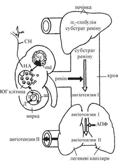
Fig. 15. Schematic of AII formation: aa — afferent arteriole; SN — sympathetic nerve; md — macula densa; NE — norepinephrine; D — wall tension; r — radius; T — tone; JG Cell — juxtaglomerular cell.
To give an example, 1 molecule of chymase converts 160 molecules of AI into AII per second, whereas other ACE pathways convert 8.3 molecules. Chymase is located in the myocardial Extracellular matrix and is closely associated with AII receptors on cardiomyocytes (M.P. Kiknadze, 1995; H. Urata et al., 1994). It is also found in specific secretory vesicles of coronary endothelial cells (the contents of these vesicles are released preferentially into adjacent cells rather than the vascular lumen), as well as in other Organs and systems maintaining cardiovascular Homeostasis, such as the vascular adventitia, atria, ventricles, and Adrenal Glands. Chymase activity has also been detected in the Uterus, Tonsils, Esophagus, Stomach, intestines, and skin.
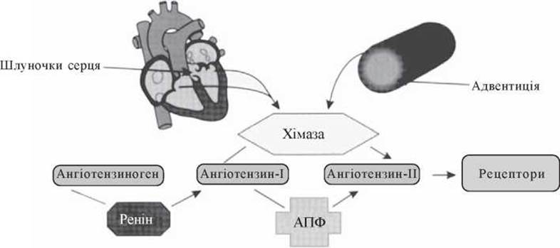
Fig. 16. Pathway of AII formation via chymase.
To put it briefly, the non-enzymatic pathway of Ang II formation cannot be blocked by the administration of ACE inhibitors.
Roughly 70% of the Ang II found in the arterial wall is produced via CAGE. Furthermore, enzymes that catalyze the direct synthesis of Ang II from angiotensinogen—bypassing The formation of Ang I altogether—have been identified.
Recent studies have also demonstrated that RAAS genes and messengers, including its Polypeptides and enzymes, are present in the Brain, the cardiac conduction system and autonomic nerves, reproductive organs, leukocytes, alveolar macrophages, and monocytes. This is a major breakthrough of recent years, as it points to the existence of local RAAS In addition to the systemic RAAS (E.H. Nesukai, 1998; H. Grobecker et al., 1993), highlighting the role of local Ang II synthesis in maintaining adequate Regional Blood Flow and metabolism independently (or more precisely, almost independently) of systemic Ang II levels.
In Tissues, Ang II increases the vascular concentration of potential vasoconstrictors and agonists of vascular smooth muscle cell proliferation and migration. In endothelial cells, it stimulates The production of endothelium-derived contracting factors (EDCFs)—endothelin-1, -2, and -3—which are potent pressor agents with proliferative properties. Local Ang II production is also recognized as a key driver in stimulating various cytokines, such as selectin, and macrophage migration into the endothelium, where they subsequently transform into foam cells. Locally synthesized Ang II acts as a catalyst for Lipid Peroxidation, a stimulus for enhanced Collagen synthesis in the vessel wall, and a factor increasing tissue thrombogenic potential—all of which are critical elements in the pathogenesis of atherosclerosis (C.M. Ferrario et al., 1996).
The cellular effect of Ang II is The stimulation of genes that control cell mass, leading to hypertrophy and proliferation, whereas the increase in intracellular calcium concentration results in vasoconstriction.
An important target for Ang II is the Kidneys. Known renal effects of Ang II include (Yu.M. Sirenko, 1995; F.N. Hutchison et al., 1992; P. Emsberger et al., 1996):
- vasoconstriction of the renal vessels, particularly the glomerular efferent arteriole, which leads to a decrease in cortical blood flow and an increase in renal vascular resistance. This results in reduced effective renal plasma flow coupled with increased intraglomerular pressure, sodium retention, and the onset of proteinuria;
- contraction of glomerular mesangial cells, leading to a reduced glomerular surface area and a lower ultrafiltration coefficient (Kf);
- a direct tubular effect—stimulation of the Na+/H+ exchanger in proximal tubule cells, resulting in increased sodium reabsorption and H+ ion secretion;
- stimulation of aldosterone production.
To summarize the above, the primary mechanisms by which Ang II regulates systemic BP are: peripheral vasoconstriction, hyperproduction of catecholamines, norepinephrine, and antidiuretic hormone, chronotropic and inotropic effects on the myocardium, hypertrophy of vascular smooth muscle cells and cardiomyocytes, and renal effects (Fig. 17).
This new understanding of RAAS biochemistry has also substantiated the system's role as a key indicator and driver of Essential Hypertension (L.M. Ruilope et al., 2001). This represents another major advance in recent years, as physicians have moved past the fear of the renin theory as the underlying pathogenesis of essential hypertension. Thus, the fundamental difference between essential hypertension and renovascular hypertension (RVH) lies solely in the timing of RAAS involvement in the pathogenesis of arterial hypertension—from the very beginning in RVH, and somewhat later in essential hypertension (J. Robertson et al., 1997).
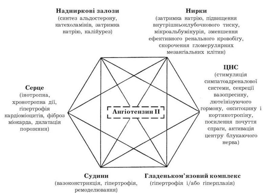
Fig. P7. The role of Ang II in the Pathophysiology of arterial hypertension.
The effects of Ang II are mediated through its binding to specific receptors. Overall, four Types of Membrane-bound Ang II receptors are distinguished.
Type 1 receptors have two subtypes (1a and 1b). Type 1a receptors predominate in the heart, lungs, kidneys, and Arteries, whereas type 2 receptors are mainly found in the adrenal glands and Pituitary Gland. The expression of the type 2 subtype is stimulated by hyponatremia, and the expression of both subtypes is regulated differently in The Heart and adrenal glands. Such differences in tissue distribution and expression mechanisms of both receptor subtypes may be important for modulating the tissue-specific effects of Ang II, although the individual specifics of their functioning are pharmacologically difficult to isolate (M.I. Oliverio et al., 1997).
The pathophysiological effects of Ang II outlined above are mediated exclusively through its action on type 1 Membrane Receptors, providing the rationale for their therapeutic inhibition (K.E. Bernstein et al., 1993; P.B.M.W.M. Timmermans et al., 1993; V.J. Dzau et al., 1994; J.Y. Bauer et al., 1995). Without delving into the details of other Ang II receptor types, It is worth noting the opposing effects—such as antiproliferation, reduction in cell mass, and vasodilation—that occur when Ang II binds to type 2 receptors. From a physiological standpoint, type 2 receptors act as a counterbalance to type 1 receptors (a "yin-yang" effect), performing a crucial counter-regulatory role.
Accumulated data suggest a hypothesis regarding a physiological cardioprotective function for type 2 Ang II receptors. A pivotal role in this function belongs to the kinin-independent stimulation of nitric oxide and, possibly, eicosanoid synthesis mediated by these receptors. It is also hypothesized that this receptor type regulates apoptosis—the genetically programmed cell death of cardiomyocytes during critical myocardial hypertrophy (P. Janiak et al., 1992; P.W. Anderson et al., 1993; H. Gavras, 1994; K. Lindpainter, 1994).
The functions of type 3 and type 4 receptors are currently under investigation. It is believed that the activation of type 3 receptors is predominantly associated with aldosterone hyperproduction, while type 4 receptor activation leads to vasodilation. In recent years, data have emerged suggesting that type 4 receptors may also be responsible for cellular memory processes.
Kallikrein-kinin system. Kinins (bradykinin, kallidin) exhibit vasodilatory, diuretic, and natriuretic effects. These are so-called tissue hormones whose activity is manifested primarily at the site of synthesis, i.e., in all organs and tissues, but predominantly in the kidneys.
All kinins are derived from a precursor—kininogen—whose site of synthesis in the kidneys has not been definitively established. It may be synthesized by the epithelial cells of the distal tubules, although renal uptake from the bloodstream is also plausible. The Synthesis of the primary enzyme, kallikrein, takes place in the epithelial portion of the nephron.
Other components of this system, including bradykinin, are also produced in the kidneys. All of them belong to compounds with a short half-life (about 30 seconds) and are degraded by kininases, with ACE being one such kininase. The Conversion of the inactive precursor kallikreinogen into the active form—bradykinin—occurs under the influence of Hageman factor, which in turn is activated by stimuli that disrupt cell membrane integrity (trauma, toxins, radiation, allergic reactions, lipid peroxidation products, and middle-molecular-weight peptides).
The Physiological Role of bradykinin boils down to altering vascular tone and permeability. Tissue bradykinin plays a key role in the inflammatory response, which represents a sanogenetic mechanism rather than a pathogenic one. One such mechanism is its hemodynamic effect: this factor is capable of dilating coronary and peripheral blood vessels, thereby improving myocardial Blood supply and reducing vascular resistance. Bradykinin stimulates the synthesis of PGE2 and PGI2, the release of endothelium-derived relaxing factors (EDRF, nitric oxide) and hyperpolarizing factors (EDHF) from endothelial cells, inhibits platelet aggregation, reduces their (and tissue) thrombogenic potential, and slows down cardiomyocyte hypertrophy. Only an inappropriate activation of this system, such as in cardiogenic shock, can lead to circulatory collapse and fatal hypotension.
Prostaglandins. Much like kinins, PGs are synthesized in all organs. Circulating PGs effectively influence systemic hemodynamics, while local PGs affect regional blood flow. These substances are synthesized from arachidonic acid by the renal interstitial tissue (with the exception of PGI2—prostacyclin, which is produced in the endothelium of renal arterioles).
The primary stimulators of their synthesis are kinins, Ang II, and catecholamines. The vasodilatory action of PGs extends to all microcirculatory vessels. In addition, PGE2 directly inhibits sodium reabsorption.
It should be noted that The Effect of PGs on hemodynamics is not uniform—some PGs, such as PGF2α, exhibit vasoconstrictor properties and even increase The activity of the sympathoadrenal system.
Overall, the effect of PGs on blood pressure can be summarized as follows:
- vasodilation (PGI2, PGE2);
- vasoconstriction (PGF2α);
- antagonism with the sympathoadrenal system at the vascular alpha-adrenoceptor level (PGI2, PGE2);
- stimulation of the sympathoadrenal system (PGF2α);
- inhibition of norepinephrine release from nerve synapses (PGE2);
- increased natriuresis and diuresis.
The important role of PGs in regulating systemic blood pressure is evidenced by the so-called Bartter syndrome—an autosomal recessive pathology characterized by high RAAS activity, pronounced electrolyte shifts (hypernatremia, hypokalemia), and metabolic alkalosis.
The leading clinical symptoms include adynamia, severe muscle weakness due to hypokalemia, and headaches. Polydipsia and polyuria are also observed.
However, unlike Conn's syndrome, patients with Bartter syndrome do not exhibit arterial hypertension. It has been shown that along with hyperplasia and Hypertrophy of the juxtaglomerular apparatus, hyperplasia also develops in the renal medullary interstitium, which produces large amounts of cyclooxygenase-2 (COX-2)-derived prostaglandins (M. Kommhoff, 1997; Y. Guan et al., 1997; F. Nantel et al., 1999). Therefore, despite high renin activity and excessive aldosterone production, the powerful vasodilatory effect of PGs prevents The Development of arterial hypertension. Consequently, the therapeutic approach in Bartter syndrome involves not Adrenal gland resection, but the administration of PG synthesis inhibitors—nonsteroidal anti-inflammatory drugs (NSAIDs) such as indomethacin (V.A. Yakovlev et al., 1987) and, notably, the latest generation of selective COX-2 inhibitors like celecoxib and rofecoxib (M. Kommhoff, 2001).
Natriuretic peptide (ANP, auriculin, atrial natriuretic factor), brain natriuretic peptide (BNP), C-type natriuretic peptide (CNP), and urodilatin form a group of structurally related polypeptides united by their natriuretic and diuretic effects. In 1954, so-called stretch receptors activated by the stretching of myocardial fibers were discovered in the atrial myocardium, revealing an increase in diuresis and thus leading to the Discovery of the atrial hormone. Until recently, it was believed that the atrial hormone is synthesized and stored in the atrial endocardium and released into plasma upon contraction. It was not until 1984 that its presence was also proven in Blood Plasma, the hypothalamus, kidneys, vascular walls, and adrenal chromaffin cells (R. Lang et al., 1985; M. Sakamoto et al., 1985). Specific receptor Proteins for the atrial hormone have been found in target organs, located on The Plasma Membrane of cells in the aorta, intestines, and adrenal glands.
The Physiological effects of the atrial hormone primarily involve The regulation of vascular tone and renal hemodynamics, accompanied by an increased Glomerular Filtration rate (GFR), enhanced diuresis and natriuresis, and the suppression of adrenal aldosterone synthesis (M. Anand-Srivastava et al., 1998). The precise localization of the receptors responsible for the renal effects of natriuretic peptides has not been definitively established; however, these receptors are believed to be located in both glomerular and tubular structures of the kidneys, particularly in the nephrocytes of the Loop of Henle (R. Keeler, 1995). The primary stimuli for natriuretic peptide production are an increase in circulating blood volume (CBV) and the overstretching of the right atrium, suggesting it acts as a protective hormone against vascular volume overload.
BNP was first isolated from porcine brain, which gave it its name. In humans, it is localized in both the ventricles and atria; the stimulus for its secretion is an increase in extracellular fluid volume, and its primary target organ is the kidneys.
CNP belongs to the paracrine Hormones Involved in the regulation of vascular tone. Its Synthesis and Secretion are attributed to the vascular endothelium.
Urodilatin, which is structurally similar to PGs, has been identified solely in urine.
The medullipin system. This substance is synthesized and secreted by the renal medulla and subsequently converted in the Liver into the highly active hormone medullipin-II, which reduces systemic and intrarenal vascular tone. This promotes an increase in GFR, diuresis, and natriuresis by enhancing effective renal blood flow (E.E. Muirhead, 1990).
Vasopressin (antidiuretic hormone, ADH) is a peptide hormone produced in the hypothalamus and stored in the neurohypophysis. The primary stimuli for its excessive release are an increase in plasma hyperosmolality and a decrease in CBV.
An essential role in the regulation of systemic and, perhaps to a greater extent, regional hemodynamics belongs to the vascular endothelium. Therefore, J. Vane's (1994) observation is quite apt: one trillion endothelial cells, covering an area equal to 6 (!) tennis courts, together with endothelium-derived vasoactive substances, act as the "maestro of the circulation."
The vascular endothelium is the site of synthesis for both vasoconstrictors and proaggregants (angiotensin II, endothelins 1, 2, and 3, thromboxane A2, PGF2α, Leukotrienes C4 and D4) as well as vasodilators and antiaggregants (bradykinin, nitric oxide, endothelium-derived hyperpolarizing factor, prostacyclin, PGE2).
Among endothelial pressor factors, endothelins play a crucial role, as mentioned above. Their pressor effect exceeds that of angiotensin II, serotonin, and norepinephrine by 10 to 100 times. The presence of these factors in peripheral Veins indicates their paracrine origin.
Nitric oxide plays a leading role among depressor endothelial factors. Recent studies have demonstrated that nitric oxide released by endothelial cells in coronary blood vessels (G.H. Gibbons, 1997) plays a key role in their autoregulation mechanism—coronary blood flow remains relatively stable despite fluctuations in systemic blood pressure (T.P. Smith et al., 1993). The involvement of nitric oxide in functional hyperemia of the myocardium and skeletal muscle vasculature in proportion to workload has also been established.
Nephrologists also share a strong interest in nitric oxide. Evidence exists (A. James Sheyman, 1997) indicating the continuous synthesis of this factor within the endothelium and smooth muscle cells of renal vessels, as well as in mesangial and tubular epithelial cells. Consequently, nitric oxide significantly influences the regulation of renal blood flow, renal excretory function, tubuloglomerular feedback, and interactions with locally produced angiotensin II and other BIOREGULATORS of renal function. Alterations in its Biosynthesis caused by Urinary Tract disorders also impact the mechanisms of mesangial proliferation, leukocyte infiltration, and the onset and stabilization of renovascular hypertension (V.G. Maidannyk et al., 1999; S. Klahr, 2001).
Currently, active research is underway regarding the role of structural changes in certain endothelial cell Organelles that may impair nitric oxide production (in the presence or absence of hypercholesterolemia). Such impairment acts as a primary factor in the development of atherosclerosis, nephroangiosclerosis, and systemic vascular tone disorders (M.M. Tkachenko, 1997).
Thus, from the foregoing, the role of depressor factors—either produced by the kidneys or acting via alterations in functional renal state—becomes clear in maintaining normal blood pressure and in the pathogenesis of renovascular hypertension. It is evident that in Kidney diseases, their depressor system is impaired much faster than in essential hypertension due to damage to the tubules and mesangial structures. This constitutes the second pathophysiological difference between essential hypertension and renal arterial hypertension.
The issue of Genetic Disorders in the pathogenesis of RAH is also actively researched, focusing on the role of renin Gene Expression, genetically determined alterations in the reception of Hormones of the kallikrein-kinin system, NO synthase enzyme activity, etc. (I.M. Kutyrina, 2000).
It is generally accepted to classify RAH into renoparenchymal (RPH, acquired or congenital), renovascular (RVH), and post-transplantation forms (M. Stimpel, 1996). RVH is further divided into intrarenal and extrarenal. Any kidney disease—unilateral or bilateral, acute or chronic—may be accompanied by the development of RAH.
From a pathophysiological perspective, RAH is subdivided into volume-dependent and volume-independent forms. Volume-dependent forms of RAH are diagnosed in 80–90% of patients and are driven by sodium and Water retention, which leads to an increased Cardiac Output and the accumulation of Calcium Ions in the Cytosol. This creates the Background for a simultaneous rise in peripheral vascular resistance. Volume-independent renal HTN is registered in 10–20% of patients. In these cases, cardiac output is either unchanged or only slightly elevated, and BP increases primarily due to changes in vascular tone.
The most frequent causes of acquired RPH include acute GN, chronic GN, acute or chronic PN, systemic lupus erythematosus, Renal Amyloidosis, diabetic nephropathy (DN), nephrocalcinosis, Renal Hypoplasia, Renal tuberculosis, and Renal Tumors (renin-producing, endothelin-producing, hypernephroid carcinoma, plasmacytoma); congenital causes include polycystic and multiple renal cysts, Hydronephrosis and Pyonephrosis, renal dystopia, and Nephroptosis (floating kidney).
RVH develops in cases of renal artery stenosis (congenital or acquired), fibromuscular Dysplasia, arteriovenous aneurysms, perirenal hematoma, extrarenal tumors, adhesions or scars compressing renal vessels, renal vein thrombosis, polyarteritis nodosa, Takayasu's disease, Leriche Syndrome, aortic dissection, neurofibromatosis (von Recklinghausen's disease), and nephroptosis.
In children, the most frequent causes of RVH—which accounts for 58.5% of all pediatric hypertension cases—are non-specific aortoarteritis, fibromuscular dysplasia, renal artery or kidney hypoplasia, and mesenchymal dysplasia with multiorgan manifestations (A.V. Pokrovsky et al., 1985; A.V. Chuprova et al., 1998). Clinical, anamestic, and biochemical markers of elevated BP in children have also been identified, including hyperuricemia, macrosomatic constitution, family history predisposing to hypertension, urolithiasis, Gout, and DM (Yu.I. Rovda et al., 1998).
In renoparenchymal kidney diseases, alongside the crucial role of glomerular ischemia with subsequent RAAS activation and elevated endothelin levels (O.V. Sinyachenko, 2001), direct impairment of renal function—such as a decrease in GFR, effective renal plasma flow, and increased sodium and water reabsorption—plays a significant role in the genesis of RAH. Consequently, hypervolemia develops, and sodium content in the vascular wall increases, leading to its hypersensitivity to the pressor effects of AT-II, endothelin, and catecholamines. Concurrently, calcium accumulation in the vascular wall is observed, which also results in elevated peripheral vascular resistance (resistance hypertension).
The main mechanism of BP elevation in RVH is sustained activation of the RAAS resulting from marked glomerular ischemia accompanied by a relatively rapid depletion of depressor systems. Therefore, while the severity of RPH correlates somewhat with the phase of the renal disease (exacerbation, remission), the clinical course of RVH is typically characterized by persistently high BP values. It should be noted that alongside the high resilience of the juxtaglomerular apparatus, which retains its renin-secreting capacity for a prolonged period, an equally important role in stabilizing BP levels in RVH is played by its reserve mechanisms: hypertrophy of myocytes in the afferent arteriole wall and their metaplasia into epithelioid cells, as well as mesangiocyte hyperplasia. In the late stages of RVH development, concurrent with the exhaustion of the juxtaglomerular apparatus on the stenotic side, activation of this apparatus occurs in the contralateral kidney, serving as a vital mechanism in maintaining hypertension.
Unilateral renal ischemia is characterized by the so-called hypertensive-hyponatremic syndrome. The history of this subject is linked to experimental studies (H. Goldblatt et al., 1934), which clearly established that renal ischemia is accompanied by the development of sustained hypertension.
In 1952, Bauer et al. described a case of malignant hypertension and hyponatremia (blood sodium level of 116 mmol/L) in a 42-year-old man who was post-mortem diagnosed with severe atheromatosis of the left renal artery. Subsequent similar reports led to the realization that certain patients with unilateral renal ischemia present with a syndrome characterized by sudden-onset hypertension, hyponatremia, hypokalemia, symptoms of thirst, polyuria, polydipsia, and weight loss. All the aforementioned symptoms are associated with excessive activation of the RAAS in the ischemic kidney. The second (non-ischemic) kidney, confronted with a sudden and marked increase in systemic BP, induces natriuresis and diuresis despite the opposing sodium-retaining effects of AT-II and aldosterone. Thus, the presence of severe and persistent hypertension combined with persistent hyponatremia indicates unilateral renal ischemia.
Recognizing this syndrome among Other forms of RVH is crucial for several reasons:
- antihypertensive agents that do not block the RAAS will fail to provide the proper clinical effect;
- ACE inhibitors as a conservative therapy in these situations should be prescribed with caution to avoid a drastic drop in BP and even more profound renal ischemia. Therefore, a combination of ACE inhibitors with intravenous sodium chloride infusions is recommended in this scenario;
- the treatment of choice is the administration of type 1 angiotensin II receptor blockers;
- other therapeutic modalities for this condition (balloon angioplasty, vascular grafting, nephrectomy) should also be considered.
Overall, the Clinical presentation of RAH and its complications does not differ from the symptomatology of hypertension syndromes of other origins. At the same time, a complete parallelism with The Nature and duration of the underlying kidney disease is not observed, although the prognosis of RAH with persistently high diastolic BP is worse than that of essential hypertension with identical BP values. In fact, sustained diastolic hypertension rapidly leads to hypertensive heart disease with characteristic changes in electrocardiographic and echocardiographic parameters, fundus vessels, etc.
Last update: 08/08/2026
Editorial and Educational Adaptation: This material has been compiled based on the primary/original source text. The project team performed an editorial review, corrected technical inaccuracies, structured sections, and adapted the content for an educational format.
What was processed:
- elimination of formatting defects (OCR errors, structural breaks, corrupted characters);
- editorial organization of content;
- standardization of terminology in accordance with academic sources;
- verification of factual statements against the original source text.
All mentions of the author, publication year, and origin of the primary text have been preserved in accordance with the source.