Nephrology for the Family Physician - O.I. Bakaliuk 2003
Certain Urological Disorders in Therapeutic Practice
Renal Tumors
Adult renal tumors are predominantly malignant, accounting for 1.5% to 3% of all tumor localizations. Men are affected twice as often as women, with the peak incidence occurring between the ages of 40 and 50. Clear Cell carcinoma (Grawitz tumor) is the most common histological type, accounting for 70-90% of cases, and presents in two forms: typical and atypical hypernephromid tumors.
The typical hypernephromid tumor is characterized by the slow development of a spherical mass encased in a thin Connective Tissue capsule, which progressively displaces the renal parenchyma as it grows.
In contrast, the atypical hypernephromid tumor lacks clear demarcation from the healthy parenchyma, tending instead toward diffuse infiltration of the Kidney. It fills the calyces and tubules and invades the Veins. This form is associated with higher malignancy and rapid metastasis to the Lungs, Brain, bones, and Liver.
Other renal neoplasms include non-hypernephroid carcinoma (8-20%) and Sarcoma (2-5%).
Occasionally, a renal tumor (known as Wilms' Tumor) is associated with other congenital anomalies, as seen in Miller syndrome.
Renal tumors are classified into four Selection/3.html">Stages of development: Stage I—tumor confined within the renal capsule; Stage II—involvement of the vascular pedicle or perirenal adipose tissue; Stage III—regional Lymph node involvement; Stage IV—presence of distant metastases (S.I. Ryabov, 2000).
For the timely Diagnosis of renal tumors, it is essential to distinguish between two groups of symptoms: renal and extrarenal.
Renal symptoms include lower back pain, Hematuria, and proteinuria.
Pain occurs in 3-56% of cases depending on the stage of the disease. It typically manifests as a sensation of heaviness in the lumbar region, or occasionally as Renal Colic occurring after (!) the onset of macrohematuria (unlike Nephrolithiasis, where pain precedes hematuria), or as lumboischialgia (when the tumor extends beyond the kidney). Guyon's sign (F.J.C. Guyon) is often positive, revealing pathological mobility (ballottement) of the enlarged kidney.
Hematuria (microhematuria with unchanged erythrocytes) is almost constant, and transient macrohematuria of unapparent origin is sometimes observed.
The latter may be a single episode, lasting from several hours to 1-2 days, and disappearing as suddenly as it appeared. Therefore, when macrohematuria occurs, its source (right or left kidney) should be determined via cystoscopy prior to the administration of hemostatic agents.
Proteinuria in renal tumors is moderate, and cylindruria is uncommon.
When the tumor invades the v. renalis, it may cause symptomatic Varicocele (8.6% in Stage I and 14.2% in Stage IV) or a collateral vascular network on the Skin of the lumbar region and abdomen.
Extrarenal symptoms (J. Kernion, 1986; T. Tsukamoto et al., 1991) include fever, elevated ESR, anemia or erythrocytosis, polycythemia, amyloidosis, paraneoplastic syndromes, enteropathies, and cachexia.
Among these manifestations, the most clinically significant are unexplained elevated ESR (55-92%), iron deficiency anemia (8-64%), arterial Hypertension (11-40%), fever without a specific pattern (15-59%), and weight loss (15-30% of cases).
Various clinical "masks" of renal tumors may also occur, including joint syndrome, lymphadenopathy, ulcerative-necrotic skin purpura, alopecia, neuromyopathic syndrome, polyneuropathy, and thromboembolism.
Stauffer's syndrome (M.H. Stauffer) is a notable tumor mask representing nephrogenic hepatic dysfunction (manifested by anorexia, weakness, hypotension, hepatosplenomegaly, significantly elevated serum levels of alpha2-globulins, alkaline phosphatase, and Class A IMMUNOGLOBULINS, along with hypoalbuminemia). Notably, this syndrome of nephrogenic hepatic dysfunction typically precedes the onset of other classic tumor symptoms, which carries significant diagnostic value (A.Yu. Nikolaev et al., 1984).
Laboratory findings may also include hypercalcemia, hyperhaptoglobinemia, elevated activity of phosphomonoesterase-1, Lactate dehydrogenase and its Isoenzymes, as well as decreased cholinesterase activity and transferrin levels (S.I. Ryabov, 2000).
The diagnostic workup for renal tumors utilizes a full range of laboratory and instrumental techniques, starting with routine Methods (Ultrasonography, isotope scanning, radiocontrast studies (Fig. 100)) and advancing to angiography (Fig. 101), computed tomography (Fig. 102), and renal biopsy.
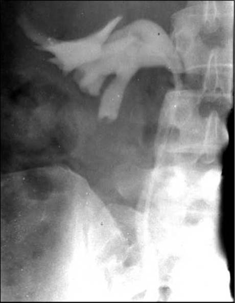
Fig. 100. Right renal tumor: retrograde pyelography (the "wilted flower" sign—downward Displacement of the upper calyces by a dense tumor mass).
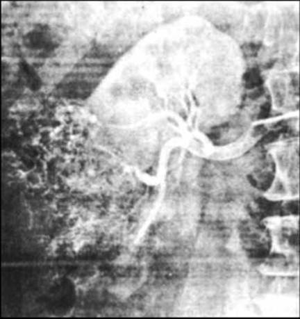
Fig. 101. Selective renal angiogram in right renal tumor (increased hypervascularity in the lower pole of the right kidney).
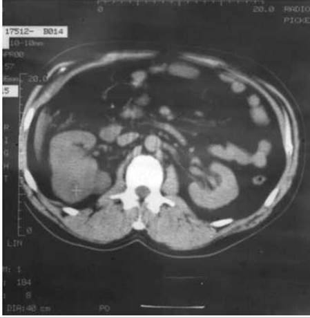
Fig. 102. Computed tomogram of a right renal tumor.
Last update: 08/08/2026
Editorial and Educational Adaptation: This material has been compiled based on the primary/original source text. The project team performed an editorial review, corrected technical inaccuracies, structured sections, and adapted the content for an educational format.
What was processed:
- elimination of formatting defects (OCR errors, structural breaks, corrupted characters);
- editorial organization of content;
- standardization of terminology in accordance with academic sources;
- verification of factual statements against the original source text.
All mentions of the author, publication year, and origin of the primary text have been preserved in accordance with the source.