Nephrology for the Family Physician - O.I. Bakaliuk 2003
Certain Urological Diseases in Therapeutic Practice
Polycystic Kidney Disease
Polycystic Kidney Disease (polycystic renal degeneration) is a hereditary congenital anomaly characterized by The formation of multiple cysts within the Kidneys, interspersed with isolated elements of the renal parenchyma. The underlying pathology stems from impaired normal embryonic development of renal structures, marked by an abnormal connection between the secretory and excretory apparatus of the kidneys—specifically, the metanephrogenic blastema system and the outgrowths of the Wolffian body.
Depending on their origin, cysts can be glomerular (lacking a connection to the tubular excretory system), tubular (developing from elements of the proximal or distal convoluted tubules), and excretory (developing from elements of the convoluted tubules).
The congenital nature of this pathology was established long ago, adhering to an Autosomal dominant inheritance pattern. Therefore, it is correct to classify this condition among congenital nephropathies, which frequently manifest only in adulthood. Modern diagnostic Methods allow such developmental anomalies to be detected as early as the antenatal period, providing appropriate recommendations regarding Pregnancy termination. Thus, it is only out of respect for tradition that we discuss this topic alongside other issues in "adult" nephrology.
In 90% of cases, the defective Gene is located on the short arm of chromosome 16 and is designated as ADPKD-1. The Pathogenesis of the disease remains unknown, although several theories have been proposed. According to one theory (A.P. Evan et al., 1987), the primary cause of polycystic kidney disease is excessive proliferation of epithelial Cells (potentially driven by growth factors), which can lead to intra-nephron obstruction and proximal cystic dilatation. Another theory (F.A. Carone et al., 1992) attributes cyst formation to a defect in Extracellular matrix production, resulting in alterations of the tubular basement membrane accompanied by the formation of diverticula and cysts.
Morphologically, polycystic kidneys are deformed, enlarged, and contain bilateral cysts ranging in size from a bean to an orange; the renal pelves and calyces are deformed and elongated. Occasionally, kidneys may weigh up to 2 kg, and their length can reach 40 cm. The cystic fluid resembles primary urine (watery, yellowish, with a low specific gravity). Polycystically altered kidneys are particularly susceptible to secondary conditions—tuberculosis, renal failure, Hydronephrosis, Pyonephrosis, and urolithiasis. In a significant number of cases, hemorrhages into the cysts occur, followed by suppuration (Fig. 95).
In 10% of cases, polycystic kidney disease is associated with polycystic changes in other Organs (Liver, Pancreas).
The Clinical presentation and course of polycystic kidney disease depend on the age of onset of its initial manifestations. Early childhood polycystic kidney disease progresses rapidly; renal failure develops in 100% of cases, culminating in anuria and irreversible renal failure. In adults, the clinical picture typically manifests between the ages of 40 and 45, frequently triggered by The Development of complications or secondary infection.
Class="center">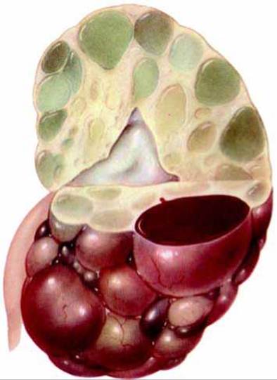
Fig. 95. Hemorrhage into cysts in polycystic kidney disease.
Polycystic kidney disease lacks specific symptoms; patients may experience thirst, polyuria, nocturia, asymptomatic dysuria, a dull ache, and a feeling of heaviness in the lumbar region, particularly after physical exertion involving body jolting (jumping, running). The pain frequently radiates to the epigastric region, creating additional diagnostic challenges (Fig. 96).
The disease may present with transient or prolonged macrohematuria accompanied by Typical symptoms of Renal Colic, hyposthenuria, or isosthenuria. Occasionally, patients can palpate their enlarged kidneys themselves.
Urinalysis reveals moderate proteinuria and microhematuria; casts are typically absent from the urinary sediment.
During the subcompensated phase, patients periodically experience general deterioration, fatigue, thirst, impaired liver and gastrointestinal function, elevated Blood pressure, hyponatremia, hypocalcemia, hyperphosphatemia, and acidosis. Glomerular Filtration and the concentration capacity of the kidneys are reduced.
Decompensation is characterized by signs of Chronic Kidney Disease (CKD) and Clinical symptoms of complications (severe arterial Hypertension, acidosis, renal failure, hydronephrosis). It is worth noting that polycystic kidney disease is virtually the only condition in which anemia does not develop, even in the terminal stage of CKD, which is attributed to the preserved ability of polycystic kidneys to produce Erythropoietin (T.B. Alekseyeva et al., 1999).
The life expectancy from the onset of clinical signs of renal impairment in such patients averages 10 years.
Diagnosing polycystic kidney disease is generally straightforward when based on history (family history), clinical findings (bilateral renal enlargement, uneven surface upon Palpation), and instrumental methods (ultrasound, excretory urography, transfemoral angiography, computed tomography).
Ultrasound reveals cysts of various sizes in both (!) kidneys (Fig. 97).
Excretory urography demonstrates altered contours of the kidneys and the pelvicalyceal system, with characteristic hyper-ramification of the calyces (increased spacing between them, Fig. 98).
Computed tomography shows varying degrees of renal enlargement accompanied by the presence of cysts (Fig. 99).
Arteriography reveals avascular zones in regions containing large cysts.
Frequent complications include urolithiasis, pyonephrosis, chronic kidney disease, hyperuricemia, and hypocalcemia, along with their corresponding clinical symptoms.
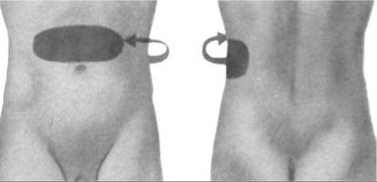
Fig. 96. Typical pain radiation in polycystic kidney disease.
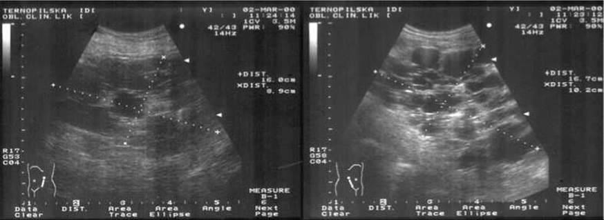
Fig. 97. Polycystic kidney disease (ultrasound).

Fig. 98. Polycystic kidney disease (excretory urography).
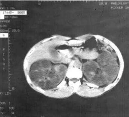
Fig. 99. Polycystic kidney disease (computed tomography).
A variant of polycystic kidney disease is the so-called Cacchi-Ricci disease (R. Cacchi, V. Ricci, 1948) – medullary sponge kidney.
In this form of pathology, small cysts (1–4 mm) are located exclusively in the medullary layer of the kidneys. A characteristic feature is the complete absence of clinical symptoms and functional renal changes, although chronic recurrent leukocyturia is observed as early as infancy. At an older age, Nephrolithiasis and renal colic develop. The pathology can occasionally be unilateral. The Diagnosis is established based on typical changes revealed by uro-X-ray Examination ("bouquet of flowers" appearance in the medullary pyramid zone). The inheritance pattern is autosomal dominant. The overall prognosis is more favorable than that of polycystic kidney disease.
Congenital polycystic kidney disease is most commonly differentiated from acquired cystic kidney disease.
Acquired cystic kidney disease is a complication of various renal disorders accompanied by varying degrees of nephrosclerosis. The exact Etiology of this condition remains unclear, although it is most frequently encountered in patients undergoing maintenance hemodialysis. According to J.J. Grantham (1991), in advanced chronic kidney disease (CKD), in response to the loss of renal parenchyma, the body produces an unidentified factor (presumably resembling numerous growth factors) that is not eliminated by hemodialysis. The diagnostic criterion is the presence of 5 or more cysts of varying sizes (from a few millimeters to 2–3 centimeters) located throughout all layers of the kidneys. In 20% of cases, potentially malignant papillary tumors are found within the cysts (W.Y. Ghung et al., 1992). Acquired cystic disease is typically asymptomatic; however, it may manifest upon tumor progression and metastasis or following cyst rupture (retroperitoneal hematoma, gross Hematuria).
Last update: 08/08/2026
Editorial and Educational Adaptation: This material has been compiled based on the primary/original source text. The project team performed an editorial review, corrected technical inaccuracies, structured sections, and adapted the content for an educational format.
What was processed:
- elimination of formatting defects (OCR errors, structural breaks, corrupted characters);
- editorial organization of content;
- standardization of terminology in accordance with academic sources;
- verification of factual statements against the original source text.
All mentions of the author, publication year, and origin of the primary text have been preserved in accordance with the source.