Human Biochemistry, Volume 1 - Murray R. 1993
Protein and Amino Acid Metabolism
Porphyrins and Bile Pigments
Hyperbilirubinemia
When Blood bilirubin levels exceed 1 mg/100 mL (17.1 µmol∙L-1), the condition is referred to as hyperbilirubinemia. Hyperbilirubinemia may result from bilirubin production exceeding the normal Liver's capacity for excretion, or from liver damage that impairs The excretion of normal amounts of bilirubin. In addition to intrinsic liver injury, hyperbilirubinemia can be caused by an obstruction of the intrahepatic Bile ducts that prevents the release of bilirubin. In all these scenarios, bilirubin accumulates in the bloodstream and, upon reaching certain concentrations, diffuses into Tissues, imparting a yellow discoloration. This condition is known as jaundice.
During the clinical evaluation of patients with jaundice, determining serum bilirubin levels provides valuable diagnostic information. Quantitative serum bilirubin analysis was first introduced by van den Bergh, building upon Ehrlich's method for measuring urinary bilirubin. Ehrlich's reaction relies on diazosulfanilic acid (Ehrlich's diazo reagent), which reacts with bilirubin to form a reddish-purple azopigments. Ehrlich's original protocol utilized methanol, which dissolves both bilirubin and the diazo reagent. While analyzing a patient's bile for bile pigments, van den Bergh inadvertently omitted methanol and was surprised to observe that color development proceeded via a 'direct' pathway even without it. Subsequently, this form of bilirubin, capable of reacting without The addition of methanol, was designated as the 'direct-reacting' form. The same direct reaction was later identified in the serum of patients with jaundice caused by bile duct obstruction. However, detecting bilirubin in normal serum still requires the addition of methanol; the same holds true for analyzing excess serum bilirubin in hemolytic jaundice unrelated to bile duct obstruction. The form of bilirubin that reacts only after the addition of methanol was termed the 'indirect-reacting' form.
It is now understood that 'indirect' bilirubin represents 'free' (unconjugated) bilirubin transported to the liver from reticuloendothelial tissues, where it is generated through The breakdown of hemoporphyrins. Because this form of bilirubin is Water-insoluble, the addition of methanol is required for its interaction with the diazo reagent. Conjugation of bilirubin with glucuronic acid takes place in the liver, yielding bilirubin glucuronide, which can then be secreted into the bile. Conjugated bilirubin is water-soluble and reacts directly with the diazo reagent; consequently, van den Bergh's 'direct bilirubin' is actually a bilirubin conjugate (bilirubin glucuronide).
Depending on whether the predominant type of bilirubin in plasma is unconjugated or conjugated, hyperbilirubinemia is classified as posthepatic (unconjugated) or regurgitation (conjugated) hyperbilirubinemia, respectively.
Only unconjugated bilirubin can cross the blood-Brain barrier to enter the Central Nervous system; therefore, encephalopathy resulting from hyperbilirubinemia (kernicterus) can only be caused by posthepatic hyperbilirubinemia. Conversely, only conjugated bilirubin can be excreted in the urine. Accordingly, choluric jaundice is observed exclusively in regurgitation hyperbilirubinemia, whereas acholuric jaundice occurs solely when there is an excess of unconjugated bilirubin.
Unconjugated Hyperbilirubinemia
Even in cases of significant hemolysis, unconjugated hyperbilirubinemia is usually quite mild (less than 4 mg/100 mL, or less than 68.4 µmol/L) due to the liver's robust capacity for bilirubin conjugation. If, however, hepatic bilirubin conjugation is impaired due to acquired or hereditary factors, pronounced unconjugated hyperbilirubinemia may ensue.
The most common form of unconjugated hyperbilirubinemia is 'physiological jaundice,' which occurs as a transient condition in newborns. This type of hyperbilirubinemia is driven by accelerated hemolysis coupled with the immaturity of the hepatic systems responsible for bilirubin uptake, conjugation, and secretion. Not only is The activity of UDP-glucuronyl transferase reduced, but the synthesis of its substrate, UDP-glucuronic acid, is likely also insufficient. Because the accumulating bilirubin remains unconjugated, it can cross the blood-brain barrier once its plasma concentration exceeds the saturation threshold of high-affinity albumin-binding sites (20–25 mg/100 mL). This can lead to hyperbilirubinemic toxic encephalopathy (kernicterus). Given that the bilirubin conjugation system is inducible, the administration of phenobarbital has proven effective in newborns with physiological jaundice. Furthermore, exposure to visible light can facilitate the hepatic clearance of unconjugated bilirubin (via an unknown mechanism), converting a portion of the pigment into derivatives that are excreted in the urine.
A. Crigler-Najjar Syndrome, Type I; Congenital Nonhemolytic Jaundice. Crigler-Najjar syndrome type I is a metabolic disorder of bilirubin conjugation. The condition is characterized as severe congenital jaundice resulting from an inherited deficiency of bilirubin-UDP-glucuronyltransferase activity in hepatic tissue. The disease is typically fatal within the first 15 months of life, though cases presenting as late as adolescence have been documented. Affected individuals have been treated with phototherapy, which achieves a moderate reduction in plasma bilirubin levels. Phenobarbital and other pharmacological agents that induce bilirubin METABOLISM in a normal liver have no effect on bilirubin glucuronide formation in Crigler-Najjar syndrome type I. Without Treatment, serum bilirubin levels generally exceed 20 mg/100 mL.
B. Crigler-Najjar Syndrome, Type II. This rare inherited disorder presumably stems from less severe defects in the bilirubin conjugation system and follows a more benign clinical course. Serum concentrations of bilirubin typically do not exceed 20 mg/100 mL, and all accumulating bilirubin is of the unconjugated type. Surprisingly, the bile of these patients does contain bilirubin glucuronide; this has led to the hypothesis that the genetic defect may affect hepatic UDP-glucuronyltransferase, which catalyzes the addition of the second glucuronyl group to bilirubin monoglucuronide.
Patients with this syndrome have been shown to respond favorably to high doses of phenobarbital. The drug-induced reduction in hyperbilirubinemia appears to result from the Induction of the entire bilirubin metabolic pathway rather than merely The stimulation of bilirubin conjugation.
In several instances, putative heterozygotes for this disorder have exhibited mild unconjugated hyperbilirubinemia indistinguishable from that seen in Gilbert's syndrome (see below). It is entirely possible that the autosomal recessive Crigler-Najjar syndrome type II represents the homozygous state for a defect that, in the heterozygous state, manifests as the mild, chronic hyperbilirubinemia of Gilbert's disease.
C. Gilbert's Disease. This condition constitutes a heterogeneous group of disorders, many of which are now known to result from compensated hemolysis coupled with unconjugated hyperbilirubinemia. There also appear to be cases caused by an abnormal reduction in bilirubin uptake by hepatic parenchymal Cells. Additionally, hepatic bilirubin-UDP-glucuronyltransferase activity is reduced in these patients.
It is worth noting that the benign disorders collectively known as 'Gilbert's disease' appear to follow an autosomal dominant mode of inheritance.
Class="center">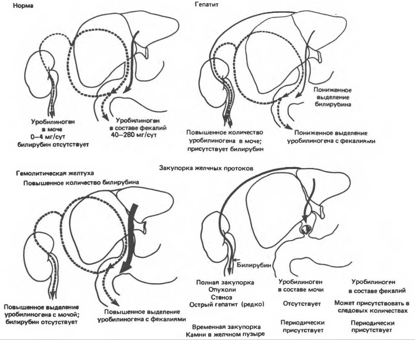
Fig. 33.16. The bilirubin-urobilinogen cycle. Solid arrows indicate The pathway of bilirubin diglucuronide, and dashed arrows indicate the pathway of urobilinogen. (Reproduced, with permission, from Krupp M. A. et al., Physician's Handbook, 21st ed. Lange, 1985.)
D. Toxic Hyperbilirubinemia. Unconjugated hyperbilirubinemia can result from hepatic dysfunction induced by the Toxic effects of various substances (chloroform, arsphenamines, carbon tetrachloride, acetaminophen), as well as from Viral Hepatitis, cirrhosis, and poisoning by *Amanita* mushrooms. Most of these acquired disorders stem from injury to hepatic parenchymal cells; obstruction of the intrahepatic biliary tree is also frequently observed, leading to conjugated hyperbilirubinemia.
Conjugated Hyperbilirubinemia
Because conjugated bilirubin is water-soluble, it is detectable in the urine of most patients with conjugated hyperbilirubinemia; consequently, the condition is frequently referred to as choluric jaundice.
A. Chronic Idiopathic Jaundice (Dubin-Johnson Syndrome). This autosomal recessive disorder manifests as conjugated hyperbilirubinemia in both children and adults. The underlying cause is believed to be impaired hepatic secretion of conjugated bilirubin into the bile. However, this secretory defect is not restricted to bilirubin alone; it also affects the excretion of several other conjugated compounds, notably estrogens and diagnostic Dyes such as sulfobromophthalein. The impaired clearance of conjugated sulfobromophthalein causes it to reflux into the bloodstream, resulting in a secondary rise in its plasma concentration—a phenomenon that is pathognomonic for Dubin-Johnson syndrome. When Other Compounds whose elimination does not require conjugation, such as indocyanine green or rose bengal, are used for testing, no secondary rebound in plasma concentration is observed in these patients. Thus, the defect in this condition involves a secretory mechanism normally specific for conjugated compounds, including conjugated bilirubins. Interestingly, hepatocytes in the centrilobular Zones of the liver in patients with Dubin-Johnson syndrome contain an unusual and as-yet-unidentified pigment.
Bile Duct Obstruction. Conjugated hyperbilirubinemia also develops when the hepatic or common bile ducts are obstructed. In this situation, bile pigments passing from the blood into the liver cells reportedly cannot be excreted. As a result, conjugated bilirubin regurgitates into the hepatic Veins and Lymphatic vessels.
The term cholestatic jaundice is applied to all forms of jaundice associated with extrahepatic obstruction, as well as to certain forms of parenchymal jaundice characterized by conjugated hyperbilirubinemia.
Urinary Urobilinogen
Normally, urobilinogen is present in the urine only in trace amounts (averaging 0.64 mg per 24-hour collection; levels up to 4 mg are still considered within the normal range). In complete bile duct obstruction, urinary urobilinogen is absent because bilirubin cannot reach the intestinal lumen, where it would otherwise be converted into urobilinogen. In this scenario, the presence of urinary bilirubin coupled with the absence of urobilinogen points to obstructive (mechanical) jaundice, whether of intrahepatic or extrahepatic origin.
In hemolytic jaundice, accelerated bilirubin production leads to increased urobilinogen synthesis, which then appears in the urine in large amounts. Bilirubin is typically undetectable in the urine of patients with hemolytic jaundice; thus, elevated urinary urobilinogen combined with an absence of urinary bilirubin is indicative of hemolytic jaundice. Enhanced destruction of Blood Cells driven by various causes (such as pernicious anemia) likewise leads to increased urinary urobilinogen levels. Furthermore, when bacterial infection invades the biliary tract, urinary urobilinogen can be elevated even in the absence of liver dysfunction due to the reductive activity of the infecting Bacteria.
The diagram in Fig. 33.16 summarizes data on The transport of bilirubin and urobilinogen from the liver to the intestine and Kidneys under normal conditions, as well as in hemolytic jaundice, hepatitis, or obstructive jaundice caused by bile duct blockage.
Battersby A. R. et al. Biosynthesis of the pigments of life: Formation of the macrocycle, Nature, 1980, 285, 17.
Berk P. D. et al. Disorders of bilirubin metabolism. In: Metabolic control and Disease, 8th ed., Bondy P. K., Rosenberg L. E. (eds), Saunders, 1980.
Kappas A., Sassa S., Anderson K.E. The Porphyrias. In: The Metabolic Basis of Inherited Disease, 5th ed., Stanbury, J. B. et al. (eds), McGraw-Hill, 1983.
Lemberg R., Legge J. W. Hematin Compounds and Bile Pigments, Interscience, 1949.
Schmid R., McDonough A. F. Formation and METABOLISM OF BILE pigments in vivo. In: The Porphyrins, Dolphin D. (ed.), Academic Press, 1978.
Tschudy D. P., Lamon J. M. Porphyrin metabolism and the porphyrias. In: Metabolic Control and Disease, 8th ed., Bondy P. K., Rosenberg L. E. (eds), Saunders, 1980.
Watson C. J. Gold from dross: The first century of the urobilinoids, Ann. Intern. Med., 1969, 70, 839.
Last update: 06/08/2026
Editorial and Educational Adaptation: This material has been compiled based on the primary/original source text. The project team performed an editorial review, corrected technical inaccuracies, structured sections, and adapted the content for an educational format.
What was processed:
- elimination of formatting defects (OCR errors, structural breaks, corrupted characters);
- editorial organization of content;
- standardization of terminology in accordance with academic sources;
- verification of factual statements against the original source text.
All mentions of the author, publication year, and origin of the primary text have been preserved in accordance with the source.