Antibiotics (Properties, Applications, Interactions) - M.P. Cherenko 1999
Hemorrhage, blood loss, hemostasis
Hemorrhage is one of the major challenges in modern surgery. Every open injury and the vast majority of surgical Procedures are accompanied by it.
What do we mean by the term "hemorrhage"? The word "hemorrhage" (haemorrhagia) comes from two Greek words: haema, meaning Blood, and rhein, meaning to flow. It is the escape of blood from Blood Vessels or The Heart chambers into Tissues, Body Cavities (such as the pleural, peritoneal, cranial, or joint cavities), hollow Organs (such as The Stomach, intestines, Uterus, or Urinary Bladder), or into the external environment. A hemorrhage is always the result of a specific factor acting upon the body; spontaneous bleeding without a cause does not occur.
What causes blood to escape from the vascular bed or heart chambers? There are several causes.
Hemorrhage due to vascular wall rupture (haemorrhagia per rhexin). This occurs As a result of any vascular injury that completely or partially opens the vessel lumen. Causes may include stab wounds, gunshot wounds, blunt force trauma, and others. In such cases, acute blood loss is more common.
Hemorrhage due to vascular wall erosion (haemorrhagia per diabrosin corrosionem). In this case, vascular destruction is the consequence of a pathological process developing in the tissues adjacent to the vessel wall, such as a disintegrating tumor invading the vessel, an inflammatory infiltrate, an ulcer, or sclerosis of the vessel wall. Sometimes, the disruption of the vessel wall integrity is caused by a foreign body (such as a bullet or shrapnel) that exerts constant pressure on the vessel.
Hemorrhage by diapedesis (haemorrhagia per diapedesui). This is bleeding in which there is no macroscopic vascular injury; instead, blood escapes through a seemingly intact wall due to increased permeability. Typically, such hemorrhages are not massive. Blood appears to ooze through a vessel wall that is functionally, rather than mechanically, compromised and loose, and this does not require immediate surgical intervention. Functional vascular permeability can be caused by various pathogenic factors: Bacterial toxins (in scarlet fever, smallpox), septic conditions, poisoning with arsenic or phosphorus, changes in blood composition (cholemia, leukemia, malignant anemia), vitamin deficiencies, etc.
Thus, based on their Pathogenesis, all hemorrhages can be divided into two major groups: I — mechanical hemorrhages, caused by direct local destruction of the vessel wall resulting from trauma (rupture) or a pathological process (inflammation, tumor, atherosclerosis, ulcer); II — neurotrophic (as defined by V.I. Struchkov) or dysmetabolic hemorrhages, caused by systemic disturbances in the MICROSTRUCTURE OF THE microcirculatory vessels that render them permeable to blood elements (diapedesis), along with systemic changes in blood chemistry and clotting ability.
Classification OF HEMORRHAGES
Depending on the type of damaged vessel, hemorrhages are classified as arterial, venous, capillary, and parenchymal.
Arterial hemorrhages are the most dangerous because blood in the Arteries flows under high pressure (120 mmHg). It is forcefully expelled from the vessel or heart chamber, causing the body to lose blood rapidly. Arterial blood typically flows in a high-pressure jet and is bright red in color; however, this is not always the case. First, in states of low blood pressure (such as Shock or collapse), arterial blood may flow out smoothly. Second, under certain pathological conditions where large amounts of carbon dioxide accumulate in the blood (Hypoxia), it may appear dark. The stream of blood escaping from an artery is synchronous with the pulse wave. In cases of complete transection or rupture, bleeding occurs from both the central and the peripheral ends of the vessel. The outflow of blood from an artery is often accompanied by a hissing sound. Spontaneous cessation of bleeding is possible only in small-caliber arteries. Hemorrhage from larger arteries and the heart rarely stops on its own because blood pressure is high and vascular constriction is insufficient.
In venous hemorrhage, blood typically flows from the vessel in a steady, slow stream and is dark in color. In some cases where tissues are unable to utilize oxygen (tissue hypoxia), large amounts of oxygen pass from the arteries into the Veins, and venous blood may take on a bright red appearance resembling arterial blood.
When venous pressure is elevated, blood may escape from a vein in a jet. Bleeding most commonly occurs from the peripheral end of the vessel, but in cases of venous congestion and a sufficient collateral network, blood may also flow from the central segment.
Hemorrhage from veins located close to the heart (such as the brachiocephalic, subclavian, and internal jugular veins) may occur in pulses due to decreased pressure during inspiration and increased pressure during expiration. Veins whose walls do not collapse because they are fixed to adjacent tissues (such as the subclavian, bone, and hepatic veins) bleed profusely.
When intrathoracic or intra-abdominal pressure rises (due to coughing, vomiting, shouting, or sneezing), venous hemorrhage intensifies. Applying pressure to the vein proximal to the bleeding site increases the hemorrhage, whereas applying pressure distally may help stop it.
Overall, venous hemorrhages are less life-threatening than arterial ones; however, injuries to large veins located closest to the heart pose a severe danger not only from blood loss but also due to the risk of air entering the vessel lumen (air embolism).
Capillary hemorrhage is of a mixed type. It is generally not life-threatening and stops spontaneously under a pressure bandage. In this type of bleeding, individual bleeding vessels are not visible; instead, blood oozes from the tissues like from a sponge. The color of the blood is dark red. Anything that enhances arterial inflow and impedes venous outflow increases the bleeding. In healthy tissues, capillary hemorrhage usually stops on its own. However, in tissues deficient in elastic elements or in conditions associated with impaired blood clotting (such as hemophilia or cholemia), capillary bleeding can also become life-threatening.
Parenchymal hemorrhage is observed when parenchymal organs are injured. It can sometimes be quite substantial. The danger lies in the difficulty of stopping it, because the vessels of parenchymal organs are fixed to the organ stroma and cannot collapse. It has a mixed character, as it originates from small arteries, veins, and capillaries.
Hemorrhage from the Liver, Spleen, Kidneys, Lungs, and other similar organs is particularly severe and dangerous. Other hazardous hemorrhages of a parenchymal nature occur in the Tongue, the spongy bone substance, and organs with erectile tissue structures (such as the corpora cavernosa of the Penis and Clitoris).
Depending on the direction of blood flow, hemorrhages are divided into internal, external, and interstitial. In internal hemorrhage, blood is poured into the natural cavities of the body (peritoneal, pleural, cranial, joint) or into hollow organs (Esophagus, stomach, intestines, urinary bladder). Eventually, blood from hollow organs is discharged to the exterior, yet these hemorrhages are still classified as internal.
Hemorrhages into various cavities and hollow organs have specific medical terms: into the peritoneal cavity — haemoperitoneum, pleural cavity — haemothorax, joint cavity — haemarthrosis, pericardial sac — haemopericardium, cranial cavity — haemocranion, from the Nose — epistaxis, from the lungs — haemoptysis, Urinary Tract — haematuria, FEMALE REPRODUCTIVE ORGANS — metrorrhagia, rectum — haemorrhoids (or rectal hemorrhage), stomach — haematemesis.
The intensity of bleeding into cavities and hollow organs depends on the caliber of the damaged vessel, the size of the rupture, blood pressure, and the duration of the hemorrhage. The largest volumes of blood accumulate in the peritoneal and pleural cavities, or in the stomach and intestines—reaching up to 2–3 liters. These hemorrhages are sometimes so massive that they require immediate surgical intervention.
In external hemorrhage, blood escapes from a wound in the Skin or mucous membranes into the external environment.
Recognizing internal hemorrhages is more difficult than identifying external ones. When bleeding occurs into the lumen of hollow organs, blood is discharged to the exterior through the body's natural orifices. However, determining the source of even such overt internal bleeding is not always easy. For instance, bleeding from the Mouth may be related to pathology in the Oral Cavity itself, or to conditions in the lungs, esophagus, stomach, duodenum, or nasopharynx. Bloody stools can result from bleeding anywhere along the gastrointestinal tract, as well as from the lungs if blood is swallowed.
In interstitial (intratissue) hemorrhage, blood either accumulates within the tissues (forming a hematoma — haematoma) or infiltrates them. Depending on the size and shape of such hemorrhages, they are classified as: petechiae — petechiae (minute pinpoint hemorrhages); vibices — vibices (streak-like hemorrhages); ecchymoses — ecchymoses (larger hemorrhagic patches); and bruises/suggillations — suggilatio s. suffusio (massive hemorrhages with irregular borders).
Hemorrhages may be overt (manifest) or occult (hidden). In the former case, blood can be seen with the naked eye; in the latter, special Laboratory tests are required for its detection.
Bleeding can be primary or secondary. Primary bleeding occurs at the moment of injury or surgery and is a direct consequence of trauma. Secondary bleeding is subdivided into early and late types. Early secondary bleeding occurs within 2 days of injury and results from the dislodgement of a blood clot from a vessel, the slipping of a ligature, or the cessation of vascular spasm (for example, the wearing off of adrenaline administered with an anesthetic during tooth extraction). Causes of ligature slippage may include tying the knot too loosely on the vessel, accidentally including adjacent tissues in the ligature along with the vessel, excessive use of vessel twisting or electrocoagulation to stop bleeding, or overlooking and underestimating minor hemorrhages.
Causes of late secondary bleeding may include: displacement of a blood clot due to secondary trauma (dressing changes, premature transportation), external pressure on a vessel leading to necrosis of its wall and rupture (such pressure can be caused by sharp bone fragments in fractures, foreign bodies, or rubber drainage tubes placed close to the vessel), or purulent dissolution of the clot or the vessel wall itself in the presence of pyogenic or putrefactive wound infection.
FACTORS CONTRIBUTING TO BLEEDING
The onset of bleeding can be influenced by various pathological conditions of the body. Blood pressure fluctuations toward Hypertension play a certain role in this regard. When combined with pathological Changes in the arterial walls (sclerosis), this can lead to vessel rupture and sudden hemorrhage (such as cerebrovascular accidents in elderly individuals).
A significant role in the occurrence of bleeding is played by a decrease in the blood's clotting properties.
In surgical practice, physicians more frequently encounter patients with conditions that impair Blood Coagulation (hemophilia, Fibrinolysis, cholemia, acholia).
Hemophilia is characterized by a drastic slowing of blood clotting, which leads to The Development of dangerous hemorrhages. It is caused by a deficiency in certain clotting factors: antihemophilic globulin (AHG) — clotting factor VIII; plasma thromboplastin component — clotting factor IX; and clotting factor X.
Hemophilia affects males. Females remain unaffected while transmitting the disease down the male line ("conductors" or carriers).
Bleeding can occur in various organs and tissues, leading to The formation of large hematomas and hemorrhages under the skin, in Muscles, and in the Brain. Most frequently (76%), hemorrhages occur in joints, as well as bleeding from the nose and Gums, and less commonly from the gastrointestinal tract and urinary pathways.
Bleeding in hemophilia can occur periodically, a fact that must be taken into account when examining patients. During periods of disease exacerbation, bleeding is triggered by traumas that would leave a healthy body unaffected. Hemophilic patients often die in childhood from recurrent hemorrhages.
During remission, patients generally show no changes in Hemoglobin, erythrocyte, or leukocyte levels. These parameters may decrease only during active bleeding episodes.
Typical changes in the BLOOD COAGULATION SYSTEM associated with hemophilia include a prolongation of the clotting time to 2–3 hours, and sometimes up to a full day (the normal range being 30 minutes). Bleeding lasts 5–10 min (normally 2–3 min), the platelet count drops to 30–35% (normal: 300,000), and blood clot retraction decreases to 3 units (normal: 0.3–0.5). These changes are variable, and the blood composition quickly normalizes.
Diagnosing hemophilia in typical cases is not difficult. Characteristic features include a history of bleeding tendencies since childhood: from the umbilical cord, the lingual frenulum, during the shedding and extraction of milk Teeth, from traumas and injuries, as well as spontaneous nosebleeds, gum bleeding, and especially joint hemorrhages. Upon examination, one's attention is drawn to the patient's stunted physical development, Muscle weakness and atrophy, and particularly the deformation of large joints (knees, elbows) accompanied by impaired function. Laboratory data indicate decreased blood coagulation indices.
In most cases, to make a correct Diagnosis, the physician must first keep the possibility of hemophilia in mind, after which a more comprehensive and, above all, targeted examination will reveal many key signs of the condition.
Therapeutic measures for patients with hemophilia should be aimed at:
1) replenishing missing Components of the blood coagulation system in the patient (transfusions of antihemophilic plasma, human antihemophilic globulin concentrates—maintaining blood levels at 30–40% of normal, and direct blood transfusion);
2) eliminating post-hemorrhagic anemia;
3) preventing and treating joint contractures and ankylosis, as well as purulent-septic complications of the disease;
4) creating conditions for performing vital surgical interventions in hemophilic patients and implementing measures aimed at preventing bleeding.
Less common than hemophilia is Christmas disease, which is characterized by a deficiency of factor IX in the blood. It has a more favorable clinical course. Treatment consists of transfusing fresh frozen plasma or factor IX concentrate.
Cholemia is a pathological condition that develops in liver diseases complicated by jaundice, which in turn leads to impaired blood coagulation. It is believed that the cause of hypocoagulation is insufficient production of prothrombin and thrombokinase. Additionally, patients with jaundice exhibit fragility and increased permeability of vessel walls. Given the high risk of postoperative hemorrhage, patients with jaundice are prepared for surgery very meticulously (using calcium preparations, vicasol, and ascorbic acid, along with repeated blood transfusions).
Acholic hemorrhages are even more dangerous. In acholia, Bile fails to enter the duodenum and instead escapes outward through a fistula. Under such conditions, blood coagulability is significantly reduced. Recommended prophylactic measures include: oral administration of the lost bile, prescribing a vitamin-rich diet, and blood or plasma transfusions. Above all, however, the underlying cause of bile loss must be eliminated.
In the postoperative period, hemorrhages sometimes occur due to a sharp depletion of fibrinogen in the blood—a condition known as fibrinolysis. Important factors in its development include hypoxia caused by anesthesia, preoperative anxiety, surgical trauma, pronounced blood loss, and liver cirrhosis. An effective hemostatic measure in such patients is a direct blood transfusion. To suppress fibrinolytic processes and increase blood coagulability, physicians use a fibrinogen solution, 5% epsilon-aminocaproic acid solution (50–100 ml), 10% calcium chloride solution (10 ml), trasylol (20,000 IU intravenously), gelatin, and vicasol.
The onset of bleeding may be facilitated by pathological platelet conditions: thrombocytopenia and thrombocytopathy. As is well known, peripheral Blood Platelets are fragments of a parent Cell—the megakaryocyte—which breaks down in the Bone Marrow into 3,000–4,000 small, oval-shaped particles known as blood platelets, or thrombocytes. A thrombocyte lacks a Nucleus and most subcellular structures.
Acquired thrombocytopenia may result from a shortage of megakaryocytes in the bone marrow. This is observed during the administration of certain medications, radiation exposure, oncological diseases (leukemia, myeloma), megaloblastic anemia resulting from a deficiency of vitamin B12 or Folic acid in the body, as well as toxico-infectious processes (uremia, Sepsis, brucellosis, etc.).
Thrombocytopenia may be caused by increased platelet resistance resulting from the deposition of immune complexes and fibrinolytic degradation products on the platelet membrane, a decrease in (or absence of) certain platelet factors, or impaired platelet METABOLISM.
Bleeding is sometimes facilitated by massive blood transfusions. The reason is that stored blood contains few viable platelets. Consequently, transfusing large volumes of blood can lead to dilutional thrombocytopenia and hemorrhage.
Hemorrhages can also be promoted by a plasma deficiency of blood coagulation factors.
In some cases, coagulation disorders may be associated with disseminated intravascular coagulation (DIC) syndrome. This condition is fundamentally driven by the formation of platelet-fibrin microthrombi in the microvasculature, leading to thrombocytopenia, a shortage of clotting factors, and secondary activation of the fibrinolytic system. The syndrome occurs following traumatic surgeries, blood loss, decaying tumors, allergic reactions, liver damage, leukemia, massive blood transfusions, shock, sepsis, and other conditions.
The direct trigger for DIC syndrome is damaged tissue, which exhibits high coagulant activity and promotes blood clotting.
DIC syndrome can proceed in two consecutive phases, depending on whether coagulation or anticoagulation mechanisms predominate. The initial hypercoagulable phase is characterized by the formation of numerous thrombi in the microvasculature. These thrombi deplete coagulation factors, leading to hypocoagulability, where the fibrinolytic process prevails and manifests as increased tissue bleeding.
Treatment of DIC syndrome depends on its current phase.
Other, less common disorders accompanied by increased bleeding tendencies include:
1) Werlhof's disease (idiopathic thrombocytopenic purpura), caused by qualitative or quantitative platelet deficiency and functional weakness of capillary walls;
2) Schönlein–Henoch purpura, characterized by vascular wall damage without significant impairment of blood coagulation processes;
3) scurvy;
4) epidemic typhus.
Increased bleeding tendencies may also result from The Use of certain medications (anticoagulants, butadion, rheopyrin, acetylsalicylic acid).
These conditions must be known to every physician performing surgeries or invasive procedures.
Clinical presentation of BLEEDING
The clinical presentation of hemorrhage is characterized by local and systemic manifestations.
Local symptoms depend on the type of bleeding (external or internal) and the specific organ or cavity into which the blood is shed. The Clinical Features of these hemorrhages are described in detail in separate chapters of specialized surgery.
Systemic manifestations are identical for both external and internal bleeding, depending primarily on the volume of blood lost. A healthy individual can lose 500 mL of blood without noticeable consequences. The loss of 1–2 L of blood, if promptly compensated by volume replacement (using crystalloid or colloid solutions), may also avoid irreversible hypotension. Massive blood loss is characterized by pallor, cold sweats, dyspnea, rapid thready pulse, decreased blood pressure and central venous pressure, and sometimes syncope. Patients may experience dizziness, spots before the eyes, dry mouth, thirst, nausea, weakness, irritability, excessive sweating, and oliguria. Blood tests reveal decreased erythrocyte counts, hemoglobin levels, hematocrit, and relative density.
The last three parameters are of particular importance for assessing blood loss volume. For instance, with a blood relative density of 1.057–1.054, Hb of 65–62 g/L, and a hematocrit of 40–44%, the patient has lost up to 500 mL of blood; with a relative density of 1.049–1.044, Hb of 53–38 g/L, and a hematocrit of 30–23%, the loss exceeds 1000 mL.
Admittedly, due to compensatory and adaptive mechanisms, hematological parameters may remain within the normal range During the first few hours following blood loss, which can sometimes mislead the physician.
Determining the magnitude of intraoperative blood loss and total circulating blood volume (CBV) is of vital importance. Methods for Assessing intraoperative blood loss are divided into Direct and Indirect. The direct methods include the colorimetric method, which is based on washing blood out of soaked Materials, determining the concentration of its components, and calculating the total volume of lost blood. The gravimetric method is indirect; it is based on the assumption that 1 mL of blood equals 1 g. There are two modifications of this method: weighing the patient or surgical materials before and after the surgical intervention.
Modern Methods for determining CBV are based on the dilution principle. The circulating blood acts as a solvent in which the concentration of introduced substances is measured. The CBV is then calculated using a specific formula. The volume of circulating erythrocytes and plasma can be measured separately by injecting a specific amount of labeled radioactive substances (such as radioactive chromium or radioactive 131I into the bloodstream).
OUTCOMES OF BLEEDING
The outcomes of hemorrhage can take two paths: either bleeding stops (spontaneously or via surgical intervention), typically leading to the patient's recovery, or it persists, leading to exsanguination and death.
Sometimes, bleeding even from major arteries can stop spontaneously. This is influenced by several factors:
1) drop in blood pressure due to ongoing bleeding;
2) generalized vascular spasm as a compensatory mechanism;
3) retraction of the injured vessel, especially after complete transection;
4) inward rolling of the tunica intima and media at the rupture site.
These factors promote thrombus formation within the vessel, which may be dislodged following a rise in blood pressure or during improper patient transport. Therefore, spontaneous cessation of bleeding cannot be considered reliable or lasting.
Venous bleeding stops spontaneously in much the same way, with the only difference that a vein—having few elastic fibers (and smooth muscle)—retracts poorly, yet the thrombus formed within its lumen holds much more firmly.
The outcome of bleeding depends on multiple factors: the site of injury, The Nature of the bleeding vessel, the patient's age, The rate of blood loss, general physiological status, physical and Chemical properties of the blood, activation of compensatory mechanisms, and the promptness of medical care.
Thus, external hemorrhages are more dangerous than internal ones. Unimpeded by resistance, blood escapes the vascular bed faster and in greater quantities. Arterial hemorrhages are the most hazardous. When large-caliber vessels are involved, spontaneous hemostasis occurs slowly or fails to occur at all.
Children and the elderly tolerate Hemorrhage and Blood Loss much more poorly due to underdeveloped adaptive mechanisms. Additionally, in elderly individuals, blood vessels are sclerotic and fail to collapse upon injury, which significantly delays the thrombotic process.
Women and Donors tolerate blood loss better, as menstrual blood loss and regular blood donations help prime their compensatory mechanisms.
The consequences of hemorrhage often depend on the patient's baseline health, presence of emaciation, starvation, avitaminosis, infectious diseases, and the functional state of various organs and systems.
For instance, if the heart is healthy, The Cardiovascular system adapts to blood loss more easily and rapidly. In the presence of pathological changes in the heart (myocardial degeneration) or blood vessels (atherosclerosis), the body is deprived of these physiological adaptations, and the patient may perish even from minimal blood loss. This frequently occurs in individuals with vascular sclerosis who suffer from bleeding gastric ulcers. The greater a person's blood reserve, the easier they cope with blood loss.
The conditions under which bleeding occurs can also influence its outcomes.
The faster the body loses blood, the more poorly it tolerates this condition. This is directly related to the recruitment capacity of compensatory mechanisms. During acute blood loss, these mechanisms lack the time to develop, and if the hemorrhage is not promptly arrested, death may ensue. In chronic hemorrhage, when blood is lost over an extended period, compensatory mechanisms are successfully engaged. These include accelerated heart rate and Respiration, peripheral vasoconstriction to redirect blood flow to vital organs (centralization of Circulation), mobilization of blood from physiological depots, fluid transudation from tissues into the vascular bed, and enhanced activity of Hematopoietic organs.
The Physical and Chemical properties of blood, particularly its clotting ability, significantly influence the outcome of bleeding.
Naturally, the consequences of hemorrhage also depend on the timeliness of treatment. The sooner the bleeding is stopped, the fewer complications will arise and the better the prognosis.
Hemorrhage may be accompanied by a range of complications. Specifically, the loss of a substantial volume of blood leads to the development of hemorrhagic shock, in the pathogenesis of which the reduction of circulating blood volume (CBV), rather than the erythrocyte count, plays the leading role. While the loss of 50% of red Blood Cells may cause no noticeable impairment, a 1-third reduction in CBV triggers severe disorders due to cerebral ischemia and the dysfunction of vital centers.
Acute loss of 25% of CBV is compensated quite rapidly. A 50% loss of CBV typically leads to circulatory failure due to inadequate venous return to the heart. However, chronic, slow blood loss—although leading to anemia—results in a significant reduction in Blood Plasma volume and may not cause severe circulatory disturbances.
Hemorrhages resulting from stab or gunshot wounds are particularly dangerous. A small zone of tissue destruction, a narrow wound tract, and the coverage of entry and exit openings by skin and soft tissues cause the escaping blood to accumulate within the damaged tissues adjacent to the vascular defect. It dissects the tissue planes and forms a hematoma. Tense muscles tamponade the hematoma, preventing its enlargement. Communicating with the vessel through the wound opening, the hematoma begins to pulsate synchronously with it. Over time, the outer wall of the hematoma thickens and becomes encapsulated, forming a so-called pulsatile hematoma that evolves into a false aneurysm (Fig. 23). It is termed "false" because, unlike a true aneurysm, the actual vessel wall does not participate in forming the aneurysmal sac. The Contents of the sac consist of liquid blood (in the center) and organized thrombi (at the periphery). Foreign bodies (such as bullets or shrapnel) can sometimes be found within the aneurysmal sac.
When the lateral defect of the vessel wall is small, peripheral circulation remains largely unimpeded. Only a minor amount of blood enters the aneurysmal sac, while the remainder flows into the distal arterial segment and the periphery. In cases of extensive vascular wall damage, peripheral pulses are diminished or entirely absent.
Depending on whether only the artery or both the artery and vein are injured, aneurysms are classified as arterial or arteriovenous.
In an arteriovenous aneurysm, a fistula forms between the two vessels. Blood escapes from the artery into both the peripheral artery and vein, which usually keeps the aneurysmal sac small. Furthermore, the bruit over the sac is continuous rather than systolic, as seen in arterial aneurysms. Arteriovenous aneurysms cause a series of cardiovascular complications due to the development of hypoxemia.
Class="center">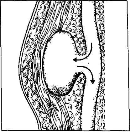
Fig. 23. False aneurysm
Early diagnosis of an aneurysm is critical as it prevents severe complications.
Sometimes a pulsating aneurysm is misdiagnosed as a Phlegmon and incised, resulting in life-threatening bleeding.
The manifestations of a traumatic aneurysm depend on its Location, the time elapsed since the injury, the severity of nerve trunk damage, and other factors. Patients complain of restricted limb movement, pain, a 'pins and needles' sensation, and a coldness in the limb.
Upon examination, an indistinctly bordered Swelling can be detected in the area of the injured vessel, which gradually increases in size and reaches its maximum within 6–10 days. The skin over the swelling becomes taut. In the case of an arterial aneurysm, Auscultation over the swelling reveals a systolic murmur synchronous with the cardiac systole. Placing a hand over the swelling allows pulsation to be felt.
The pulse at the peripheral end of the vessel becomes weaker or disappears.
If the hematoma is large, the peripheral segment of the limb may exhibit decreased skin sensitivity, motor dysfunction, and joint contracture.
The symptoms of an arteriovenous aneurysm differ somewhat from those of an arterial one. First of all, the swelling is much smaller, pulsation is sometimes barely noticeable, and the murmur has a systolic-diastolic, i.e., continuous character. The pulse in the periphery of the limb is weaker, and edema, venous congestion, and cyanosis appear. I. Rufanov notes that arteriovenous aneurysms are characterized by the 'slowdown sign', which means that after compressing the afferent part of the arterial trunk, the heart rate per minute decreases by 10–15 beats or more.
Angiography is an essential diagnostic method for patients with traumatic aneurysms.
A formidable complication of a false aneurysm is its rupture. This is accompanied by an increase in its size or profuse bleeding from the wound. Sometimes the wound becomes infected, and the aneurysm suppurates. Blood clots can detach from the aneurysmal sac and cause thromboembolism.
The treatment of traumatic aneurysms is surgical. There are three groups of operations:
1) vessel ligation with removal of the aneurysmal sac;
2) intrasaccular vessel suturing after opening the aneurysmal sac, with additional over-sewing of its cavity down to the vessel lumen;
3) circulation-restoring operations (removal of the aneurysm and suturing the ends of the vessel, or replacing the damaged segment with a graft in case of a large defect). In modern vascular surgery, these latter operations occupy a leading place in the complex of treatments for traumatic aneurysms.
In arteriovenous aneurysms, the operation consists of separating the artery and vein by eliminating the sac or channel between them while preserving the integrity of both vessels.
A dangerous complication of vascular injury is air embolism, which is the penetration of air into a blood vessel. This complication is more commonly observed in injuries to large-caliber veins (jugular, subclavian, axillary) and is explained by two factors: a) the presence of negative pressure in these veins; b) the fixation of their walls to adjacent tissues, which prevents them from collapsing upon injury. The exact amount of air entering the bloodstream that causes a critical condition in the patient has not been precisely established. Typically, a small amount of air that slowly enters the bloodstream does not cause fatal complications (the air is pushed from the right atrium into the lungs, where it may have minor consequences). If air enters the blood rapidly, even in a small amount, it can block the pulmonary artery or pulmonary capillaries, leading to death from asphyxia. If a significant amount of air enters the vessel, death occurs from cardiac paralysis due to overexpansion of the right side of The Heart and tricuspid valve insufficiency.
The clinical picture of air embolism includes unconsciousness, rapidly progressing skin pallor, heart rhythm and respiratory disturbances, and convulsions. With a slower onset, dyspnea, cold sweat, a feeling of dread, decreased heart rate, pupil dilation, and fainting are observed. If veins close to the heart are injured, a whistling sound appears. Frothy blood is released from the wound upon exhalation.
If this complication occurs, the wound opening must be tightly tamponaded or compressed with a finger, and subsequently, both ends of the ruptured vein must be ligated.
To prevent this complication during neck surgery, especially tumor removals, extreme caution must be exercised when dissecting veins by pre-placing a ligature underneath them.
Air embolism can also be arterial if air enters the VEINS OF THE Pulmonary Circulation and penetrates the systemic Arterial System through the left side of the heart. This occurs most frequently during Operations on the lungs and Pleura.
The patient should be placed in a HEAD-down position. If the operation is performed under local anesthesia, the patient is advised to hold their breath and strain. Intubation is performed, and the patient is switched to controlled ventilation with increased airway and pulmonary pressure. If the patient's condition does not improve after these maneuvers, a puncture of the right side of the heart must be performed to aspirate the air.
HEMOSTASIS
Bleeding can stop in two ways: spontaneously, as happens in A number of cases, and through surgical intervention.
The process of spontaneous hemostasis involves the following components: the blood vessel wall, plasma blood-clotting factors, and clotting factors derived from formed blood elements (platelets, erythrocytes, leukocytes).
There are two periods of spontaneous hemostasis: vascular-platelet (primary hemostasis) and coagulation (secondary hemostasis).
Primary hemostasis is responsible for stopping bleeding from small vessels. In case of damage to larger vessels, it is incapable of stopping the bleeding on its own.
Vascular-platelet hemostasis occurs in several stages. Immediately after vessel wall injury, a reflex spasm occurs as a result of vasoactive compounds formed in the damaged areas. Vasoconstrictive substances (serotonin, adrenaline, thromboxane) also take part in this process; they are released during platelet destruction. The vascular spasm is short-lived, quickly subsides, and bleeding may resume. Reliable hemostasis requires the action of other factors.
Immediately after an injury, platelets adhere to the damaged tissues (platelet adhesion). Upon doing so, the platelets change shape and extend long, thread-like projections known as pseudopodia. This promotes platelet aggregation (clumping). A crucial role in this process is played by adrenaline, adenosine triphosphate, arachidonic acid, and Prostaglandins released from ruptured platelets. Driven by these substances, a primary—so-called white—thrombus forms, sealing the injury site. This thrombus is loose, non-dense, and permeable to plasma.
Vascular spasm and the platelet plug contribute to the initial closure of the damaged vessel wall area.
Concurrently with vascular-platelet hemostasis, coagulation hemostasis develops, giving the platelet plug the necessary mechanical strength. This facilitates the formation of a secondary (true) thrombus. The primary mechanism here is The conversion of soluble fibrinogen into insoluble fibrin, creating a mesh of fibers that entraps blood formed elements. The enzyme Thrombin catalyzes the transformation of fibrinogen into fibrin. Normally, thrombin is absent from the blood, existing instead as its precursor, prothrombin. The conversion of prothrombin into thrombin is mediated by another enzyme, prothrombinase, which exists in two forms: tissue and blood. Tissue prothrombinase appears in the blood rapidly, within 5–10 s of injury, whereas blood prothrombinase appears later, after 7–10 min.
Within 5–7 s of its formation, prothrombinase adsorbs prothrombin onto its surface and converts it into thrombin. The latter possesses specific proteolytic properties that allow it to cleave fibrinopeptides A and B from the fibrinogen molecule, thereby transforming them into fibrin.
The subsequent stage in the Evolution of the blood clot is retraction. Serum and a portion of the formed elements are expelled, causing the clot to contract and become compact. Retraction is completed within 2–3 hours. The culmination of the blood clot transformation process is the formation of a dense thrombus.
The process of restoring the damaged area concludes with fibroblasts sprouting into the blood clot.
Some time after the secondary thrombus forms, a reverse process occurs—The breakdown of fibrin, known as fibrinolysis. This is driven by the active enzyme plasmin, which is generated from inactive plasminogen. As a result of this process, the thrombus dissolves and is replaced by regenerative elements, such as epithelium, neoinitima, and granulation Connective Tissue. Thus, fibrinolysis represents The final stage of hemostasis.
The processes of fibrinolysis and coagulation are closely interrelated, facilitated by the presence of the anticoagulant system in the blood. Under normal conditions, a dynamic equilibrium exists between the coagulation and anticoagulation systems, maintaining blood in a fluid state.
Hypocoagulation is mediated by several factors: the smooth surface of the vascular endothelium, the negative electrical charge of the vessel walls and formed elements (which causes them to repel one another), the presence of a thin fibrin layer on the vessel walls that actively adsorbs clotting factors (especially thrombin), a high blood flow velocity that prevents blood-clotting factors from reaching the required local concentration, and, most importantly, the presence of anticoagulation inhibitors in the blood. Physiological inhibitors include heparin, antithrombins, and antithromboplastins.
The most potent inhibitor is heparin, which is synthesized by heparinocytes located in various organs and tissues, particularly in the liver, lungs, and muscles. Heparin acts as an inhibitor across all phases of blood coagulation.
Among the six antithrombins, antithrombin III is the most versatile in terms of action and activity. It inactivates thrombin as well as nearly all other active clotting factors.
Antithromboplastins block the early phase of the blood coagulation process.
If primary, unassisted hemostasis proves insufficient and bleeding persists, surgical hemostasis is applied.
In turn, surgical hemostasis is categorized as temporary (provisional) and definitive.
Temporary hemostasis is performed immediately following trauma and constitutes one of the methods of emergency care.
The general public should also be trained in self-aid and mutual aid methods, as the timeliness and quality of such intervention can sometimes determine the victim's survival.
Definitive arrest of bleeding can only be achieved in a clinical Setting, specifically in an operating room or a dressing station.
Methods of temporary bleeding cessation are classified as follows.
Application of a tight, sterile pressure bandage to the bleeding site. This method is feasible only when sterile materials are readily available to the practitioner. The sole exception involves life-threatening hemorrhages; however, even in such cases, a pressure bandage is frequently insufficient.
Digital compression of arteries. This method can be utilized for hemorrhages from large blood vessels. Its principle involves compressing the artery (its proximal segment) against adjacent bony prominences near the bleeding site. For instance, the carotid artery can be compressed against the transverse processes of the cervical vertebrae; the Subclavian Artery against the first rib along the upper border of the middle third of the clavicle; the axillary artery within the armpit against the head of the humerus; the brachial artery against the humerus; the femoral artery directly above the inguinal ligament against the superior horizontal ramus of the pubis; and the Abdominal Aorta against THE Vertebral Column.
The artery is compressed against the bone using the two thumbs of both hands placed one over the other, or the thumb of a single hand. As fatigue sets in, the hands are alternated.
Digital arterial compression has certain drawbacks: prolonged pressure causes pain (and may compress neural trunks), the compressing fingers fatigue rapidly—potentially leading to the resumption of bleeding—and logistical challenges arise during patient transport.
Digital compression of the vessel directly within the wound. After donning a sterile glove or sanitizing the hands with alcohol, alcoholic iodine solution, chlorhexidine, or another antiseptic agent, the surgeon compresses the bleeding vessel inside the wound, thereby halting the hemorrhage.
Maximum flexion or hyperextension of a limb at a joint (Adelman's maneuver). Maximal flexion or hyperextension proximal to the injury site compresses the supplying artery. Flexion can be employed at the elbow, hip, and knee joints, whereas hyperextension is applicable at the shoulder and hip joints. These techniques are indicated for injuries to the subclavian, axillary, brachial, femoral, and tibial arteries. Adelman's method is unreliable for injuries to other arteries.
To achieve maximal flexion, a cotton roll is placed in the flexural crease of the joint, the limb is flexed, and it is secured in this position using a bandage (Fig. 24, b).
To achieve hyperextension, both elbows with bent forearms are brought together behind the back (almost until they Touch) and secured in this position with bandages. This method causes pain and is uncomfortable for the patient (Fig. 24, a).
Elevated position of the limb. This is achieved by placing cushions underneath, raising the head of the bed, or simply suspending the limb. As a result, blood pressure in the arteries and veins of the limb drops significantly, and the Temperature of the elevated part decreases by several degrees, which helps to stop the bleeding. This method is particularly effective for stopping venous bleeding.
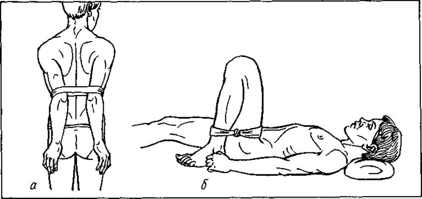
Fig. 24. Fixation of arms in THE POSITION OF maximum hyperextension (a) and maximum flexion (b) to stop bleeding
Circular compression of the limb proximal to the bleeding site is performed using an Esmarch tourniquet. This is the most commonly used method for temporary hemostasis. If a standard tourniquet is unavailable, bandages, improvised windlasses, belts, pneumatic cuffs, and other auxiliary means can be used.
The application of this method in surgery has a long history, dating back to the surgeons of Alexandria. Ambroise Paré refined it, and in 1873 Esmarch proposed using an elastic tourniquet for circular limb compression—a sturdy rubber tube 1.5 m long and 1.5 cm wide, featuring a hook on one end and a small chain on the other. Bier used a thick rubber band 2 cm wide with a button on one end and holes on the other for the same purpose.
In addition to rubber tourniquets, fabric ones have also been proposed. Elastic bandages are used for this purpose as well, as they exert a softer pressure. They are most commonly applied to the shoulder and in children.
When applying a tourniquet, the following rules must be observed.
1. Do not apply the tourniquet to bare skin to prevent pinching.
2. Before applying the tourniquet, the limb should be elevated and held in this position for 1–2 minutes so that the blood present in the vessels drains out and does not participate in the general circulation.
3. Apply the tourniquet proximal to the bleeding site in a stretched state.
4. Compress the limb until the pulse in the peripheral artery disappears. A loosely applied tourniquet may only compress the veins, thereby increasing bleeding.
5. The tourniquet should not be applied too tightly to avoid compressing nerve trunks (this is especially important during manipulations in the middle third of the arm, where the radial nerve can be pressed against the bone).
6. Each successive turn of the tourniquet should be applied with less tension than the preceding one.
7. The duration of tourniquet compression should not exceed 1.5 hours for the upper limb and 2 hours for the lower limb. Prolonged compression can lead to ischemic limb contracture resulting from the degeneration and breakdown of poorly perfused muscle fibers. If the tourniquet needs to be kept in place longer, it is periodically released after temporarily digitalizing (compressing with a finger) the artery proximal to the bleeding site.
8. A tag indicating the time of application is attached to the tourniquet.
9. The tourniquet must remain visible during patient transport.
Contraindications to applying a tourniquet include inflammation of the limb's blood vessels (phlebitis, arteritis, thrombophlebitis). This can cause a thrombus to dislodge, leading to Pulmonary Embolism or its branches. If an infection is present in the vessels, removing the tourniquet can cause it to enter the general bloodstream. In elderly patients, as well as those with alcoholism, Diabetes Mellitus, Syphilis, or Vascular Diseases, vascular compression with a tourniquet can cause severe trauma.
Potential errors and associated complications when using a tourniquet include: the development of paralysis and paresis (due to excessive compression); skin necrosis (applying the tourniquet to bare skin); limb gangrene (prolonged compression exceeding 2–2.5 hours); reduced tissue resistance to infection; and an increased risk of gas gangrene (due to the cessation of oxygen delivery to the tissues).
Temporary vascular shunting is usually performed under conditions close to those of an operating room. Both ends of the damaged artery are connected using a tight-elastic tube, around which the vessel wall is secured with ligatures. The advantage is that Blood Circulation is preserved in the body area distal to the injury. The shunt can function for up to several days until conditions are established for definitive hemostasis.
Clamping the bleeding vessel with a forceps. If a sterile forceps is available, it can be used to clamp the bleeding vessel, taking care not to capture adjacent nerve trunks.
Definitive hemostasis can be performed directly within the wound or proximally, at a certain distance from it.
There are 4 types of definitive hemostasis: mechanical, physical, chemical, and biological.
Mechanical hemostasis can be performed directly in the wound or proximally, at a certain distance from it.
In some cases, temporary hemostasis methods are used for definitive control: a pressure bandage (for capillary, venous, and minor arterial bleeding); a slightly elevated limb (for bleeding from a ruptured varicose vein of the lower extremities).
Minor capillary or parenchymal bleeding can be stopped by tightly packing the wound with gauze strips. The inserted pack exerts pressure on the wound walls and bleeding vessels, thereby slowing blood flow and creating favorable conditions for thrombus formation.
Hemostasis by packing can be used for capillary bleeding from the rectum, Vagina, Nasal cavity, parenchymal organs, Dural Venous Sinuses, and bone cavities.
This method has its drawbacks. Packing inhibits tissue regeneration; packing fresh wounds carries a risk of infection; and premature removal of the pack can cause secondary hemorrhage.
Packs are made of white, highly absorbent, sterile gauze—either dry or soaked in an antiseptic solution or warm isotonic sodium chloride solution.
It is recommended to remove the pack no earlier than 6–7 days. This Procedure is often painful and should therefore be performed under anesthesia.
Nevertheless, surgical intervention is most frequently resorted to for definitive hemostasis, involving various operative maneuvers (Fig. 25).
Fig. 25. Mechanical methods of hemostasis: a — ligation; b — electrocoagulation; c — vessel ligation using a Deschamps needle; d — ligation in continuity; e — suture ligation
Ligation of a bleeding vessel (using silk, catgut, synthetic threads, etc.) is performed as follows. The vessel is grasped with a specialized hemostatic clamp and ligated just below it. Prior to this, it is advisable to isolate the vessel from surrounding tissues. Before clamping the bleeding vessel, one must inspect the area carefully to avoid including adjacent tissues and structures (nerves, intestines, etc.) in the clamp. Various types of knots can be used for tying the ligature (Fig. 26): a reef (square) knot, in which the ends in the First and Second loops are passed in opposite directions; a surgeon's knot, which is a reef knot with a double twist on the first loop; and a granny knot, in which the ends in the first and second loops are passed in the same direction. The surgeon's knot is more secure and is therefore used when ligating large vessels. A reef knot suffices for medium and small vessels. The granny knot is unreliable and prone to slipping, so it is rarely used.
To prevent retrograde bleeding, both ends of the vessel (proximal and distal) must be ligated.
In some cases, a lateral ligature is applied to a vessel (rarely an artery, more often a vein). Such a ligature does not occlude the vessel lumen, but only narrows it slightly.
When applying a ligature to a vessel, the first throw of the thread is tightened while the clamp is still in place, and the second throw is tightened after the clamp is removed.
Suture ligation (transfixion). This method is used when There is a risk of the ligature slipping off the vessel. The clamp holding the vessel is lifted slightly, a needle with a thread is passed through the soft tissues and the vessel just beneath the tip of the clamp, and they are tied on both sides of it.
A drawback of the method described above is that the needle and thread are passed blindly, creating a risk of puncturing deeper-lying vessels and causing additional hemorrhage.
Circumferential suture (purse-string suture) of a bleeding vessel is used when it is difficult to grasp the bleeding site in the vessel with a clamp or when it is not visible in the wound. Using a needle, the thread is passed through the soft tissues around and near the vessel (in a purse-string fashion) and tied. In this way, the vessel is compressed along with the soft tissues. This method is used when ligating Vessels of the scalp, dura mater, fascia, certain muscles, the omentum, and the mesentery.
Fig. 16. Surgical knots:
a — simple; b — double surgical; c — reef
A disadvantage of this method, as with the previous one, is that the suturing is performed blindly.
Forced mechanical methods of definitive hemostasis include leaving a clamp in the wound (on the clamped vessel) for 4–7 days (a demeure) until thrombosis occurs. This method is unreliable and is used only as a last resort in cases of deep vascular injuries where other hemostatic techniques cannot be applied.
In cases of bleeding from a vessel located within a septic, purulent wound or a disintegrating tumor, when it is technically impossible to use any of the aforementioned methods, proximal ligation of the vessel is performed in healthy tissue upstream from the bleeding site. Indications for such ligation include the inability to ligate the bleeding vessel within the wound due to its friability, putrefactive breakdown, or tissue crushing, which makes any orientation within the wound impossible. Additionally, this method can be used as a preliminary stage before certain surgeries (ligation of the External Carotid Artery prior to jaw resection, or ligation of the lingual artery prior to glossectomy). This method also has drawbacks: if collateral circulation is well-developed, bleeding may persist, whereas if collaterals are poorly developed, tissue necrosis may occur in the area supplied by the ligated artery.
Artificial vascular embolization is most commonly used for bleeding from pulmonary, bronchial, cerebral, and gastric vessels. The method involves advancing a special catheter under fluoroscopic guidance into the bleeding vessel to deliver emboli that occlude its lumen. These are typically gelatin, silicone, or polystyrene microspheres. Subsequently, a blood clot forms at the site of the embolus.
The ideal mechanical method for definitive hemostasis is vascular anastomosis (suture) or replacement of the damaged vascular segment with a graft (preserved vessel or synthetic graft). Every surgeon should be proficient in performing vascular anastomoses. A major advantage of this method is the complete restoration of BLOOD FLOW IN the injured vascular bed. Large vessels can be anastomosed without magnification, whereas small ones require microsurgical techniques. The following conditions must be met when performing a vascular suture:
a) strict asepsis and antisepsis, as the success of vascular repair largely depends on the condition of the wound;
b) mandatory apposition of the vessels by their intimal lining;
c) availability of appropriate suturing instruments — fine anatomical forceps, atraumatic needles, needle holders (in recent years, special devices have been proposed for connecting vessels using tantalum staples or special medical glue);
d) avoiding trauma and extensive Separation of the vessel from its outer sheath (denudation) over a significant distance, as this may damage the vessels supplying its wall.
In cases of complete vessel transection, a circular suture is applied; for lateral wounds, a lateral suture is used.
To prevent thrombosis at the suture site, heparin is administered into and around the vessel lumen.
Physical (predominantly thermal) methods of hemorrhage control are based on the application of high or low temperatures. Low temperatures cause vasospasm, while high temperatures induce protein coagulation and thrombus formation. Cold therapy is used to stop gastric or duodenal bleeding of ulcer origin as part of conservative management. Patients are instructed to swallow ice or have an ice pack applied to the epigastric region. The Effect of cold on deeply situated bleeding vessels is reflex-mediated. Ice is also used for intradermal or subcutaneous hemorrhages. It must be borne in mind that using cold to stop bleeding is unreliable. Prolonged exposure to cold, combined with impaired tissue Nutrition, can lead to skin necrosis in the affected area.
High temperatures used for hemostasis may include: hot (56–60 °C) isotonic sodium chloride solution
(for bleeding from muscles, parenchymatous organs, or Bone tissue); electrocautery (a tip heated by an electric current that generates high temperatures upon contact with tissues, thereby coagulating blood Proteins); Shamrayevsky biactive electrodes — biactive scissors and forceps (when the blades of the forceps or scissors are compressed, an electric current is generated between them, which also raises the tissue temperature and promotes thrombus formation).
Improper use of an electrosurgical knife (diathermy or biactive electrodes) can result in a wide zone of tissue necrosis.
In recent years, cryosurgery (local tissue freezing) and laser photocoagulation have been introduced into clinical practice for hemostasis. The latter method offers several advantages over electrocoagulation: there is no direct contact between the electrode and the tissues; the necrobiotic zone in the coagulation area is small; and it provides a better view of the coagulated vessel since it is not obscured by the electrode.
Chemical methods of hemostasis are used either in combination with mechanical methods or independently. They rely on vasospasm or enhanced blood clotting. Hemostatic chemicals are classified into topical (external) and systemic (internal).
Among topical agents, adrenaline — the active hormone of the Adrenal Glands — is most commonly used today. Applied locally, it causes vasoconstriction and thrombosis. It is typically used in combination with local anesthetics, most frequently in dental practice. A drawback of this method is that once the effect of adrenaline wears off during or after the postoperative period, the vessels may dilate, leading to recurrent bleeding.
Hydrogen peroxide (H2O2) can be used for bleeding from mucous membranes (nose, gums, tongue, tooth extraction socket) and bone tissue. Within tissues, hydrogen peroxide breaks down into Water and oxygen. The wound surface becomes covered with foam, and the blood clots.
Systemic hemostatics are divided into two subgroups: agents that cause vascular constriction and agents that enhance blood coagulation.
Substances of the first subgroup include: ergot extract (20 drops 3 times daily), adrenaline solution (1:1000; 0.5 ml subcutaneously or 10–20 drops 3 times daily), and adrenoxon (a stabilized product of adrenaline oxidation, used to stop capillary and parenchymal bleeding via gauze tampons soaked in a 0.075% solution, or via intramuscular or subcutaneous administration of 1 ml of a 0.075% solution 2–3 times daily).
Substances of the second subgroup include calcium chloride, administered intravenously in a 10 ml volume at a 10% concentration. A hemostatic effect is also achieved by the intravenous administration of a 5% sodium chloride solution and a 40% glucose solution. Other notable preparations include epsilon-aminocaproic acid (used to control bleeding associated with fibrinolysis, liver disease, burn disease, hemophilia, etc.). It is administered intravenously as a 5% solution (50–100 ml) or orally at 0.1 g per 1 kg of body weight every 4 hours.
Biological methods of hemostasis are based on the capacity of biological tissues to promote blood clotting. Today, they are widely used in surgical practice. Biological agents are divided into two groups: for local and general application. Preparations of the first group include fibrin film, various sera, hemophobin, and thrombin (sprinkled onto the bleeding wound or used to saturate inserted tampons).
Favorable outcomes are achieved using the sponge introduced by L. Bogomolova in 1948. It is manufactured from human native blood plasma and thromboplastin. Upon contact with blood, it forms a film that seals the lumen of small bleeding vessels.
Hemostatic sponge can be combined with Antibiotics.
Dentists use bioplastic — a preparation introduced by L. Bogomolova in 1957 — for packing tooth sockets.
Dry thrombin is a powder readily soluble in isotonic sodium chloride solution. Tampons are moistened with the thrombin solution and inserted into the wound for 10–20 minutes. If the effect is insufficient, the procedure can be repeated. Thrombin solution can also be instilled into the urinary bladder and stomach if bleeding originates from these organs.
The preferred local hemostatic method, particularly for parenchymatous organs, is packing the bleeding wound with the patient's own tissues rich in thromboplastin. For this purpose, a pedicled greater omentum, muscle, or subcutaneous adipose tissue can be used. Flaps of such tissues are applied as a tampon to the bleeding site or secured with sutures. This method is convenient because the "living tampon" remains permanently in the body, allowing such wounds to be closed tightly.
Among systemic hemostatic agents, top priority is given to whole blood stored for no longer than 6 hours. Fresh whole blood contains a range of components that facilitate blood clotting (prothrombin, calcium salts, vitamin K, etc.). Concurrently with blood, native, fresh-frozen, antihemophilic plasma and serum are transfused. Fibrinogen has found widespread application for hemostasis. It is supplied as a sterile powder in 250–500 ml vials (containing 1–2 g, respectively). Before use, the powder is dissolved in isotonic sodium chloride solution and administered intravenously.
Fibrinolysis inhibitors, which reduce the fibrinolytic activity of blood (trasylol, kontrikal, iniprol), also exhibit hemostatic properties.
Other biological hemostatic agents include vikasol, ascorbic acid, animal blood serum, or its derivatives (hemostol, autohemostol, vivicol). Fresh serum (stored for 12–15 days) is used to stop bleeding. Sera with longer storage periods have the opposite effect — they reduce blood clotting. Serum is administered subcutaneously in doses of 20–40 ml or used to moisten tampons prior to their insertion into the wound.
In clinical practice, the physician employs the method that is optimal for the given situation. Sometimes this is a single method, but more often it is a combination of them. However, in every case, it must be remembered that stopping the hemorrhage is only half the battle in treating patients with vascular injuries and blood loss. Immediately following (or even concurrently with) hemostasis, measures must be taken to restore lost blood volume.
Last update: 08/08/2026
Editorial and Educational Adaptation: This material has been compiled based on the primary/original source text. The project team performed an editorial review, corrected technical inaccuracies, structured sections, and adapted the content for an educational format.
What was processed:
- elimination of formatting defects (OCR errors, structural breaks, corrupted characters);
- editorial organization of content;
- standardization of terminology in accordance with academic sources;
- verification of factual statements against the original source text.
All mentions of the author, publication year, and origin of the primary text have been preserved in accordance with the source.