Military Surgery with Emergency Surgery - V.Ya. Bilyi 2004
Hemorrhage and Blood Loss
Introduction. Bleeding and Blood loss are among the most frequent and life-threatening consequences of combat trauma in wartime, as well as a major cause of mass casualties during natural disasters and man-made catastrophes in peacetime.
Bleeding and blood loss occur in both open and closed injuries. They are a frequent cause of death among the wounded on the battlefield, at forward medical evacuation echelons, and in mass casualty incidents during peacetime disasters.
Bleeding and blood loss accounted for 34.6% of immediate battlefield deaths during World War II, and 24% during the nine-year war in Afghanistan. In medical facilities within combat zones, blood loss, Hemorrhage, and Shock caused the death of 30% of the wounded during World War II and 46% in Afghanistan. In peacetime, following the earthquake in Armenia, blood loss and shock were observed in 25.7% of the victims, resulting in a mortality rate of 4.6% among all casualties.
Characteristics and Classification of Hemorrhage. Hemorrhage is the escape of blood from the lumen of Blood Vessels due to trauma or increased permeability of their walls.
Hemorrhages are classified According to the time of onset, The Nature and caliber of the damaged blood vessels, and the site of blood flow. A distinction is made between actual hemorrhage, focal hemorrhage (hemorrhagic infiltration), and hematoma.
Hemorrhage refers to the escape of blood from blood vessels either externally, into a hollow organ, or into Body Cavities.
Hemorrhagic infiltration is the diffuse permeation of Tissues by blood (tissue imbibition).
A hematoma is the infiltration of body tissues by blood accompanied by its accumulation in newly formed cavities.
When a hematoma communicates with the lumen of a medium- or large-caliber artery, a pulsatile hematoma develops, which may result in a false aneurysm.
Hemorrhages are distinguished into Primary and secondary. Primary hemorrhages occur immediately after injury. Recurrent hemorrhages are subdivided into early and late. Early recurrent hemorrhages occur before a thrombus is formed.
Classification of Hemorrhages.
1. Anatomic classification: arterial, venous, capillary, and parenchymal.
2. By mechanism of occurrence:
- hemorrhage resulting from mechanical injury or rupture of a vessel;
- hemorrhage resulting from erosion of the vessel wall (due to purulent-septic processes, necrosis, or tumors);
- hemorrhage resulting from microscopic-level Impairment of the vascular wall permeability (avitaminosis, hemorrhagic vasculitis, Sepsis, uremia).
3. By time of onset:
- primary hemorrhage, associated with direct vascular injury during trauma, occurring immediately after the injury is inflicted;
- secondary hemorrhage, which can be either early or late.
Early secondary hemorrhage predominantly occurs from a few hours up to 3–5 days after injury. There are two main causes of early secondary hemorrhage: slippage of a ligature from a vessel due to insecure ligation, and dislodgement of a thrombus from the vascular lumen upon stabilization of blood pressure.
Late secondary hemorrhages (after 4–5 days) are typically erosive in nature, associated with Necrosis of the vessel wall or the thrombus As a result of a purulent-septic process.
4. By relation to the external environment:
- external, when blood escapes outwardly from the wound;
- internal, when blood flows into the lumen of hollow Organs or internal body cavities (hemoperitoneum into the Abdominal cavity; hemothorax into the pleural cavity; Hemopericardium into the pericardial cavity; hemarthrosis into the joint cavity).
Etiology AND Pathogenesis of Blood Loss. The clinical manifestations of bleeding depend on the volume of blood lost.
Blood loss is a pathological condition resulting from hemorrhage, characterized by a series of adaptive and pathological reactions.
Two key factors determine the outcome of hemorrhage: the volume and The rate of blood loss. The highest rate of blood loss is observed in arterial bleeding, with both volume and rate depending on the vessel caliber and The Nature of the injury. A single loss of approximately 40% of the circulating blood volume is incompatible with life.
Blood loss is classified according to both its volume and the severity of physiological alterations. A distinction is made between the absolute volume of blood loss and the severity of post-hemorrhagic disorders, which are primarily assessed based on the degree of hypovolemia caused by the reduction in circulating blood volume (CBV).
The magnitude of blood loss is evaluated from three Perspectives: the reduction in fluid filling the vascular bed; the loss of erythrocytes, which are responsible for Oxygen transport; and the loss of plasma, which plays a crucial role in tissue METABOLISM.
The primary driver in the pathogenesis and thanatogenesis of blood loss remains the reduction of the CBV filling the vascular bed, leading to hemodynamic instability. Another critical factor is the alteration of the body's oxygen balance. Both hemodynamic andemic factors trigger the body's defense mechanisms, which may lead to the compensation of blood loss. Compensation occurs through the shift of extracellular fluid into the vascular bed (hemodilution); increased Lymph flow; regulation of vascular tone, known as the "centralization of Circulation"; increased Heart rate; and enhanced tissue oxygen extraction. Compensation is achieved more easily when less blood is lost and bleeding occurs more slowly. Conversely, when compensatory mechanisms fail—and even more so during decompensation—blood loss transitions into a critical condition.
The so-called threshold of death is determined not by the volume of bleeding, but by the number of erythrocytes remaining in circulation. This critical reserve equals 30% of the erythrocyte volume and only 70% of the plasma volume. The body can survive the loss of 2/3 of its erythrocyte volume, but cannot tolerate the loss of 1/3 of its plasma volume. This approach to evaluating blood loss allows for a more comprehensive Assessment of the body's compensatory processes.
Classification of blood loss. There are three degrees of severity of blood loss: mild, severe, and extremely severe (massive):
1. Mild - loss of up to 20% of CBV (up to 1000 mL).
2. Severe - loss of 20% to 30% of CBV (1000–1500 mL).
3. Extremely severe — loss of more than 30% of CBV (more than 1500 mL).
Determining the degree of blood loss is fundamental for establishing the appropriate course of
transfusion replacement therapy.
Clinical manifestations of severe hypovolemia appear starting from a blood loss of 20–30% of CBV.
There are three stages of severe hypovolemia:
1st stage — compensated (reversible);
2nd stage — provisionally decompensated (provisionally reversible);
3rd stage — decompensated (irreversible).
Compensated hypovolemia — the volume of blood loss is successfully managed by the patient's intrinsic compensatory mechanisms.
Provisionally decompensated hypovolemia — characterized by profound Circulatory Disorders, where arteriolar spasm can no longer compensatorily maintain central hemodynamics and normal blood pressure. Decentralization of circulation develops as a consequence of metabolite accumulation in tissues and capillary bed paresis.
Decompensated hypovolemia — characterized by prolonged (exceeding 12 hours) refractory hypotension, unresponsive Transfusion Therapy, and The Development of multiple organ dysfunction syndrome.
Diagnosis of hemorrhage and blood loss.
1. Local symptoms (external or internal bleeding).
2. General symptoms of bleeding (pallor of the Skin; alterations in consciousness and blood pressure; central venous pressure; heart rate; hourly diuresis; body Temperature; Hemoglobin and hematocrit levels; blood oxygen tension).
3. Special diagnostic Methods (diagnostic puncture; endoscopy; laparocentesis, laparoscopy; thoracentesis, thoracoscopy; angiography; ultrasound; radiography; computed tomography, MRI).
Determining the magnitude of blood loss. Assessing blood loss during wartime presents specific challenges due to the lack of a sufficiently informative and rapid evaluation method, requiring physicians to rely on a combination of clinical signs and laboratory data.
In combat settings, 4 groups of indicators are used to determine the volume of blood loss:
1. By the localization of the injury and the extent of damaged tissue.
2. By hemodynamic parameters (shock index, systolic BP).
3. By blood concentration parameters (hematocrit, hemoglobin).
4. By changes in BCC.
When providing aid to a casualty, the volume of blood loss can be roughly estimated based on the localization of the injury: for severe chest trauma, it amounts to 1.5–2.5 L; abdominal trauma, up to 2 L; multiple pelvic bone fractures, 2.5–3.5 L; open femoral fracture, 1.5–1.8 L; closed femoral fracture, 2 L; lower leg fracture, up to 0.8 L; humeral fracture, 0.6 L; and forearm fracture, 0.5 L. These data are usually sufficient when administering initial medical care. One can also use the standardized measure of injured tissue volume by taking the casualty's palm as a unit of measurement, which corresponds to approximately 0.5 L of blood loss.
Accordingly, all wounds are divided into 4 groups:
1. Minor wounds — the area of damage is smaller than the palm. Blood loss is up to 10% of BCC.
2. Medium-sized wounds — the area of damage does not exceed the area of 2 palms. Blood loss — up to 30% of BCC.
3. Large wounds — the area is larger than 3 palms, but does not exceed 5 palms. Average blood loss — about 40% of BCC.
4. Massive wounds — the area is larger than 5 palms. Blood loss — about 50% of BCC.
Under any conditions, the volume of blood loss can be determined using a hemodynamic parameter — the shock index. In essence, this is the first crucial objective indicator that allows for an approximate assessment of both the severity of the casualty's condition and The amount of blood lost.
The shock index represents The ratio of heart rate to systolic blood pressure. Normally, this indicator is 0.5. Each subsequent increase of 0.1 corresponds to a loss of 0.2 L of blood, or 4% of BCC. An increase in this indicator to 1.0 corresponds to a loss of 1 L of blood (20% of BCC), to 1.5 — 1.5 L (30% of BCC), and to 2 — 2 L (40% of BCC).
To determine acute blood loss, it is more appropriate to use hematocrit or hemoglobin levels. The hematocrit method is the most widely used and is represented by the following formula:
BL = BCCn X (Hctn -Hctf)/Hctn,
where: BL — blood loss, L; BCCn — normal BCC; Hctn — normal hematocrit, which is 45 g/L for men and 42 g/L for women; Hctf — actual hematocrit determined in the casualty after cessation of bleeding and hemodynamic stabilization. In this formula, hemoglobin levels can be used instead of hematocrit, assuming a normal level of 150 g/L. The determination of blood loss in trauma based on hemodynamic parameters is presented in Table 2.
However, concentration-based Methods for determining blood loss relying on hematocrit and hemoglobin values can only be recommended for calculations in slow blood loss, because their true values become realistic only after complete hemodilution, which occurs in the body within 2–3 days. In all cases of severe blood loss, the diagnosis is established using the simplest and least time-consuming methods, because due to a lack of time, any additional examinations that delay surgical intervention are unacceptable at this stage. We are referring to the complex of necessary diagnostic methods for a casualty with massive blood loss admitted to the hospital.
There are 2 priority tiers for examination, which correspond to the choice of tactical decisions regarding surgical intervention (emergency, delayed, or early). The primary objective of the examination is to determine the severity of the casualty's condition, the CHARACTERISTICS OF THE bleeding, and the volume of blood loss, culminating in a clear Conclusion regarding further management tactics.
First-tier diagnostic Procedures include:
1. A rapid external examination of the casualty, skin, and mucous membranes.
2. Determination of heart rate and measurement of BP.
3. Assessment of consciousness.
4. Inspection and Auscultation of the chest, and Palpation of the abdomen.
5. Determination of blood loss volume using the shock index.
6. Performing X-ray Examination.
7. Clinical assessment of the severity of hypovolemia using the capillary refill test (if the pale spot formed after pressing on the forehead skin disappears within 1.5 seconds, the blood volume deficit is at least 20%; in severe hypovolemia, this test is unfeasible).
8. ECG recording.
9. Insertion of a catheter into a major vein and blood sampling to determine hematocrit, hemoglobin, blood group, acid-base balance, and blood gases; under appropriate conditions, injection of an indicator to assess blood volume; initiation (or continuation) of infusion therapy.
10. Urinary Bladder catheterization with hourly diuresis measurement.
11. Making a decision on emergency surgery or the tactics of further examination and Treatment. If the casualty is transferred to the operating room, a catheter is inserted through subclavian vein puncture into the right Chambers of the heart to measure central venous pressure (CVP).
Subsequent second-stage measures:
1. Thorough examination of the injury and bleeding site. A conclusion regarding the continuation or cessation of bleeding is mandatory. This involves the full range of additional instrumental examinations (fibrogastroscopy, laparocentesis, diagnostic puncture of body cavities).
2. X-ray examination.
3. Assessment of CVP.
4. Determination of the severity of hypovolemia and the deficit of major blood volume components.
5. Repeat testing of peripheral blood hematocrit to evaluate changes in hematological parameters.
6. Examination of blood biochemical parameters and the body's fibrinolytic system.
7. Conclusion on the severity of the casualty's condition and determination of further treatment tactics (conservative or surgical, involving delayed or early surgery).
8. Calculation of the required volume of plasma substitutes to restore blood loss.
Thus, The Scope of examination depends on the severity of the casualty's condition and the objectives at hand. First and foremost, casualties arriving in an extremely severe condition with a clearly identified source of bleeding are examined, where treatment begins literally at the moment of examination and postponing the decision for surgery is impossible. In unclear cases, when it is necessary to identify the source of bleeding and comprehensively assess the severity of changes occurring in the body, the examination is repeated. Subsequent measures serve to refine the diagnosis, are often carried out under dynamic monitoring, and also conclude with a decision on further treatment tactics.
Treatment of bleeding and blood loss at the medical evacuation stage. Therapeutic tactics consist of stopping the bleeding and timely replenishment of lost blood volume. Methods of arresting hemorrhage are described in the chapter on injuries to major vessels. At pre-hospital medical evacuation stages, temporary hemostasis is performed, while definitive hemostasis is carried out at the stage of qualified and specialized care.
First aid on the battlefield (in the disaster area):
1. Temporary arrest of bleeding using one of the following methods: a tourniquet, a pressure bandage, tight wound packing, maximum flexion of the extremity, digital compression of the bleeding vessel in the wound and proximally, application of a clamp to the damaged vessel, elevation of the limb, or temporary shunting.
2. Intramuscular administration of analgesics.
3. Transport immobilization of the injured extremity.
First-aid/Pre-hospital care. Inspection and adjustment of bandages and tourniquets, and improvement of transport immobilization. In case of severe blood loss, infusion therapy (Ringer's solution, rheosorbilact, sorbilact) and cardiovascular agents (caffeine, cordiamin, etc.) are administered. If necessary, administration of analgesics can be repeated. Immediate evacuation.
First medical care. During medical sorting, the following groups of casualties are identified:
Group I — casualties with ongoing external bleeding, with a previously applied tourniquet, and with mild-to-moderate or severe blood loss are sent to the dressing room;
Group II — casualties with arrested bleeding and mild-to-moderate blood loss; they receive medical care in the sorting and evacuation tent or on the sorting platform and are promptly evacuated to the next medical evacuation stage.
In the dressing room, bleeding must be stopped as a matter of priority.
For every wounded patient with a tourniquet in place, it is necessary to determine whether a major blood vessel is indeed injured and, if so, whether it is possible to replace the tourniquet with an alternative means of temporary hemorrhage control (tight wound packing; application of a hemostatic clamp to a visible vessel in the wound; ligation; vessel suturing; intravenous infusion of plasma expanders).
In cases of extensive wounds, the injured vessel may be readily visible and accessible for clamping or ligation. Occasionally, wound edges can be retracted with hooks to facilitate vessel localization.
When a clamp is applied to a vessel within the wound, it must be securely anchored using a bandage passed through its ring. Such precautionary measures are necessary not only because a pulsating clamp can cause severe discomfort, but also to prevent it from shifting or dislodging during patient evacuation.
The retention of a clamp or pack within the wound must be documented in the medical record.
During the provision of initial medical aid, the physician checks the tourniquet in the dressing room, having previously ensured that manual compression of the vessel proximal to the wound can be performed immediately if needed. If bleeding resumes, the tourniquet must be reapplied. Before re-clamping the vessel, the extremity is allowed a recovery period: digital pressure is applied to the vessel for 10–15 minutes; Blood Circulation in the limb is partially restored through collateral pathways during this time, thereby reducing the risk of irreversible ischemia.
Reapplication of the tourniquet should be performed after placing a piece of plywood splint or dense cardboard beneath the tourniquet on the aspect of the limb opposite the vascular bundle projection. This technique largely preserves collateral circulation and makes the tourniquet much better tolerated by the casualty.
For bleeding from wounds in the gluteal region or popliteal fossa, tight wound packing may be used. To prevent the pack from being expelled from the wound, the skin over the inserted pack can be secured with a few sutures.
All casualties with vascular injuries are administered Antibiotics, tetanus toxoid, and analgesics.
During evacuation, measures must be taken to prevent hypothermia or frostbite in the limb compressed by the tourniquet, which can occur even at relatively high ambient temperatures. In mass casualty scenarios, the scope of care for patients with vascular injuries during initial medical aid is inevitably reduced to life-saving interventions only and is limited to hemorrhage control using tourniquets or pressure dressings. Infusion of plasma expanders is performed solely in cases of life-threatening blood loss.
Qualified medical care. Approximately 60% of casualties with bleeding and blood loss arrive in severe condition. During medical sorting, casualties should be divided into 5 groups:
1) those in urgent need of Surgical treatment: ongoing hemorrhage, external bleeding previously stopped by a tourniquet, tight wound packing, or an expanding hematoma;
2) those requiring delayed surgical treatment: presence of wounds along the projection of major vessels, subcompensated limb ischemia, and other signs suggesting major vascular injury; in specific situations and partially during a high influx of casualties, evacuation of this category to a specialized vascular unit is necessary;
3) casualties with grade III contusion of a major limb trunk accompanied by a critical post-traumatic time (exceeding 6 hours) and decompensated limb ischemia, requiring surgical intervention;
4) casualties with grade I–II contusion of a major limb trunk presenting with early signs of compensated ischemia; these patients are evacuated to a specialized vascular unit for evaluation and treatment;
5) casualties in a terminal state; admitted for palliative and supportive care.
Class="center">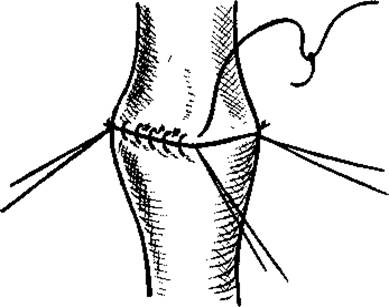
Fig. 11. Circular vascular anastomosis.
The surgeon must perform definitive hemostasis for all casualties who require it on life-saving grounds. The following emergency surgical measures are applied: ligation of both vessel ends within the wound; ligation in continuity; application of a lateral or circular suture to the vessel (Fig. 11).
Temporary vascular shunting or endoprosthesis should be performed for temporary hemostasis and restoration of main blood flow.
In wartime, the choice of definitive hemorrhage control method is determined not only by medical indications, but also by the tactical situation, the feasibility of a comprehensive examination, the surgeon's qualifications, the availability of necessary instruments, and time for dynamic postoperative monitoring.
In modern warfare, at the stage of qualified surgical care, reconstructive vascular procedures that require relatively significant time expenditure may only be undertaken if surgeons are not overwhelmed with life-saving emergency care. Therefore, despite modern surgical capabilities, vessel ligation remains the primary method of bleeding control at this stage.
In modern vascular surgery, vessel ligation is a forced Procedure performed under extreme combat and medical conditions. The frequent development of acute ischemia following major vessel ligation necessitates subsequent temporary shunting or grafting with early definitive repair at hospital-base medical facilities.
Qualified surgical care for vascular injuries includes the following measures: surgical intervention for definitive hemostasis; Management of acute blood loss; Prevention of wound infection complications; rapid evacuation of the casualty to a specialized care facility.
Surgical intervention should be performed not as a exploratory wound revision to locate the bleeding source, but rather to explore the vessel indicated by the presenting symptoms. The operation must be performed under anesthesia, beginning with the exposure of the vessel along its entire course and its control with a soft atraumatic vascular clamp. Surgical access to the vessel is achieved through a standard incision corresponding to the TOPOGRAPHIC AND ANATOMICAL Features of the given area and the suspected level of arterial injury. Access to the vessel through the wound itself should be used only when the wound lies directly over the PROJECTION OF THE vascular bundle.
In cases of partial tears and lateral vascular injuries, ligatures should be placed as close to the injury site as possible, with the most conservative trimming of the damaged arterial segment.
The most effective method for permanently stopping bleeding is the ligation of both ends of the damaged vessel directly within the wound. Applying ligatures to only the proximal end of the artery, let alone ligating it along its entire course (the Hunterian method), results in a 30 to 50% recurrence rate of hemorrhage and therefore should only be used under unavoidable circumstances.
Ligation of both vessel ends in the wound is advisable in cases of extensive soft tissue damage and large vascular defects, multiple injuries to a single vessel, severe inflammatory processes in the wound, delayed admission of casualties where viability is maintained via collateral circulation, or traumatic limb amputations.
In instances where ligating the ends of the damaged vessel in the wound is impossible, vessel ligation along its entire course is performed. This intervention is recommended for recurrent hemorrhage, the presence of purulent-septic complications in the wound area, or arterial injuries in anatomically inaccessible locations.
Fasciotomy is mandatory in INJURIES OF THE popliteal artery or vein, or when surgical treatment is delayed by more than 6-8 hours following the injury. Fasciotomy must always be performed upon the appearance of tissue edema and neurological deficits in the distal segments of the limb. For this purpose, two longitudinal skin and subcutaneous incisions, 5 cm in length, are made in the upper and lower thirds of the leg and forearm, through which the fascia is incised using opposing longitudinal cuts.
On the leg, it is necessary to open the fascial compartment of the Posterior Muscle group and the anterior tibial space; on the forearm, the fascial spaces of the wrist and finger flexors and extensors. Following fasciotomy, limb edema subsides, elevated intercompartmental pressure decreases, and both main and collateral circulation improve. Skin wounds are sutured after 7-8 days.
Vascular surgery requires the utmost tissue gentleness, alongside the perivascular administration of 10-15 ml of a 2% novocain solution. Upon identifying the site of vascular injury, 2000-3000 IU of heparin dissolved in 40-60 ml of a 0.25% novocain solution is injected into the distal segment, which relieves peripheral arterial spasm and prevents thrombosis.
A lateral vascular suture is indicated solely for marginal longitudinal injuries of large vessels with a defect not exceeding 1/3 of the vessel's diameter (Fig. 12).
If necessary, an autologous patch harvested from an adjacent vein can be utilized; however, in massive blast or gunshot wounds, such a vein may be absent due to severe tissue destruction. Sutures are placed in a transverse direction.
Temporary endoprosthetic repair of a damaged artery involves preparing the arterial ends, inserting an endoprosthesis of appropriate length first into the peripheral and then into the central vascular segment, and securing it with individual ligatures. The most common length of endovascular tubes used is 65-75 mm. A standard blood transfusion tubing is frequently employed as an endoprosthesis (see insert, Fig. 13). The shunt can function effectively from several hours up to 2 days.

Fig. 12. Lateral vascular suture.
Naturally, the smaller the tube diameter, the higher the probability and frequency of thrombosis; however, this process develops gradually with The formation of a layered thrombus. Consequently, There is a sufficiently large time margin to transport the casualty to a specialized medical facility and perform reconstructive restorative surgery.
If a casualty arrives with a tourniquet applied and signs of limb necrosis, removing the tourniquet is strictly contraindicated. In such cases, when providing limited qualified surgical care, a limb amputation must be performed directly above the tourniquet level without removing it.
In casualties whose vascular injuries follow a relatively favorable course, surgical intervention can be postponed until arrival at a specialized care facility. Such injuries include hematomas that show no tendency to expand and carry no threat of external rupture. Anticipating potential bleeding, a provisional (un tightened) tourniquet is applied to each of these casualties prior to evacuation.
During surgery, the following measures may be implemented to prevent and combat acute ischemia:
- transfusion of packed red Blood Cells and blood substitutes that elevate blood pressure, thereby enhancing collateral function;
- novocain blockade of the neurovascular bundle or compartment anesthesia of the limb proximal to the injury level.
Following surgery on major vessels, the casualty requires close monitoring for 6-12 hours to verify adequate limb perfusion. If perfusion is sufficient and the patient's general condition is satisfactory, the casualty should be evacuated During the first 24 hours using appropriate medical transport. During evacuation, transport immobilization and a provisional tourniquet must be applied.
In the postoperative period, maintaining blood pressure at an adequately high level (no lower than 100 mm Hg) is crucial for preventing and treating acute ischemia. Permitting circumstances allow, it is advisable to perform a novocain blockade of the stellate ganglion or lumbar sympathetic ganglia, repeat the neurovascular bundle blockade, and administer antispasmodic agents intramuscularly. Warming the ischemic limb is strictly contraindicated.
Under favorable conditions, the scope of qualified care for vascular injuries can be expanded. The fundamental principle of modern management in acute vascular trauma is the restoration of the anatomical integrity and function of the injured vessel. Achieving this requires that every surgeon be capable of arresting hemorrhage and restoring vascular continuity.
To determine the rational strategy when providing qualified care to casualties with vascular injuries, one must be guided by the degree of ischemia in the affected limb:
- in compensated ischemia (preserved active movements and sensation distal to the injury site) — reconstructive vascular surgery is not indicated; both ends of the damaged vessel may be ligated within the wound;
- in subcompensated ischemia (loss of active movements and sensation distal to the injury site, with preserved passive joint movements) — preserving the limb requires restoring the patency of the main artery via temporary shunting at the qualified care stage, followed by reconstructive surgery in a specialized hospital;
- in decompensated ischemia (rigor mortis of the limb Muscles: muscles are firm, passive movements impossible) — restoring blood circulation is life-threatening; amputation is indicated. Primary amputations for major vessel trauma are indicated in cases of massive destruction of the vessels themselves, as well as soft tissues, bones, and nerves. These indications expand in the presence of combined radiation injuries.
Specialized surgical care. When specialized medical care is delivered in a vascular surgery unit, the majority of casualties with major vessel injuries arrive within the first few hours to 24 hours post-injury (via air or helicopter transport). Following clinical and radiological evaluation (angiography), various reconstructive and restorative vascular procedures are performed: vascular suturing for marginal injuries, autogenous vein grafting (see insert, Fig. 14), alloplastic grafting, circular vascular anastomosis, aneurysm resection with restoration of blood flow, etc.
Specialized care includes the following measures: vascular restoration combined with thorough surgical wound debridement; management of acute blood loss resulting from the injury; prevention and treatment of acute ischemia and other complications associated with acute vascular trauma; and prevention of wound infection.
A crucial aspect of emergency blood loss management at the stages of qualified and specialized medical care is determining the BCC, assessing its quantitative and qualitative parameters, and evaluating the efficacy of the ongoing infusion therapy.
If infusion therapy is delayed, the required volume of solutions and their composition change. Specifically, for every hour following the first 60 minutes after the injury, the transfusion volume is increased by 15–20%. If the infusion is initiated 2–4 hours after trauma or wounding, colloidal solutions are added to the infusion regimen at a 1:1 ratio with crystalloids.
The primary goal of infusion therapy using plasma expanders is to restore the BCC to 95–100% of the individual normal value, achieve an acceptable degree of hemodilution (hemoglobin 70 g/L, hematocrit 0.20), and reach safer target parameters (100 g/L and 0.30, respectively).
An important consideration in compensating for blood loss is the accurate assessment of extracellular fluid in maintaining a stable BCC. To uphold the principles of adequate blood replacement therapy, its volume should be 170–180% of the blood loss volume, while THE RED BLOOD Cell mass should account for 50%, ensuring that tissue oxygen transport is not compromised. In the settings of qualified and specialized medical care, backed by a wide array of plasma expanders and blood components, a strict protocol for acute blood loss compensation must be followed across five distinct grades (Table 3).
Last update: 08/08/2026
Editorial and Educational Adaptation: This material has been compiled based on the primary/original source text. The project team performed an editorial review, corrected technical inaccuracies, structured sections, and adapted the content for an educational format.
What was processed:
- elimination of formatting defects (OCR errors, structural breaks, corrupted characters);
- editorial organization of content;
- standardization of terminology in accordance with academic sources;
- verification of factual statements against the original source text.
All mentions of the author, publication year, and origin of the primary text have been preserved in accordance with the source.