Military Surgery with Emergency Surgery - V.Ya. Bilyi 2004
Modern Gunshot Wound and Wound Disease
Introduction. The issue of gunshot wounds remains one of the most pressing challenges in military surgery.
Despite the accumulated extensive experience from Major and minor wars, the outbreak of armed conflicts has consistently been accompanied by recurring errors in surgical care, particularly regarding the technique of primary surgical wound debridement. This is attributed to insufficient knowledge among most surgeons regarding the specifics of gunshot wounds, wound ballistics theory, The Structure of gunshot wounds, and an individualized approach to their general and local management. Ultimately, this leads to unfavorable Treatment outcomes for casualties.
Over the past decade, substantial experience has been gained in treating injuries inflicted by modern firearms. New experimental data have been obtained in light of weapon advancements, shedding light on the Anatomical and morphological changes characteristic of gunshot wounds as well as the optimal tactics for surgical debridement.
Cytology/cytology/25.html">General characteristics and Classification of Gunshot Wounds. A wound is defined as a mechanical injury accompanied by a disruption in the integrity of external integumentary Tissues, primarily the Skin. In general terms, a wound constitutes an open mechanical trauma.
A gunshot wound is an injury to tissues and Organs characterized by a breach in the integrity of their covering (skin, mucous membrane, or serosa) caused by firearms and marked by a primary necrosis zone and alterations that prompt the Formation of secondary necrosis foci in surrounding tissues, along with inevitable primary microbial contamination, which significantly heightens the risk of wound infection.
Depending on the wounding projectile, gunshot wounds are categorized into bullet and shrapnel (fragment) wounds (see insert, Fig. 5). The damaging properties of these projectiles are determined by the Specific characteristics of the weapons and ammunition.
In accordance with International Humanitarian Law, upheld by the armed forces of most civilized countries, military engagement against enemy manpower must employ only means that disable combatants and deprive them of combat readiness without inflicting superfluous suffering or aiming to cause inevitable death. It is worth noting that the foundation of International Humanitarian Law norms was laid by the Saint Petersburg Declaration of 1868, the first international agreement prohibiting the use against personnel of explosive bullets and other projectiles designed to inflict additional suffering, exceed the scope required to achieve the primary military objective, and disable the maximum number of enemy combatants.
Nevertheless, despite all adopted international agreements, the severity of combat gunshot injuries has increased significantly in recent years. This is driven by general scientific and technological progress, as well as changing conditions and forms of modern warfare. Wars have become more dynamic, expanding into aquatic and aerial domains. This gave rise to the demand for lightweight firearm ammunition that could be transported over long distances at minimal cost while retaining its lethal effect. Consequently, automatic rifles utilizing small-caliber 5.56 and 5.45 mm bullets were developed. Such bullets possess a high initial muzzle velocity (990 m/s) and are currently standard issue in most armies.
The increase in the kinetic energy of bullets, compensating for the reduced caliber of automatic small arms, has altered wound ballistics and increased injury severity. Upon entering The Human Body, a small-caliber bullet rapidly loses energy, transfers it to surrounding tissues, yaws, and creates deviations (deviations of the wound tract). This increases the volume of Primary and secondary tissue necrosis and complicates the PROJECTION OF THE wound channel. Furthermore, certain bullets (such as the 5.56 mm M-855 bullet used in the American M-16A rifle) frequently fragment when fired from a distance of up to 10 m, creating additional prerequisites for more severe injuries.
During the Great Patriotic War, explosive injuries were typically not segregated into a separate category of gunshot wounds, as their primary driver—shrapnel wounds—closely resembled general gunshot trauma. Recently, the situation has shifted: the drive to preserve the destructive power of explosive weapons (primarily shells and mines) and extend their effective radius, despite modern body armor, has led to severe, combined, and multi-factor explosive injuries.
Particularly severe forms of trauma emerge during localized internal conflicts, where the civilian population, lacking personal body armor, is inevitably drawn into the combat zone. This necessitated classifying explosive injuries as an independent category of modern combat trauma, the majority of medical aspects of which must be addressed as blast injuries (see insert, Fig. 6). However, when shrapnel wounds constitute the primary or even sole component of an explosive injury, it is more appropriate to classify them under gunshot wounds due to the similarity of their damaging factors.
These circumstances create substantial difficulties in developing a unified classification of modern gunshot wounds.
Classification of Gunshot Wounds:
I. By The Nature of the wounding projectiles: bullet wounds, shrapnel wounds (irregular fragments; standard fragmentation elements such as flechettes, spherical balls, etc.).
II. By the Nature of the wound: blind (penetrating without exit), through-and-through, tangential (Fig. 7).
III. In relation to Body Cavities: penetrating, non-penetrating.
IV. By quantitative characteristics: single, multiple.
V. By localization: isolated (HEAD, neck, chest, abdomen, pelvis, spine, limbs); Polytrauma/combined (involving 2 or more anatomical regions).
VI. By aggravating consequences, accompanied by:
- massive Hemorrhage (including injury to major Blood Vessels);
- acute regional tissue ischemia;
- injury to vital organs and anatomical structures;
- Bone and joint trauma;
VII. By the clinical course of the wound process: complicated, uncomplicated.
The specified classification option for modern gunshot wounds makes it possible to implement a differentiated approach to their Diagnosis and treatment at the stages of medical evacuation. In severe gunshot wounds of various body regions, this classification should include additional classification criteria based on the requirements of specialized treatment.
Ballistic and pathomorphological characteristics of gunshot wounds. The severity of wounds caused by modern low-caliber 5.56 mm and 5.45 mm bullets is associated with their high initial velocity and deviation from a straight-line trajectory within tissues. The combination of a wide range of ballistic characteristics of modern bullets (velocity, mass, caliber, shape, tissue motion) with the diverse Properties of the damaged tissues (density, elasticity, consistency) creates A number of Structural Features of gunshot wounds (Fig. 7).
Class="center">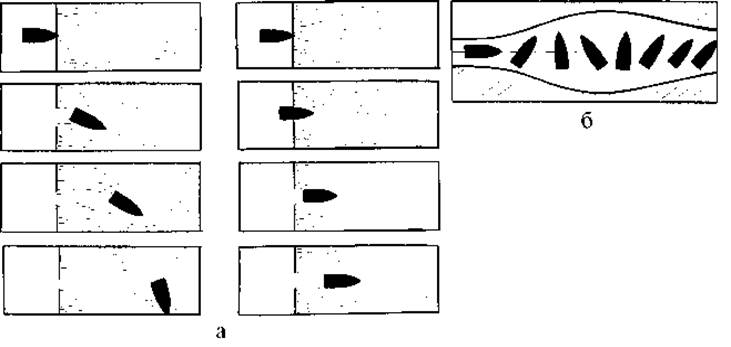
Fig. 7. Pulse (microsecond) diagram of bullet trajectory through the wounding object: a - 7.62 mm bullets; b - 5.56 mm bullets.
The occurrence of a temporary pulsating cavity in tissues at the moment of wounding is a specific feature of gunshot wounds.
Currently, the Main characteristics of a gunshot wound, especially the severity of Organ and tissue damage far beyond the wound tract, are associated with The formation of a pulsating cavity. This is due to the fact that the temporary pulsating cavity arises mainly from the lateral shock force of the bullet's energy directed away from the wound tract.
The dimensions of the temporary pulsating cavity significantly exceed the caliber of the wounding projectile, and the duration of its existence is 500 times greater than the time it takes for the projectile to pass through the wounding object. The width of tissue damage along the circumference of the wound tract is directly dependent on the dimensions of the temporary cavity and the duration of its existence. In turn, the parameters of the temporary pulsating cavity depend on the ballistic CHARACTERISTICS OF THE wounding projectile (flight velocity, kinetic energy prior to wounding, the fraction of energy absorbed by tissues as the projectile passes, energy distribution in tissues along the projectile's path and laterally away from the wound tract). The greater the kinetic energy of the bullet, the more pronounced the pulsation of the temporary cavity and the longer its duration. It is precisely these factors that explain the extensive tissue damage along the wound tract and the formation of intra-tissue hematomas, vascular, nerve, and even bone injuries far from the wound tract (Fig. 8).
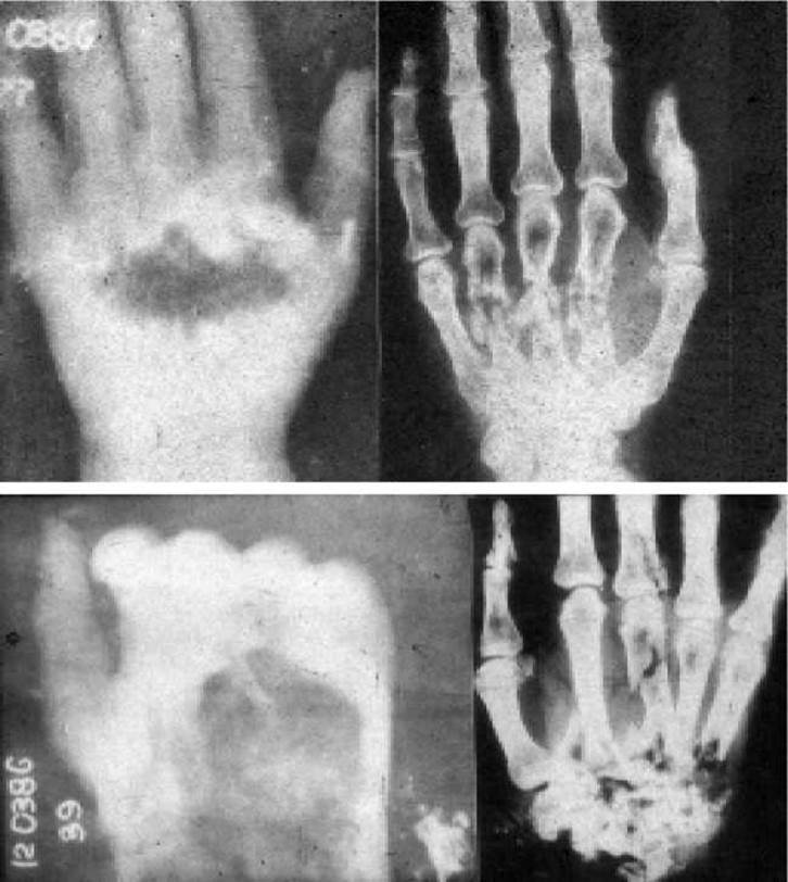
Fig. 8. Comparative characteristics of the temporary pulsating cavity effect and degrees of hand destruction (based on radiographs). High-speed imaging.
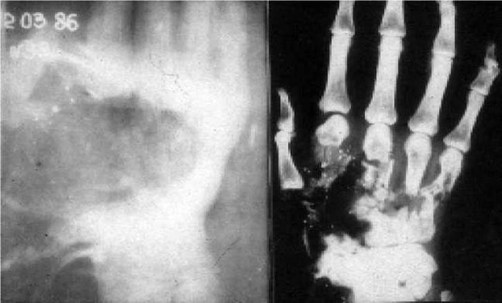
Fig. 8 (continued). Comparative characteristics of the temporary pulsating cavity effect and degrees of hand destruction (based on radiographs). High-speed imaging.
The dimensions of the temporary cavity and the extent of tissue damage also depend on the anatomical and PHYSIOLOGICAL CHARACTERISTICS OF the tissues and organs through which the bullet or shrapnel passes. For instance, when a projectile passes through the Brain, which has a soft consistency, the Formation of the temporary pulsating cavity causes it to shift laterally away from the wound tract. This is resisted by the BONES OF THE cranial vault and base, resulting in hemorrhages into the brain tissue and its ventricles at a considerable distance from the wound tract. Thus, the severity of Skull and brain injuries depends not only on the direct damage to the brain tissue by the projectile, but also on the overall deformation of the brain during the formation of the temporary pulsating cavity.
When a projectile passes through the Lungs, a small temporary cavity is formed, resulting in relatively minor damage compared to other organs and tissues. This is due to the lightweight nature of the lung tissue and the presence of A large number of elastic fibers within it.
When a projectile passes through hollow abdominal organs filled with liquid contents or gas, the pulsation of the temporary cavity leads to significant wall ruptures extending peripherally from the wound tract. Such injuries occur due to The transfer of the projectile's energy to the organ walls via their contents. This explains the negligible size of the necrosis zone (up to 0.2-0.3 cm) in the area of major organ wall ruptures, which must be taken into account during the surgical debridement of abdominal gunshot wounds.
When parenchymal organs are injured, the projectile's Energy is transferred directly to the tissue. Here, along the path of the emerging temporary pulsating cavity, there is no intermediate medium in the form of liquid or gas, which is why the destruction of these organs is observed with cracks radiating in various directions. The degree of parenchymal organ damage depends on the ballistic properties of the wounding projectiles.
In the tissues surrounding the wound tract, 3 zones of damage are distinguished (Fig. 9).
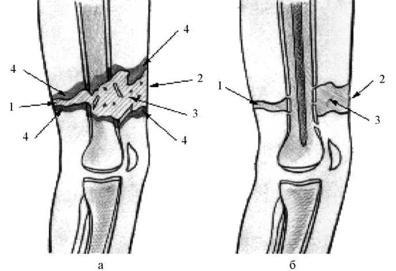
Fig. 9. Structure of a gunshot wound of the thigh: a - before surgical debridement; b - after surgical debridement; 1 - entrance wound, 2 - exit wound, 3 - wound tract, 4 - zone of primary traumatic necrosis.
The first zone is the wound tract proper, resulting from the direct destruction of tissues by the projectile. It is filled with fragments of traumatized tissues, blood clots, and wound exudate.
The second zone is the zone of contusion, or primary traumatic necrosis of tissues surrounding the wound tract.
The third zone is the zone of concussion (molecular concussion), or secondary necrosis.
The latter two zones arise As a result of the lateral action of the projectile during the formation of the temporary pulsating cavity.
One can speak with sufficient certainty only about the wound tract and the zone of primary necrosis. The third zone is distinguished only conventionally. It is not characterized by uniform morphofunctional changes in tissues identical in depth and nature. These changes depend on the elasticity and resilience of tissue structures in specific areas. Therefore, secondary necrosis is more often mosaic in nature, and its foci can be located both close to and at a considerable distance from the wound tract.
Several factors are involved in The Development of secondary necrosis. The primary ones are microcirculatory disorders and proteolysis caused by the release of Enzymes in the zone of primary necrosis. A specific third mechanism of recurrent necrosis is also distinguished. It is associated with cavitation and shock-wave damage to delicate subcellular structures. Thanks to targeted research into the wound ballistics of modern gunshot projectiles, as well as The Study of the biomolecular essence of the shock-wave mechanism's impact on tissues beyond the wound tract, METABOLISM/2.html">THE CONCEPT OF "molecular concussion" has acquired a new specific meaning.
During the formation of secondary necrosis foci, all three specified mechanisms (cavitation damage to subcellular structures, microcirculatory disorders, and proteolysis) combine and enter into a synergistic interaction.
In through-and-through wounds, the wound tract has both an entry and an exit opening, whereas in penetrating (blind) wounds, there is only an entry opening. The size of these openings depends on the ballistic properties of the projectile. High-velocity, small-caliber bullet wounds feature expanding tissue damage toward the exit opening. Conversely, flechette and needle-like bullet wounds show uniform tissue damage all the way from the entry to the exit opening. Meanwhile, wounds caused by steel spherical shrapnel, ribbed cubes, rubber and plastic balls, and shell casing fragments exhibit the most severe damage specifically in the entry zone. This occurs because the shape of these fragments causes them to lose velocity rapidly both in the air and in denser media, resulting in typically minimal tissue damage near the exit zone.
The trajectory and length of a wound tract can vary widely. In through-and-through wounds, these parameters are determined by correlating the entry and exit openings, which helps anticipate which tissues and organs might have been injured. In blind wounds, however, determining the length and direction of the wound tract through a simple physical examination of the casualty is difficult.
Even with through-and-through wounds caused by a 7.62 mm bullet, the wound tract is rarely a straight line. When a bullet strikes a bone, for instance, it may deflect, causing the wound tract to become arched or curved. These deviations are known as primary deviations of the wound tract. Furthermore, tissues directly impacted—such as skin, Muscles, and fascia—have varying elastic recoil properties, causing the inner profile of the wound tract to become wavy or jagged. These distortions are referred to as secondary deviations of the wound tract.
It is crucial to account for the Specific features of wound tracts in injuries accompanied by comminuted fractures. In such cases, bone fragments of varying sizes absorb kinetic energy from the projectile and scatter at high velocity away from the main wound tract, creating foci of secondary tissue damage.
The progression of the wound healing process is also closely tied to microbial contamination, which is an inevitable and natural consequence of trauma. However, a gunshot wound does not always lead to wound infection. Wounds can heal without complications despite the presence of microorganisms. This happens because not all microorganisms entering a wound find suitable conditions for survival, leading to a natural Selection process. For instance, anaerobes struggle to thrive in a wide-open, well-aerated wound; certain microorganisms cannot survive acidic conditions within the wound; and others are mechanically flushed out by blood and wound secretions. Additionally, microorganisms are subjected to phagocytosis and the action of humoral immunobiological factors. At the same time, gunshot wounds present several conditions that favor infection, such as closed dead spaces where air cannot penetrate, which facilitates the growth of anaerobic Bacteria. Necrotic tissues along the walls of the wound tract can serve as an ideal nutrient medium for microorganisms. Furthermore, one must consider that the wound tract is surrounded by tissues with altered reactivity and lowered resistance to infection. Additional factors favoring wound infection include massive blood loss, protein and electrolyte imbalances, vitamin deficiencies, immunodeficiency, and other adverse conditions.
The clinical manifestations of a gunshot wound depend on the caliber and configuration of the projectile; whether the wound is through-and-through or blind, single or multiple, isolated or combined; its anatomical Location; injuries to blood vessels, nerves, and other vital structures or Internal Organs; and the stage of the wound healing process. Generally, the size and shape of the skin wounds indicate the type of projectile involved (bullet, standard shrapnel, or irregularly shaped fragment). Correlating the entry and exit openings allows clinicians to anticipate injuries to various anatomical structures along the projectile's path. Pain at the site of injury, bleeding of varying intensity, and impaired function of the affected body part are constant clinical signs of a gunshot wound. The severity of systemic disorders depends on the extent and localization of the trauma.
In accordance with general phylogenetically established mechanisms of local tissue repair, gunshot wounds can heal by either primary or secondary intention.
Healing by primary intention occurs in "punctiform" wounds—most commonly through-and-through gunshot injuries—that are free from extensive tissue destruction, edema, or hemorrhage. Such wounds typically do not require surgical debridement. Self-cleansing of the wound occurs concurrently with traumatic edema, which simultaneously helps bring the wound edges together. The scab formed over punctiform wound openings acts as a natural biological dressing, facilitating scarring and subsequent epithelialization of the wound surface.
Large gunshot wounds heal by secondary intention. This is associated with the presence of necrotic tissue, where self-cleansing of the wound occurs through suppuration. In this context, suppuration is not necessarily a sign of wound infection; rather, it is replaced by the formation of granulation tissue, marking the transition to the proliferative phase of inflammation. Wound healing begins with marginal epithelialization and concludes with scar formation.
General Principles of gunshot wound management. Managing wounds across various anatomical locations is the responsibility of qualified and specialized surgical care. The standard model used to examine the principles of gunshot wound treatment is a musculoskeletal injury inflicted by modern projectiles, such as high-velocity small-caliber bullets and explosive fragmentation munitions.
The primary targets of therapeutic intervention in a gunshot wound are the zone of primary tissue destruction (necrosis), the surrounding areas of secondary necrosis, and the wound's microbial flora. From the very first hours following injury, alongside pain management and hemorrhage control, it is essential to create conditions that facilitate wound self-cleansing and limit the spread of secondary necrobiosis. Treatment begins with the application of a primary dressing, which protects the wound from adverse environmental factors and secondary microbial contamination, ensures the drainage of wound exudate, and partially removes small primary contaminants. For major gunshot injuries, immobilization of the affected body segment is necessary to protect it from further mechanical trauma.
The core component of therapeutic management is the surgical debridement of the gunshot wound. Most gunshot wounds require early surgical debridement. Depending on clinical indications, primary, delayed primary, and secondary surgical debridements are distinguished.
Primary surgical debridement (PSD) is the initial surgical intervention performed on a wound to remove non-viable tissue, prevent complications, and establish favorable conditions for healing.
Indications for PSD include: penetrating gunshot wounds of the skull, chest, abdomen, major joints, and Eyeball; ongoing hemorrhage from the wound; gunshot injuries to long tubular bones, major blood vessels, and nerve trunks; wounds contaminated with chemical or radioactive agents or soil; and wounds involving massive soft tissue damage.
Delayed or repeat surgical debridement of gunshot wounds is performed when the initial intervention was non-radical for any reason. In such cases, a repeat intervention may be necessary—typically performed before the clinical signs of infectious complications appear, based on the same primary indications. Therefore, repeat surgical debridement refers to second and subsequent surgical interventions following inadequate initial PSD, carried out before the onset of wound infection.
Secondary surgical debridement (SSD) is always performed based on secondary indications—that is, to address complications (predominantly infectious) that require additional factors for their development (such as actively proliferating pathogenic microflora in the wound), representing an indirect rather than direct consequence of the ballistic trauma. Even if a debridement performed for secondary indications is the patient's very first surgical Procedure, it remains fundamentally classified as secondary surgical debridement.
When indications for PSD are present, the procedure should be performed as early and radically as possible. Gunshot wounds that do not undergo PSD account for up to 30% of all gunshot injuries. These cases include tangential, punctiform, through-and-through, and blind soft-tissue wounds with small entry and exit diameters, no damage to major vessels or nerves, no penetration into body cavities, and an absence of fractures (with the exception of localized, punched-in fractures) or significant wound contamination.
Early PSD is performed within the first 24 hours after injury. However, in actual combat conditions, casualties are often delivered to qualified and specialized medical care facilities—where surgical intervention can be provided—at later stages following the injury. Consequently, PSD performed 24–48 hours post-injury is considered delayed, while any intervention after 48 hours is classified as late.
The radical nature of PSD is measured not so much by the extent of tissue excision as by the thorough fulfillment of all surgical objectives, which depend on the nature of the injuries and the timing of the procedure.
In summary, the general objectives of this operation are as follows:
1. Incising the wound, converting it into an open crater to provide access to deep foci of injury and establish optimal conditions for biological self-cleansing.
2. Excision of all dead and clearly non-viable tissues, which serve as a medium for the formation and spread of secondary necrosis along the periphery of the wound tract due to autocatalytic enzymatic proteolysis.
3. Ensuring meticulous hemostasis along with the removal of large intermuscular, interstitial, and subfascial hematomas.
4. Removal of large foreign bodies and free bone fragments that lack Blood supply and are capable of causing further tissue trauma.
5. Establishing optimal drainage for all Branches of the wound tract and interfascial "pockets".
6. Reconstruction of damaged similar structures (nerve or vessel suture, tendon repair, vascular or tendon grafting, application of external fixation devices in extremity gunshot wounds, and various types of skin plastic surgery).
Compliance with all these requirements determines the radicality of primary surgical debridement (PSD). During the operation, all intervention steps must be fully executed in accordance with the pathogenetic Concept of the wound process. However, this does not mean that radical surgical debridement is always definitive.
According to current military surgical doctrine, a tight primary suture is not applied after the debridement of a gunshot wound. The placement of primary sutures with continuous active drainage should be regarded as an exception, permissible only with thorough surgical debridement when the wounded person is treated in a stationary Setting under the constant Supervision of the operating surgeon. Exceptions also include wounds of the face, head, external genitalia, and chest wounds with open pneumothorax, for which primary closure is used.
In the event of a forced delay in PSD during a mass influx of casualties, measures must be taken to limit the spread of secondary necrosis and reduce the risk of infectious complications. These primarily include the correct Organization of Medical triage to identify wounded patients who require Surgical treatment in the first place: those with prolonged bleeding, applied tourniquets, traumatic amputations and severe extremity destruction, or signs of purulent or anaerobic infection. All other casualties with indications for surgical debridement receive primary surgical care to a limited extent. The main measure in this case is the infiltration of the wound periphery with a 0.25% novocaine solution and broad-spectrum antibiotic solutions.
In addition, corrective infusion therapy is administered. When indicated, extensive subcutaneous fasciotomy is performed, and the deepest wound "pockets" are drained using additional incisions.
Specifics of PSD techniques for wounds. Before surgery, the nature of the wound is thoroughly examined to determine the direction of the wound tract and the presence of damage to bones, joints, major vessels, and nerves. Depending on the anticipated scope of the operation, general or local anesthesia is used.
The skin is incised through the wound, or, in the case of perforating gunshot wounds, from the side of both the entry and exit openings. Then, clearly non-viable areas of skin are sparingly excised. The length of the skin incision must provide adequate access for debriding the wound tract (see insert, Fig. 10).
Next, the aponeurosis is incised with additional transverse cuts in the areas of the wound corners to prevent the aponeurotic sheath from compressing edematous muscles postoperatively. The wound edges are retracted with hooks, and non-viable muscles with foci of necrosis are excised layer by layer. Muscle viability is assessed by their color, bleeding capacity, contractility, and characteristic resistance (elasticity).
Viable muscles retain active bleeding and contractility upon mechanical stimulation.
When excising non-viable tissues, freely lying foreign bodies and small bone fragments are removed from the wound. One should not search for small bone fragments or projectiles away from the main wound tract, as this leads to additional tissue trauma, wound enlargement, and ultimately creates unfavorable conditions for healing. If major vessels or nerve trunks are exposed during the excision of non-viable tissues, they are carefully retracted to the side using blunt hooks. Damaged bone fragments are not debrided, except for sharp ends capable of causing repeated tissue trauma. Loose sutures are placed on muscles to cover exposed bone in order to prevent wound Osteomyelitis. Muscles should also be used to cover exposed Vessels and nerves to avoid vascular thrombosis and nerve fiber death.
The operation must be concluded by infiltrating the tissues around the debrided wound with antibiotic solutions and immobilization.
If PSD of a gunshot wound is performed in a peacetime stationary setting where the operating surgeon can maintain observation, postoperative management can follow various approaches depending on the nature of the injury, the stage of the wound healing process, The Scope of the performed surgery, and the facility's resources. Under these conditions, with the complete removal of non-viable tissues, primary wound closure can be performed combined with continuous irrigation-suction or vacuum drainage. In the latter case, a double-lumen drainage tube is used. Some medical facilities are equipped with devices for treating large wounds in a controlled abacterial environment.
Nevertheless, the most common and accessible method in military conditions is wound treatment under dressings.
The ultimate goal of wound treatment is always its healing and the restoration of integumentary tissues. Secondary healing is often a prolonged process; therefore, Surgical Methods for tissue restoration are used at various stages of treatment. 4–5 days after the operation, when the wound is clean but granulation tissue has not yet formed and there are no signs of wound infection, delayed primary sutures are applied. This is the most rational method for treating gunshot wounds in combat conditions. If the wound can be closed later (10–14 days after PSD) once granulation tissue has formed, early secondary sutures are applied.
Sometimes closing the wound is delayed because new areas of necrosis have formed within it, and their sloughing is protracted. In such cases, not only granulation tissue but also scar tissue develops in the wound, which must be excised prior to suturing. Late secondary sutures are then applied (after 15–30 days).
To close large wounds, it is necessary to resort to various plastic surgery techniques for skin coverage.
Principles of organizing care for gunshot wounds at medical treatment facilities (MTF). The combat, rear-area, and medical environment require adjustments in the provision of care for gunshot wounds. Modern weapons inflict much more severe injuries than during the Great Patriotic War of 1941–1945. This necessitates the development of simple yet reliable methods for caring for the wounded during medical evacuation support.
First aid. Provided on the battlefield near the site of the injury through self- and mutual aid, as well as by combat medics and corpsmen. The scope of first aid includes the following measures:
- temporary cessation of external bleeding (digital pressure on the vessel; application of a tourniquet, improvised tourniquet, pressure dressing; flexion or hyperextension of the extremity at the joint);
- application of a primary aseptic dressing, which protects the wound not only from secondary microbial contamination but also from recurrent minor trauma and drying, and ensures rest;
- administration of an analgesic from the syringe-tube located in the individual first-aid kit;
- transport immobilization using makeshift means; in their absence, the injured upper extremity is bandaged to the torso, and the lower extremity to the uninjured one;
- oral intake of tableted Antibiotics (0.2 g of doxycycline hydrochloride from the individual first-aid kit);
- protection against repeated trauma and adverse weather conditions, and careful evacuation of the wounded from the battlefield.
Pre-medical care. When providing pre-medical care, it is necessary to correct the shortcomings of initial first aid: verify the appropriateness of and readjust improperly placed tourniquets, aseptic dressings, and splints.
In addition, at this stage, the following Procedures are performed:
- readministration of analgesics;
- application of standard transport splints (from set B-2);
- administration, as indicated, of cardiac (caffeine 1.0) and respiratory analeptics (cordiamine 2.0);
- antishock therapy;
- intravenous infusion therapy (400 ml of Ringer's solution, or 400 ml of sorbilact, or 400 ml of rheosorbilact);
- oxygen inhalation;
- re-dispensing of oral tablet antibiotics;
- careful evacuation.
First medical aid (FMA). When providing FMA, 4 sorting and evacuation groups are distinguished:
1. Severely wounded patients requiring FMA in the dressing room.
2. Wounded patients who do not need assistance at this stage and are sent to the evacuation ward. If necessary, they can be provided with FMA.
3. Lightly wounded patients who are subject to outpatient treatment and return to their unit.
4. Agonal patients who receive symptomatic care.
Emergency FMA measures performed in the dressing room include:
- relief of asphyxia: clearing the Airways; suturing the Tongue; endotracheal intubation; tracheostomy; application of an occlusive dressing for open pneumothorax; puncture or drainage of the pleural cavity for tension pneumothorax; Oxygen therapy;
- temporary cessation of external bleeding: pressure dressing on the wound; tight wound packing with temporary skin-fixing sutures; application of a clamp to the damaged vessel; inspection and adjustment of the tourniquet;
- antishock measures for severe shock: pain management – intravenous administration of analgesics and narcotics (except morphine), auto-analgesia with the "Tringal" device; infusion therapy – Ringer's solution, rheosorbilact (10 ml/kg), sorbilact (5 ml/kg); intravenous administration of cardiac and respiratory medications; oxygen inhalation; correction of transport immobilization deficiencies contributing to shock development;
- Prevention of infectious complications: intramuscular administration of a prophylactic dose of antibacterial drugs (ofloxacin, ciprofloxacin, leflocin), subcutaneous administration of 0.5 ml of tetanus toxoid.
Other emergency care measures are performed depending on the nature of the injury.
A primary medical card is filled out for all wounded personnel.
FMA measures that can be delayed include:
- infiltration of wound edges with an antibiotic solution;
- novocaine blocks for extremity injuries without signs of traumatic shock;
- correction of transport immobilization deficiencies that do not threaten the development of traumatic shock.
Strict compliance with all aseptic and antiseptic rules is essential when providing initial medical aid.
Considering the severity of the casualties' condition, the primary indications for evacuation to the stage of qualified surgical care include:
- ongoing internal bleeding;
- presence of tourniquets on the extremities;
- head injuries accompanied by respiratory compromise;
- penetrating wounds and closed abdominal trauma;
- open and tension pneumothorax;
- pronounced blood loss and severe shock;
- contamination of wounds with radioactive and toxic agents.
Qualified Surgical Care. When providing QSC, casualties are
divided into 4 groups:
1. Casualties in need of qualified surgical care at this stage.
2. Casualties subject to evacuation.
3. The lightly wounded with a treatment period of up to 10 days.
4. Agonizing casualties.
Depending on the severity of the trauma, casualties of the first group receive qualified surgical care measures: 1) according to vital indications; 2) urgent measures in the first place; 3) urgent measures In the second place.
Life-saving interventions include emergency surgical procedures of primary wound debridement (PWD) for bleeding, PWD to suture an open pneumothorax, and secondary surgical debridement for anaerobic infection.
In a reduced scope, urgent and additional prompt surgical measures are carried out in the first place: PWD for gunshot extremity wounds with significant soft tissue damage or gunshot fractures of long bones, for wounds contaminated with toxic agents, radioactive substances, or soil, and for major open soft tissue injuries; limb amputation in cases of destruction or ischemic gangrene; PWD for penetrating hollow-organ wounds without signs of bleeding, asphyxia, or severe Peritonitis; secondary surgical debridement for wound infection.
In full scope, first-priority urgent measures and second-priority additional surgical interventions are performed (indicated PWD and repeat surgical debridement).
Following PWD, casualties are transferred from the operating and dressing unit to the temporary hospitalization ward. Depending on the localization of the wound and the extent of the surgical intervention, they receive a comprehensive treatment regimen, including the prevention of infectious complications. These measures include:
- intramuscular administration of prophylactic antibiotic doses (ofloxacin, ciprofloxacin, leflocin);
- parenteral administration of plasma expanders, blood products, and vitamin complexes;
- symptomatic treatment based on clinical indications (oxygen therapy, analgesia, cardiac and respiratory analeptics, antihistamines, and other medications).
Specialized Surgical Care. SSC must be comprehensive in nature. Its goal is to achieve a successful treatment outcome with the maximum restoration of the wounded personnel's working and combat capacity.
The entire arsenal of surgical treatment Methods for the wounded in specialized medical facilities comes down to primary, repeated, and secondary surgical debridement of gunshot wounds of all anatomical locations, performed by specialized surgeons with relevant narrow expertise and appropriate material equipment.
Postoperative management encompasses key conservative modalities such as antibacterial and infusion-Transfusion Therapy, stimulation of the body's reactivity, and wound healing acceleration. Depending on clinical indications, advanced detoxification techniques (such as lympho- and hemosorption), ultraviolet blood irradiation (UVBI), and hyperbaric oxygen therapy are employed, alongside symptomatic treatment, physical therapy, and rehabilitation exercises.
Concurrently with treatment, medical board and prognostic evaluations are carried out. If recovery occurs within the established timeframe (60–90 days) and the wounded service member is fit for duty, treatment continues in a specialized hospital until a successful outcome is achieved. If treatment exceeds the designated period or the injury is clearly incompatible with military service, casualties are evacuated to rear-area hospitals.
The primary errors in providing surgical care include:
- performing non-radical primary surgical debridement (PSD) of gunshot wounds, leaving behind non-viable tissues, foreign bodies, projectile fragments, bone shards, unincised fascial compartments, or uncontrolled bleeding;
- applying primary sutures following PSD in all cases without exception;
- excision of damaged skin in areas of multiple superficial wounds caused by small shrapnel;
- attempting to provide specialized medical care at the stage of qualified medical care, particularly for head and extremity injuries;
- failure to immobilize limbs in the presence of extensive soft tissue wounds;
- unjustified expansion of indications for emergency surgical interventions at the stage of qualified medical care for casualties with head, chest, or extremity wounds;
- performing surgical interventions on wounded patients in a state of traumatic shock (TS) without adequate anti-shock infusion-transfusion therapy.
Such errors inevitably lead to an increased rate of adverse treatment outcomes for the wounded.
Last update: 08/08/2026
Editorial and Educational Adaptation: This material has been compiled based on the primary/original source text. The project team performed an editorial review, corrected technical inaccuracies, structured sections, and adapted the content for an educational format.
What was processed:
- elimination of formatting defects (OCR errors, structural breaks, corrupted characters);
- editorial organization of content;
- standardization of terminology in accordance with academic sources;
- verification of factual statements against the original source text.
All mentions of the author, publication year, and origin of the primary text have been preserved in accordance with the source.