Orthopedics - Oleksa A.P. 2006
Osteomyelitis
Brodie's abscess
Brodie's abscess is a rare inflammatory and purulent lesion of the spongy area within the metaphysis of a long tubular bone. The most frequent site of the abscess (accounting for 80% of cases) is the proximal metaphysis of the Tibia (Fig. 399). Typically, the lesion is located in the center of the metaphysis, while in children and adolescents—prior to the closure of the epiphyseal Cartilage—it is situated beneath it.
Clinically, Brodie's abscess is asymptomatic due to its primary chronic course. It may be an incidental radiological finding, but patients more frequently seek medical attention for vague pain following physical exertion of the leg, or for knee joint synovitis caused by contact "sympathetic" inflammation of the synovial membrane.
Class="center">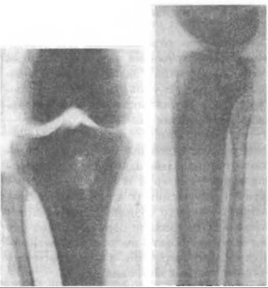
Fig. 399. Radiograph of a Brodie's abscess in the region of the upper tibial metaphysis (Reinberg S.A., 1964).
It should be noted that these clinical manifestations may subside and periodically recur, prompting patients to seek medical help only when the bone pathology becomes clearly pronounced.
Clinical examination of patients reveals no specific pathognomonic signs of the abscess; Blood tests and body Temperature are normal, and the condition can only be detected radiologically.
In the Cytology/cytology/16.html">Early stages of the disease, a slight radiolucency of the spongy Bone Structure is observed, which can be mistaken for Osteoporosis. Only computed tomography allows for the assessment of density differences between the affected and healthy bone areas, as well as the dimensions of the inflammatory focus.
Over time, the inflammatory process destroys the bone, and necrotic masses gradually resorb, which appears as a radiolucent area on radiographs. A pyogenic membrane forms around this purulent-necrotic focus, accompanied by surrounding osteosclerosis, causing the lesion to slowly expand over many years.
If the sclerotic walls of the abscess become rigid, it ceases to expand and remains unchanged for decades.
The shape of the abscess cavity is generally round, occasionally oval, and its dimensions vary, reaching up to 2 cm. No other bone changes are detected radiologically.
Depending on the duration of the process, the Contents of the abscess may consist of pus mixed with necrotic bone debris or dried detritus. Sequestra never form. Bacteriological examination OF the abscess contents typically yields no Microbial growth.
Treatment of Brodie's abscess involves Curettage of the purulent-necrotic focus. The surgery is performed under general anesthesia with a thigh tourniquet applied. A linear incision down to the bone is made along the anteromedial surface of the tibia overlying the abscess. The cortical layer of the tibia is trephined, creating a window after the removal of the cortical plate. The precise Location OF THE abscess can be verified using a Dufour needle. A Volkmann spoon is used to scrape out the lesion within healthy cancellous bone, and the cavity is irrigated with a 1:5000 furacilin solution and dried.
If the cavity is small, antibiotic powder is poured directly into it from a vial, the cortical plate is replaced, and it is covered with the elevated periosteum. The wound is closed without drainage.
In cases where a large cavity has formed, it should be filled with a cancellous bone graft harvested from the iliac crest, or preferably with an antibiotic-loaded ceramic composite (kergap).
The postoperative period is uneventful, with no wound suppuration. The patient should be considered cured, but a follow-up radiography must be performed six months later, even if the patient's condition is satisfactory.
Last update: 10/08/2026
Editorial and Educational Adaptation: This material has been compiled based on the primary/original source text. The project team performed an editorial review, corrected technical inaccuracies, structured sections, and adapted the content for an educational format.
What was processed:
- elimination of formatting defects (OCR errors, structural breaks, corrupted characters);
- editorial organization of content;
- standardization of terminology in accordance with academic sources;
- verification of factual statements against the original source text.
All mentions of the author, publication year, and origin of the primary text have been preserved in accordance with the source.