Tuberculosis - I.T. Pyatnochka 2005
Extrapulmonary tuberculosis
Bone and joint tuberculosis
In the overall Structure of bone and Joint Diseases, tuberculosis accounts for 5-7%. Within The Musculoskeletal System, the spine is affected most frequently (30-40%) (Fig. 27), followed by the hip (Fig. 28) and knee joints (20% each); the ankle, FOOT bones, and upper extremities are involved significantly less often.
Pathomorphology. Mycobacterium tuberculosis enters the Bones and joints via the hematogenous route during primary infection or As a result of late generalization due to the reactivation of healed tuberculous foci. Triggering factors may include trauma, hypothermia, etc.
The specific pathological process originates in areas of the bone with the most developed vascular network (vertebrae, metaphysis, and epiphysis of long bones). Typical tuberculous granulomas develop in bones and joints, subsequently merging to form foci of caseous necrosis. The spread of the process leads to joint involvement (exudate accumulation) as well as soft tissue damage. As a result, cold (congestive) abscesses may form, which frequently accompany spinal tuberculosis.
Clinical Features. There are three distinct phases in the clinical course of Osteoarticular Tuberculosis: Phase I, the pre-arthritic phase, is characterized by The formation of a specific focus within unchanged Bone tissue. Clinical signs of the disease are subtle, presenting with mild joint pain and slight symptoms of intoxication.
Class="center">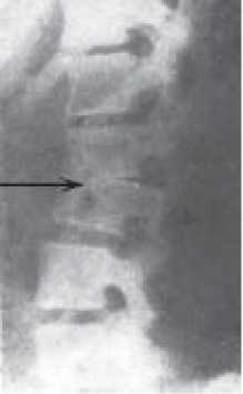
Fig. 27. Tuberculous Spondylitis
Phase II, the arthritic phase, is characterized by the spread of the process to the articular surfaces, Cartilage, and Joint Capsule. Destructive changes occur within the joint, intoxication and pain intensify, accompanied by joint Swelling, Muscle guarding, and subsequent partial muscle atrophy.
Phase III, the post-arthritic phase, is marked by the stabilization of the process accompanied by The Development of permanent skeletal deformities.
During the arthritic phase of osteoarticular tuberculosis, complications may arise, including destructive bone and joint lesions, cold (congestive) abscesses, fistulas, and secondary Amyloidosis of internal Organs. In addition to these complications, tuberculous spondylitis can lead to paresis and paralysis resulting from Spinal Cord compression.
X-ray and tomographic imaging are employed to diagnose osteoarticular tuberculosis. The Diagnosis is confirmed through histological, cytological, and MICROBIOLOGICAL EXAMINATION OF abscess contents, joint cavities, punctures, and biopsies obtained from the affected joint and bone tissue.
Differential diagnosis must be performed to rule out chronic Osteomyelitis, infectious and traumatic Arthritis, and Bone tumors.
Treatment of osteoarticular tuberculosis primarily involves prolonged (at least 8-12 months) combined antimycobacterial therapy using 4-5 drugs, supported by desensitizing, vitamin, and general restorative therapy. In cases of delayed diagnosis, radical or reconstructive-restorative surgeries on the joints and spine are frequently required. Orthopedic measures are crucial for successful treatment, most notably the immobilization of the affected organ in a physiological position until the tuberculous process stabilizes.
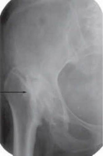
Fig. 28. Tuberculous coxitis
Last update: 10/08/2026
Editorial and Educational Adaptation: This material has been compiled based on the primary/original source text. The project team performed an editorial review, corrected technical inaccuracies, structured sections, and adapted the content for an educational format.
What was processed:
- elimination of formatting defects (OCR errors, structural breaks, corrupted characters);
- editorial organization of content;
- standardization of terminology in accordance with academic sources;
- verification of factual statements against the original source text.
All mentions of the author, publication year, and origin of the primary text have been preserved in accordance with the source.