Orthopedics - Oleksa A.P. 2006
Bone and Joint Tuberculosis
Tuberculous Spondylitis
Tuberculosis most commonly localizes in the vertebral bodies of the thoracic and lumbar spine (Fig. 382). The primary focus is typically situated near the anterior or lateral wall of the vertebra or the intervertebral disc. As Bone tissue is destroyed, an abscess may rupture beneath the periosteum and the anterior longitudinal ligament, stripping them away, and spread to adjacent vertebrae or, by destroying the disc, involve the neighboring vertebra in the process. If several vertebrae are affected, the abscess breaks into the interfascial space and descends along the iliopsoas Muscle into the pelvis or below the inguinal ligament into the femoral triangle. Occasionally, it may extend laterally and be palpable above the iliac crest in Petit's triangle (Fig. 383).
Clinical symptoms depend on the phase of the process. In the prespondylitic phase, during the Formation of primary osteitis, clinical signs are mild. Symptoms of tuberculous intoxication—loss of appetite and general weakness—gradually appear. Following the destruction of the vertebral body, pain and reflex (protective) spasm of the back Muscles develop. Muscle relaxation occurs during Sleep, causing children to cry out from pain, which is a characteristic sign of spondylitis. Body Temperature is subfebrile, the ESR is elevated, and lymphocytosis increases. Tuberculin tests are typically positive. In the spondylitic phase, all these signs become markedly pronounced. The patient holds the spine rigid through intense contraction of the back muscles, known as Kornev's "reins" sign. Spinal mobility is restricted, giving rise to another characteristic sign: to pick up an object from the floor, the patient does not bend over, but instead squats down while supporting themselves with the other hand on the thigh.
Class="center">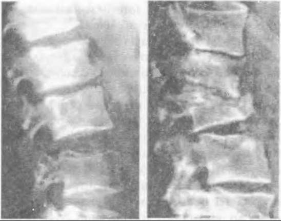
Fig. 382. Radiographs of tuberculous spondylitis.
Percussion over the spinous processes in the affected vertebral region elicits pain. Pain sharply increases upon axial loading (pressing down on the HEAD), though this should be avoided to prevent compression of the affected vertebra. Significant destruction and wedge-shaped deformation of the vertebral body make the protruding spinous process palpable. If multiple vertebrae are involved, a characteristic gibbus (hump) deformity develops. In this phase, cold cold/psoas abscesses may already be present.

Fig. 383. Tuberculous spondylitis of Th4-Th5 with a cold abscess.
Radiological examination is decisive in the Diagnosis of tuberculous spondylitis. Gross destructive Changes in the vertebra are visible on standard radiographs in two projections. Following plain radiography, tomography is performed to determine the size and depth of the foci. Computed tomography enables the detection of even the smallest foci of primary osteitis. In the presence of a cold abscess, contrast abscessography sometimes helps trace its pathway to the affected vertebra (Fig. 384). Undeniably, Magnetic Resonance imaging provides even greater diagnostic value in such cases.
As the inflammatory process subsides, spinal changes stabilize, and the patient's general condition and laboratory parameters improve. Proliferative processes predominate in the spine, leading to scarring, encapsulation of isolated foci, and, in cases of severe destruction and deformity of the vertebrae, their gradual ankylosis.
Treatment includes general tonic measures, antibacterial therapy, and orthopedic interventions aimed at immobilizing the affected area and offloading the spine using a brace or a plaster bed with a special cotton-gauze corrective pad to prevent The Development of a gibbus deformity.
Last update: 10/08/2026
Editorial and Educational Adaptation: This material has been compiled based on the primary/original source text. The project team performed an editorial review, corrected technical inaccuracies, structured sections, and adapted the content for an educational format.
What was processed:
- elimination of formatting defects (OCR errors, structural breaks, corrupted characters);
- editorial organization of content;
- standardization of terminology in accordance with academic sources;
- verification of factual statements against the original source text.
All mentions of the author, publication year, and origin of the primary text have been preserved in accordance with the source.