Orthopedics - Oleksa A.P. 2006
Bone and Joint Tuberculosis
Surgical Treatment of Tuberculous Spondylitis
If the tuberculous osteitis of a vertebral body progresses and fails to respond to conservative therapy, surgical intervention is indicated.
Spinal surgeries are performed under general anesthesia with proper Anesthetic Management. The surgical approach depends on the level and localization of the tuberculous lesion.
The vertebral body has four terminal Arteries, so the Koch's bacillus can settle in any quadrant of its body and cause primary osteitis. Most commonly, the tuberculous process is localized in the D12— L1 and D5— D9 vertebral bodies.
The easiest surgical approach to the D12— L1 vertebral bodies is the posterolateral approach with costotransversectomy. This approach has been used since 1946 by Alexander and was subsequently improved by other phthisiatric surgeons (S. Malawski, K. Rapata, and others).
Class="center">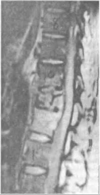
Fig. 384. Magnetic Resonance image of a tuberculous lesion in the Th11-Th12 vertebral bodies.
Surgical technique. The patient is placed on the healthy side with a support roll underneath. In the D12— L1 projection, the Skin, subcutaneous tissue, and fascia are incised layer by layer linearly, 1 cm away from the spinous processes, as this ensures the wound heals without complications. The Muscles are divided paravertebrally to expose the transverse process of the first lumbar and twelfth thoracic vertebrae along with the proximal end of the rib.
First, the transverse process is resected at its base and retracted aside with the muscles (sometimes it is removed), and then the proximal end of the twelfth rib along with its process is resected. During these manipulations, nerve roots and Blood Vessels should be identified and retracted using slings (Fig. 385).
After exposing the lateral aspect of the vertebral body, thorough hemostasis is performed. The lateral wall of the vertebra is trephined, and necrotic-caseous masses are scraped out and removed using a Volkmann spoon. The wound is flushed under pressure with a 1:5000 furacilin solution, dried, and irrigated with an antibiotic solution. As a rule, the wound is not drained and is closed tightly in layers.
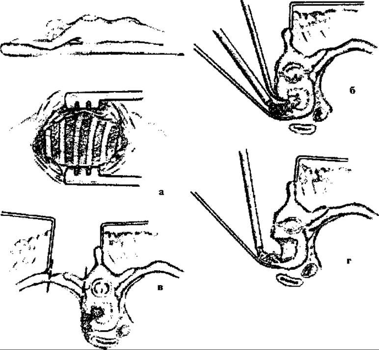
Fig. 385. Surgical approach to the body of the tuberculosis-affected vertebra with aspiration of necrotic masses: a - vertebral hump, b - exposure of paravertebral Ribs, c - resection of the ribs and transverse processes of the vertebra, d - aspiration of detritus with removal of osteonecrosis.
Postoperatively, the patient remains in bed on a firm mattress for 3–4 months, receiving antibacterial drug therapy According to the regimen.
A similar operation is performed for tuberculous osteitis of the thoracic vertebrae.
In the spondylitic phase, when two or three thoracic vertebrae are affected, the posterior approach also offers advantages because it does not open the thoracic cavity—which often poses a risk of hemopneumothorax and Pneumonia—and results in lesser surgical trauma.
The surgical technique is practically identical to the one described above, but is supplemented by the removal of the affected two or three intervertebral discs and caseous bone destructions. Depending on their size and shape, the vertebral and disc defects are filled with an autograft harvested from the ribs or the iliac wing.
When a single vertebra is affected, Rapata does not perform bone grafting following necrectomy, especially in the thoracic spine. Postoperatively, the patient continues conservative Treatment in a plaster bed.
If surgery reveals a breakthrough of pus outside the vertebra with The formation of a cold abscess, the pus is evacuated first, and if a pyogenic capsule is present, it is removed along with the granulations. The operative field is dried, and then the surgery on the vertebrae and discs is continued.
In Tuberculous Spondylitis, an abscess from the area of the affected vertebra can spread along interfascial spaces as a cold abscess into Petit's triangle, along the psoas major Muscle into the iliac region, beneath Poupart's ligament, or even down into the popliteal fossa and below. It can be detected by Palpation and puncture. In such cases, the cold abscess is punctured, the pus is aspirated, and its cavity is washed with hydrogen peroxide and a 1:5000 furacilin solution. Large abscesses are incised and drained using an open method.
Sometimes an abscess may rupture outward through the skin, resulting in a persistent fistula through which secondary microflora enters, complicating patient treatment. Previously, before the advent of Antibiotics, a fistula was considered the patient's "gateway to death," but nowadays, in the era of antibacterial therapy and surgical Procedures, this threat no longer exists.
Transthoracic approaches are most commonly used for tuberculous lesions of the D5— D9 vertebral bodies. This is the most frequent localization of spinal tuberculosis.
Transthoracic surgeries became widely available to phthisiatric surgeons following the Structure/175.html">Implementation of endotracheal anesthesia and comprehensive anesthetic management.
When vertebrae in the thoracic spine are affected, patients with lower paraplegia are operated on via a posterolateral or transthoracic approach. Surgical interventions performed under endotracheal anesthesia with proper anesthetic support facilitate a radical, complete operation and prevent complications.
Surgical technique. A linear tissue incision is made down to the rib located in the PROJECTION OF THE most affected vertebra. The rib is resected subperiosteally over a length of 15–20 cm of its proximal end; if better exposure is required, an adjacent rib can also be resected while preserving the intercostal vessels and nerve. Hemostasis is performed as the surgery progresses.
The right thoracic cavity is widely opened using a chest retractor, and the lung is retracted to provide adequate access to the affected vertebrae. The paravertebral vessels in this area are ligated in continuity and transected between the ligatures. During the surgery, the area is checked for the presence of a cold abscess. If an abscess is identified and confirmed by puncture, the parietal Pleura and fascia are incised between the ligated vessels, and odorless liquid pus and necrotic masses are removed using an electric suction device. The abscess cavity is washed with a 1:5000 furacilin solution and dried. Afterward, caseous masses and sequestra are removed from the affected vertebra using a Volkmann spoon.
If two or three vertebrae and intervertebral discs are affected, their remnants are removed simultaneously with the vertebral necrectomy. It is desirable not to leave even small tuberculous foci, operating instead within the limits of healthy vertebral bone. In cases of total destruction of the vertebral bodies, the remnants are completely removed.
Regarding the reconstruction of a defect after single-vertebral resection, it should be noted that various surgical strategies exist. Most orthopedic surgeons logically reconstruct the defect using a bone graft harvested from the resected rib, as this is considered necessary in all such cases. However, Rapata performs bone grafting of the vertebral bodies only after the resection of two vertebrae along with the affected disc, whereas for thoracic vertebral defects, he omits bone grafting altogether, stabilizing the spine via posterior spinal fusion during a second planned stage.
The surgery is concluded with hemostasis and airtight closure of the chest wall wound. Following this, any hemopneumothorax is aspirated. The patient is placed in a plaster bed for 5–6 months, and comprehensive conservative treatment is continued. Typically, in cases following lower paraplegia, signs of movement in the lower extremities appear no later than 3–4 weeks postoperatively, which warrants the continuation of rehabilitation therapy.
Last update: 10/08/2026
Editorial and Educational Adaptation: This material has been compiled based on the primary/original source text. The project team performed an editorial review, corrected technical inaccuracies, structured sections, and adapted the content for an educational format.
What was processed:
- elimination of formatting defects (OCR errors, structural breaks, corrupted characters);
- editorial organization of content;
- standardization of terminology in accordance with academic sources;
- verification of factual statements against the original source text.
All mentions of the author, publication year, and origin of the primary text have been preserved in accordance with the source.