Orthopedics - Oleksa A.P. 2006
Joint Diseases
Degenerative and Dystrophic Joint Diseases
Deforming Arthrosis Following Aseptic Osteonecrosis
Osteoarthritis most commonly develops As a result of delayed Diagnosis or improper Treatment of Aseptic osteonecrosis of the femoral HEAD in adults and osteochondropathy (Perthes disease) in children; less frequently, it occurs following osteonecrosis of the lunate and scaphoid bones, among others.
In aseptic Necrosis of the femoral head in adults and children, osteoarthritis arises due to compression fractures and deformation of the pathologically altered head under physiological loads during limb weight-bearing or even normal walking.
Involvement of the femoral head may be segmental or total, and the process is very frequently bilateral. The femoral head loses its spherical shape, and its articular surface becomes incongruent with the acetabulum.
This articular surface incongruity causes pain and a restricted range of motion in the hip joint. Consequently, it triggers antalgic contracture of the thigh Muscles, which increases pressure on the femoral head and, in turn, further intensifies the pain. This creates a vicious pathological cycle accompanied by a flexion-adduction contracture of the thigh, functional shortening, and impaired limb function.
Promptly diagnosed osteonecrosis and appropriate treatment initiated in the Cytology/cytology/16.html">Early stages of the disease can prevent the onset of osteoarthritis.
Conservative treatment is primarily administered to children with minor femoral head deformation and preserved hip joint function during periods of pain exacerbation. It involves unloading the limb, applying Skin traction, administering analgesics, and utilizing physical and balneotherapy Procedures.
If pronounced deformation of the head is present, long-term success from comprehensive conservative treatment is unlikely, and surgical management is recommended.
In cases of segmental head involvement and compression of a minor area, an intertrochanteric wedge osteotomy (Fig. 341) can be performed, rotating the head into a position where the compression zone is relieved of weight-bearing. This Procedure is technically straightforward; the osteotomized Femur is fixed with hardware, eliminating The Need for a plaster cast and allowing early mobilization of the hip joint.
For severe coxarthrosis following aseptic necrosis in adults, manifested by persistent pain, impaired limb function, and consequent loss of working capacity, surgery is indicated. In unilateral coxarthrosis in manual laborers, hip arthrodesis is performed. Following The Development of bony ankylosis, patients are often able to resume even heavy physical labor.
Class="center">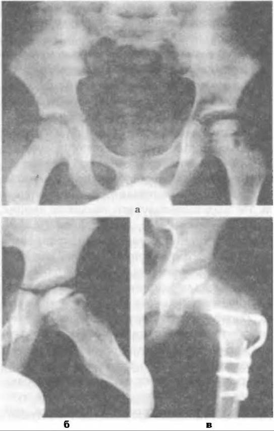
Fig. 341. Perthes disease of the left hip joint: a - radiograph of both hip joints, b - lateral radiograph of the left hip joint, c - post-operative view following intertrochanteric varus osteotomy.
Total hip replacement has become widely adopted and is performed for unilateral and bilateral coxarthrosis in adults resulting from aseptic osteonecrosis of the femoral head. The indication for arthroplasty is determined by the physician, taking into account the patient's age and occupation, while the final decision rests with the patient. For bilateral coxarthrosis in working-age individuals, the optimal strategy is considered to be arthroplasty of one joint combined with arthrodesis of the contralateral joint.
For patients engaged in sedentary or intellectual work, bilateral hip replacement is advisable. The surgical technique is detailed in the chapter covering the Surgical treatment of coxarthrosis.
Aseptic necrosis of the lunate or scaphoid bones typically occurs following fracture-dislocations or fractures complicated by compromised arterial Blood supply. Compression and deformation of the necrotic bone lead to the development of osteoarthritis. Post-traumatic aseptic necrosis is initially managed conservatively in the hope of restoring Blood supply and The Structure of the affected bone. Treatment involves immobilization of the wrist joint to eliminate motion in all planes, along with UHF therapy, reflexology, and pharmacotherapy.
Since conservative treatment is time-consuming and its success is unpredictable, surgical intervention is employed.
If deformation of the lunate bone has not yet occurred, the Matti procedure is performed. This involves Curettage of the necrotic tissue followed by packing the cavity with a healthy cancellous bone autograft, which successfully prevents the development of Arthritis.
If osteoarthritis secondary to aseptic necrosis of the lunate or scaphoid has already developed, manifesting as persistent pain, restricted mobility, and disability, arthrodesis is used for manual laborers, whereas arthroplasty is indicated for patients who will not subject the hand to heavy stress.
Surgical technique for arthrodesis. Under anesthesia with a tourniquet, a linear incision of the Tissues is made on the dorsal aspect of the wrist joint,
taking utmost care to preserve the Veins. The tendons of the finger extensors are retracted, and the dorsal ligament-capsule apparatus in the region of the lunate and scaphoid bones is incised. The articular Cartilage is excised from the radius, scaphoid, and lunate bones on the side of the radiocarpal joint. The hand is positioned in a functionally advantageous posture, aligning the resected surfaces.
Subsequently, the hand is fixed with a Bogdanov rod or a Steinmann pin, driven from the neck region of the third metacarpal bone through the carpus into the radius (Fig. 342). The resected joint is bridged with a bone autograft or decalcified matrix.
Occasionally, surgeons use a bone autograft instead of a metal pin, which fixes only the Carpal Bones to the radius. The wound is closed in layers, and a plaster cast is applied.
Some surgeons, following bone resection, forego internal rod fixation and plaster immobilization, achieving ankylosis instead by means of the Ilizarov compression apparatus.
Surgical technique for arthroplasty. Under anesthesia using a tourniquet, the lunate or scaphoid bone is exposed via a similar surgical approach and excised. The optimal size is selected from a set of silicone implants to replace the resected bone.
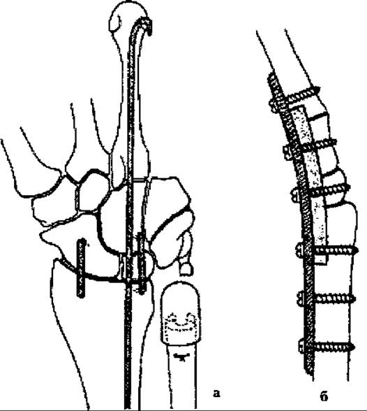
Fig. 342. Schematic illustration of wrist arthrodesis: a - fixation with a Bogdanov rod and bone autografting, b - fixation with a metal plate and autografting.
To prevent dislocation after semi-lunar bone arthroplasty, the prosthesis is secured to the adjacent bones by transfixing it with a Kirschner wire. For this purpose, scaphoid bone endoprostheses are manufactured with a peg that is inserted into a recess in the trapezium and additionally fixed with wires to the carpal bones and the radial styloid process (Fig. 343).
G. L. Voskresensky and I. A. Movshovych pressed a Dacron tape into the scaphoid endoprosthesis and sutured its ends to the adjacent tissues to prevent dislocation. The arthroplasty technique is quite straightforward, and following wound closure, the hand is immobilized for 6 weeks to allow sufficient time for The formation of dense scar tissue. The fixation wires can be removed three weeks later by pulling on their ends protruding above the skin.
After studying the outcomes of carpal bone arthroplasty, R. Smith (1985), P. Kleinert et al. (1985), G. Evans (1986), and others established that initially fully satisfactory results deteriorated in the majority of patients within 2-3 years. Patients develop pain, Swelling, and silicone synovitis resulting from chronic proliferative aseptic inflammation, as well as destructive Changes in the joints and bones adjacent to the prosthesis.
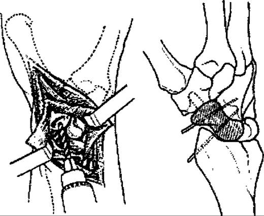
Fig. 343. Endoprosthetic replacement of the scaphoid bone.
In such cases, the endoprosthesis is removed, and a revision reconstructive or stabilizing procedure—arthrodesis—is performed.
Last update: 10/08/2026
Editorial and Educational Adaptation: This material has been compiled based on the primary/original source text. The project team performed an editorial review, corrected technical inaccuracies, structured sections, and adapted the content for an educational format.
What was processed:
- elimination of formatting defects (OCR errors, structural breaks, corrupted characters);
- editorial organization of content;
- standardization of terminology in accordance with academic sources;
- verification of factual statements against the original source text.
All mentions of the author, publication year, and origin of the primary text have been preserved in accordance with the source.