MEDICAL BIOLOGY, HUMAN ANATOMY, PHYSIOLOGY AND PATHOLOGY - Ya.I. Fedoniuk 2010
ANATOMY, PHYSIOLOGY, PATHOLOGY
CHAPTER 2. MUSCULOSKELETAL SYSTEM (THE MECHANISM OF MOVEMENT)
GENERAL ISSUES OF THE ANATOMY AND PHYSIOLOGY OF THE MUSCULAR SYSTEM
MUSCLES AND FASCIAE OF BODY PARTS
MUSCLES AND FASCIAE OF THE NECK
TOPOGRAPHIC AND ANATOMICAL STRUCTURES OF THE NECK
The following regions are distinguished in the neck:
1. Anterior region - has the shape of a triangle bounded above by the Base of the Mandible, below by the jugular notch of the Sternum, and on the sides by the anterior borders of the sternocleidomastoid Muscles.
2. Sternocleidomastoid region - corresponds to the external surface of the Muscle of the same name.
3. Lateral region - has the shape of a triangle bounded by the posterior border of the sternocleidomastoid muscle, the clavicle, and the lateral border of the trapezius muscle.
4. Posterior region - bounded above by the superior nuchal lines, below by a transverse line passing through the spinous process of the 7th cervical vertebra, and on the sides by the lateral borders of the trapezius muscles.
Within the anterior and lateral Regions of the neck, a series of triangles of significant practical importance are identified (Fig. 2.80).
Class="center">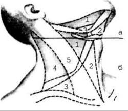
Fig. 2.80. Triangles of the neck region
In the anterior region:
1. Carotid triangle, trigonum caroticum, bounded posteriorly by the anterior border of the sternocleidomastoid muscle, superiorly by the posterior belly of the digastric muscle, and inferiorly by the superior belly of the omohyoid muscle.
2. Omotracheal triangle, trigonum omotracheale, located between the anterior border of the omohyoid muscle and the anterior median line.
3. Submandibular triangle, trigonum submandibulare, bounded by the two bellies
of the digastric muscle and the body of the mandible. The salivary gland of the same name is located within this triangle. Another triangle is also distinguished here — the lingual triangle, trigonum linguale — which is bounded by the posterior border of the mylohyoid muscle, the posterior belly of the digastric muscle, and the Hypoglossal nerve. This triangle is known as Pirogov's triangle, through which the lingual artery passes.
In the lateral region:
1. Omotrapezoid triangle, trigonum omotrapezoideum, bounded by the posterior border of the sternocleidomastoid muscle, the lateral border of the trapezius muscle, and the inferior belly of the omohyoid muscle.
2. Omoclavicular triangle, trigonum omoclaviculare, located between the clavicle, the inferior belly of the omohyoid muscle, and the posterior border of the sternocleidomastoid muscle.
Last update: 08/08/2026
Editorial and Educational Adaptation: This material has been compiled based on the primary/original source text. The project team performed an editorial review, corrected technical inaccuracies, structured sections, and adapted the content for an educational format.
What was processed:
- elimination of formatting defects (OCR errors, structural breaks, corrupted characters);
- editorial organization of content;
- standardization of terminology in accordance with academic sources;
- verification of factual statements against the original source text.
All mentions of the author, publication year, and origin of the primary text have been preserved in accordance with the source.