HUMAN MEDICAL BIOLOGY, ANATOMY, PHYSIOLOGY AND PATHOLOGY - Ya.I. Fedoniuk 2010
ANATOMY, PHYSIOLOGY, PATHOLOGY
SECTION 2. MUSCULOSKELETAL SYSTEM (THE MECHANISM OF MOVEMENT)
MUSCLES AND FASCIAE OF BODY REGIONS
MUSCLES AND FASCIAE OF THE NECK
The Muscles of the neck are divided into superficial and deep groups. In addition, a separate group of superficial muscles consists of those attached to the Hyoid bone, namely the suprahyoid and Infrahyoid muscles.
1 - temporal Muscle (cut);
2 - lateral pterygoid muscle;
3 - medial pterygoid muscle;
4 - buccinator muscle; 5 - parotid duct (cut).
SUPERFICIAL MUSCLES OF the neck (Fig. 2.77):
Class="center">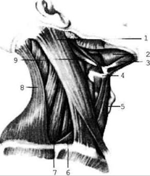
Fig. 2.77. Muscles of the neck; lateral view
1. Platysma (m. platysma) - a thin muscular sheet located beneath the Skin. It originates from the pectoral fascia below the clavicle and inserts into the region of the Mandible. Function: pulls the angle of the Mouth downward and elevates the skin of the neck.
2. Sternocleidomastoid muscle (m. sternocleidomastoideus) - originates by two heads from the clavicle and Sternum and inserts into the mastoid process of the Temporal bone. Function: unilateral contraction tilts the cervical spine to the same side while turning the HEAD in the opposite direction; bilateral contraction maintains the head in an upright position, and maximal contraction pulls it backward.
3. Suprahyoid muscles (Fig. 2.78):
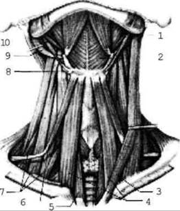
Fig. 2.78. Muscles of the neck; anterior view
a) digastric muscle: the anterior belly extends from the mandible, and the posterior belly originates from the mastoid notch of the temporal bone. A tendon connects the two bellies and is attached to the hyoid bone. Function: elevates the hyoid bone and, along with it, the Larynx; participates in the acts of swallowing and articulated speech. When the hyoid bone is fixed, this muscle depresses the mandible and takes part in mastication;
b) mylohyoid muscle extends from the mandibular arch to the hyoid bone, forming the floor (Diaphragm) of the Oral Cavity. Its function is identical to that of the previous muscle;
c) geniohyoid muscle runs from the mental spine of the mandible to the hyoid bone. Function: when the hyoid bone is fixed, it depresses the mandible; when the jaws are closed, it elevates the hyoid bone together with the larynx (mastication, swallowing, speech);
d) stylohyoid muscle runs from the styloid process of the temporal bone to the hyoid bone. Its function is similar to that of the preceding muscles, except that it does not depress the mandible.
4. Infrahyoid muscles (Fig. 2.78):
a) sternohyoid muscle originates from the sternum and inserts into the hyoid bone. It pulls the bone downward;
b) omohyoid muscle extends from the scapula to the hyoid bone, depressing it upon contraction. It consists of superior and inferior bellies;
c) the sternothyroid muscle extends from the sternum to the external surface of the thyroid Cartilage; it depresses the thyroid cartilage and, consequently, the larynx and hyoid bone;
d) the thyrohyoid muscle is a continuation of the previous one, stretching from the thyroid cartilage to the hyoid bone; when the hyoid bone is fixed, it elevates the larynx.
Deep Muscles of the Neck: these include the scalene muscles (anterior, middle, and posterior), as well as the prevertebral muscles (longus colli and longus capitis, anterior and lateral rectus capitis muscles).
The scalenus anterior is preceded by the scalene hiatus (anterior scalene space), while the interscalene space lies between the anterior and middle scalene muscles, serving as a passage for Blood Vessels and nerves.
Cervical fascia. According to recent data, There is a single cervical fascia comprising three layers, or lamina (Fig. 2.79): superficial, pretracheal, and prevertebral. The superficial layer is located deep to the platysma and forms fascial sheaths for the trapezius and sternocleidomastoid muscles. The pretracheal layer spans between the omohyoid muscles and forms sheaths for the infrahyoid muscles. The prevertebral layer covers the prevertebral and scalene muscles, forming sheaths for them.
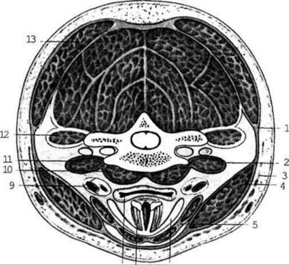
Fig. 2.79. Arrangement of the layers of the cervical fascia (transverse section at the level of The Thyroid Gland)
Last update: 08/08/2026
Editorial and Educational Adaptation: This material has been compiled based on the primary/original source text. The project team performed an editorial review, corrected technical inaccuracies, structured sections, and adapted the content for an educational format.
What was processed:
- elimination of formatting defects (OCR errors, structural breaks, corrupted characters);
- editorial organization of content;
- standardization of terminology in accordance with academic sources;
- verification of factual statements against the original source text.
All mentions of the author, publication year, and origin of the primary text have been preserved in accordance with the source.