MEDICAL BIOLOGY, HUMAN ANATOMY, PHYSIOLOGY AND PATHOLOGY - Ya.I. Fedonyuk 2010
ANATOMY, PHYSIOLOGY, PATHOLOGY
CHAPTER 2. MUSCULOSKELETAL SYSTEM (THE MECHANICS OF MOVEMENT)
MUSCLES AND FASCIAE OF BODY REGIONS
MUSCLES AND FASCIAE OF THE HEAD
The Muscles of the HEAD are divided into facial muscles (Muscles of facial expression) and masticatory muscles.
Facial muscles are thin Muscle bundles that lack fasciae, insert directly into the Skin, are typically located around the natural orifices of the face, and in some cases originate from bones.
Only the most important facial muscles are described below (Fig. 2.74).
Class="center">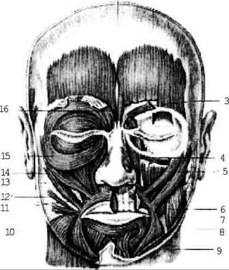
Fig. 2.74. Facial muscles
1. The occipitofrontalis muscle features a broad tendinous portion, or epicranial aponeurosis, which fuses with the skin, along with frontal and occipital bellies. Function: elevates the eyebrows, draws the scalp of the forehead upward and backward, forming transverse wrinkles ("muscle of surprise").
2. The corrugator supercilii muscle draws the eyebrows together ("muscle of anger").
3. The orbicularis oculi Muscle consists of circular muscle bundles surrounding the orbital margin and inserting into the skin of the eyelids. It dilates the lacrimal sac, closes the eyelids, pulls the eyebrows downward, and draws the cheek skin upward.
4. The orbicularis oris muscle lies as circular muscle bundles beneath and around the skin of the Lips. It closes the Mouth and purses the lips ("kissing muscle").
5. The depressor anguli oris muscle pulls the angle of the mouth downward.
6. The levator anguli oris muscle is the muscle of a pleasant (cheerful) mood ("comedy mask").
7. The buccinator muscle forms the lateral wall of the Oral Cavity, fusing with its mucosa. When contracted, it presses the cheek against the Teeth and participates in the act of sucking. It is covered by a buccal fat pad (better developed in children and women).
Other facial muscles include the zygomaticus Major and minor, risorius ("smiling muscle"), procerus, and mentalis muscles, among others.
The masticatory muscles are represented by four pairs of muscles. Their common feature is that they originate on the BONES OF THE Skull, insert onto various Regions of the Mandible, and act upon the temporomandibular joint.
1. The masseter muscle originates from the zygomatic arch and inserts into the lateral surface of the mandibular angle (Fig. 2.75). Function: elevates the mandible.
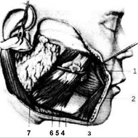
Fig. 2.75. Muscles of the face; lateral view
2. The temporalis muscle originates from the entire temporal fossa and inserts into the coronoid process of the mandible. Function: elevates the mandible; its posterior fibers pull the jaw backward (chewing action).
3. The lateral pterygoid muscle lies within the infratemporal fossa (Fig. 2.76). It originates from the pterygoid process of the Sphenoid bone and inserts into the articular process of the mandible. Function: bilateral contraction protrudes the mandible forward; unilateral contraction moves the jaw to the opposite side.
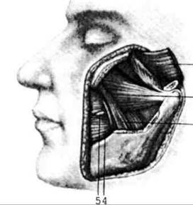
Fig. 2.76. Pterygoid muscles, left lateral view. The coronoid process of the mandible has been removed
4. The medial pterygoid muscle originates from the pterygoid fossa and inserts into the pterygoid tuberosity on the Medial surface of the mandibular angle. Function: together with the masseter muscle, it forms a functional muscular sling that allows for powerful elevation and clenching of the mandible against the Maxilla.
Last update: 08/08/2026
Editorial and Educational Adaptation: This material has been compiled based on the primary/original source text. The project team performed an editorial review, corrected technical inaccuracies, structured sections, and adapted the content for an educational format.
What was processed:
- elimination of formatting defects (OCR errors, structural breaks, corrupted characters);
- editorial organization of content;
- standardization of terminology in accordance with academic sources;
- verification of factual statements against the original source text.
All mentions of the author, publication year, and origin of the primary text have been preserved in accordance with the source.