Review of Medical Physiology - William F. Ganong 2002
Respiration
Adaptive respiratory changes in health and disease
Hypoxic hypoxia
Hypoxic Hypoxia is a common challenge for individuals at significant altitudes, as well as a complication of Pneumonia and various other respiratory disorders.
Effects of Reduced Barometric Pressure
The composition of ambient air remains constant, but total barometric pressure decreases with increasing altitude (Fig. 37-6). Consequently, PO2 also drops. At an altitude of 3000 m above sea level, alveolar PO2 is approximately 60 mm Hg, which is sufficient to stimulate peripheral chemoreceptors and increase ventilation. As one ascends higher, alveolar PO2 decreases more gradually, while alveolar PCO2 declines only slightly, because hyperventilation driven by lowered arterial PCO2 induces respiratory alkalosis.
Hypoxic Symptoms While Breathing Air
Because numerous compensatory mechanisms require time to enhance altitude tolerance (acclimatization), unacclimatized individuals often exhibit psychological symptoms, such as irritability, at altitudes of 3700 m. At 5500 m, hypoxic symptoms become severe, and above 6100 m, loss of consciousness typically occurs in most people (Fig. 37-7).
Hypoxic Symptoms While Breathing Oxygen
Total atmospheric pressure becomes the ultimate limiting factor for altitude tolerance when breathing 100% O2. The partial pressure of Water vapor in alveolar gas remains constant at 47 mm Hg; assuming a normal PCO2 of 40 mm Hg, the minimum barometric pressure required to maintain a normal alveolar PO2 of 100 mm Hg is 187 mm Hg, which corresponds to an altitude of 10,400 m. Beyond this point, the increased ventilation triggered by declining alveolar PO2 reduces PCO2 only marginally.
However, the maximum alveolar PO2 achievable while breathing 100% O2 at an ambient barometric pressure of 100 mm Hg at an altitude of 13,700 m is merely 40 mm Hg.
At an altitude of 14,000 m, an individual loses consciousness despite the administration of 100% O2 (see Fig. 37-5). Nevertheless, a synthetic atmosphere can be established around a person inside a space suit or a pressurized cabin equipped with proper O2 supply and CO2-scrubbing systems, making it possible to ascend to any height and survive in the vacuum of interplanetary space.
At an altitude of 19,200 m, barometric pressure drops to 47 mm Hg. At or below this pressure, Body Fluids "boil" at core body Temperature due to The formation of gas bubbles resulting from decreased gas solubility. This concept is largely academic, however, since any person exposed to such low pressure would succumb to hypoxia long before gas bubbles in the bloodstream could cause death.
Class="center">
Fig. 37-6. Composition of alveolar gas in individuals breathing ambient air (0–6100 m) and 100% O2 (6100–13,700 m). The minimum alveolar PO2 that an unacclimatized person can tolerate without losing consciousness is 35–40 mm Hg. Note that with increasing altitude, alveolar PCO2 decreases because hyperventilation results from the hypoxic stimulation of carotid and aortic chemoreceptors. The decrease in barometric pressure with altitude is nonlinear because air is compressible.

Fig. 37-7. Acute effects of hypoxia in individuals breathing air at various altitudes.
Delayed Effects of High Altitude
When individuals first arrive at high altitudes, many develop acute mountain sickness. This syndrome typically manifests 8 to 24 hours after arrival and lasts for four to eight days. It is characterized by headache, irritability, insomnia, dyspnea, nausea, and vomiting. Although the exact Pathophysiology remains fully undetermined, the condition is strongly linked to cerebral edema. Low altitude PO2 triggers arteriolar dilation, and if cerebral autoregulation fails to compensate, capillary pressure rises, promoting increased fluid transudation into Brain tissue.
Individuals who do not develop mountain sickness maintain normal urine output at high altitudes, whereas a decrease in urine volume is characteristic of those who do. Furthermore, Treatment with Diuretics other than acetazolamide does not prevent mountain sickness. Symptoms are alleviated if alkalosis is reduced through acetazolamide therapy or if brain edema is mitigated by administering high doses of glucocorticoids.
High-altitude pulmonary edema (HAPE) is a severe form of mountain sickness. Susceptibility is particularly high in individuals who rapidly ascend to 2500 m and engage in strenuous physical exertion During the first three days following arrival. HAPE can also occur in previously acclimatized individuals who spend two or more weeks at sea level and then return to high altitude. It occurs in the absence of underlying cardiovascular or pulmonary disease and is accompanied by marked pulmonary Hypertension, whereas left atrial pressures remain normal. The edema fluid is characterized by high permeability, elevated protein content, and cellular elements. It is hypothesized that this fluid accumulates because not all pulmonary arterioles possess sufficient smooth Muscle to constrict in response to hypoxia; consequently, capillaries supplied by these inadequately protected vessels experience the generalized rise in pulmonary arterial pressure, leading to elevated capillary pressure that disrupts their structural integrity (stress failure). In such cases, the Ca2+ channel blocker nifedipine, which lowers pulmonary arterial pressure, proves beneficial for treatment and Prevention. Pulmonary edema responds favorably to rest and Oxygen therapy, and it generally does not develop in individuals who ascend gradually and avoid physical exertion during their first few days at high altitude.
Acclimatization
Acclimatization to high altitude results from the interplay of various compensatory mechanisms. Respiratory alkalosis develops due to hyperventilation, shifting the oxyhemoglobin dissociation curve to the left, accompanied by a simultaneous increase in erythrocyte 2,3-DPG concentration, which reduces the affinity of Hemoglobin for O2. The net effect is a decrease in P50 (see Chapter 35). Lowering O2 affinity facilitates the release of O2 to the Tissues. However, the increase in P50 is constrained because if arterial PO2 rises significantly, a reduced O2 affinity would also hinder O2 uptake by hemoglobin in the Lungs.
The initial ventilatory response upon ascent to altitude is relatively modest because alkalosis counteracts the stimulatory effect of hypoxia. Nevertheless, a sustained increase in ventilation develops after four days (Fig. 37-8). Active Transport of H+ into the CEREBROSPINAL FLUID (CSF) or The Development of lactic acidosis within the brain leads to a drop in CSF pH, which enhances the ventilatory response to hypoxia. After four days, the ventilatory response begins to level off gradually, although returning to baseline levels requires years of continued residence at high altitude. Concurrently with this decline, a gradual desensitization to the stimulatory effects of hypoxia occurs.
With ascent to high altitude, Erythropoietin secretion increases rapidly (see Chapter 24) and then declines slightly over the subsequent four days as the ventilatory response intensifies and arterial PO2 rises. The resulting expansion of circulating red Blood Cell mass begins within two to three days and continues for as long as the individual remains at altitude.
Adaptive changes also occur at THE TISSUE LEVEL. The number of Mitochondria—the sites of oxidative reactions—increases, and Myoglobin levels rise (see Chapter 35), facilitating O2 diffusion into tissues. Concurrently, intracellular cytochrome c oxidase content increases.

Fig. 37-8. Effect of acclimatization on ventilatory responses at various altitudes. VE/VO2, ventilatory equivalent—The ratio of expired minute volume (VE) to O2 consumption (VO2) (reproduced with permission from Lenfant C, Sullivan K+. Adaptation to high altitude. N Engl J Med 1971;284:1298)
The efficiency of the acclimatization process is highlighted by the fact that long-term human settlements exist at altitudes of up to 5,500 m in the Andes and the Himalayas. Native populations living in these settlements exhibit a barrel-shaped chest and pronounced polycythemia. Although their alveolar PO2 values are low, other physiological parameters remain within normal limits.
Disorders Caused by Hypoxic Hypoxia
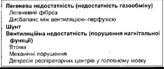
Hypoxic hypoxia is the most common form of hypoxia encountered in clinical practice. The conditions that cause it can be classified into two main categories: first, those characterized by primary failure of the gas-exchange apparatus (e.g., Congenital Heart defects where large volumes of blood bypass the Pulmonary Circulation and pass directly from the venous to the arterial side); and second, those involving ventilatory failure (Impairment of the breathing apparatus's pumping function, Table 37-1). Pulmonary insufficiency occurs when conditions such as pulmonary fibrosis cause a blockade of the alveolar-capillary barrier and ventilation-perfusion mismatch. Ventilatory failure may result from respiratory muscle weakness when the work of breathing is increased, or from various mechanical disorders, such as pneumothorax or bronchial obstruction that restricts ventilation. It can also be caused by abnormalities in the neuronal mechanisms regulating Respiration, such as the depression of respiratory Neurons in the Medulla Oblongata by morphine or other drugs.
Table 37-1. Disorders Causing Hypoxic Hypoxia
Ventilation-Perfusion Mismatch
Correcting ventilation-perfusion mismatch, which is the most frequent cause of hypoxic hypoxia, is a primary objective in clinical practice. The Physiological effects of impaired ventilation-perfusion equilibrium and their role in altering alveolar gas composition due to gravity are described in Chapter 34.
When a disease process impedes ventilation in certain alveoli, the ventilation-perfusion ratio across different Regions of the lungs is reflected by a deviation in systemic arterial PO2. If unventilated alveoli continue to be perfused, a right-to-left shunt effect occurs within that unventilated yet perfused lung region, resulting in the dumping of deoxygenated blood into the left side of The Heart. Milder degrees of ventilation-perfusion mismatch are particularly common. In the example illustrated in Fig. 37-9, unventilated alveoli (B) exhibit a low PO2, whereas hyperventilated alveoli (A) show a high alveolar PO2. Furthermore, the unsaturated hemoglobin in the blood coming from region B is not fully compensated for by the higher saturation of blood coming from region A, because hemoglobin under normal conditions is already nearly saturated in the lungs. Raising alveolar PO2 adds only a minimal amount of extra O2 to hemoglobin beyond its normal carrying capacity. As a result, the arterial blood remains unsaturated. On the other hand, the arterial CO2 content is usually normal, as excess CO2 elimination in hyperventilated areas can balance out the reduced elimination in unventilated areas.
Venoarterial Shunts
When cardiovascular anomalies, such as an atrial septal defect, allow a large volume of deoxygenated blood to bypass the pulmonary capillaries and mix with oxygenated blood in the systemic Arteries (a right-to-left shunt), chronic hypoxic hypoxia and cyanosis develop (cyanotic congenital heart disease). Administration of 100% O2 increases the O2 content in alveolar air, mitigating diffusion impairments or ventilation-perfusion imbalances (such as short-term perfusion in otherwise unventilated segments) and thereby increasing the O2 content of blood leaving the lungs. However, in patients with venoarterial shunts and otherwise normal lungs, any beneficial effect of 100% O2 is negligible and results solely from a slight increase in The amount of dissolved O2 in the blood.

Fig. 37-9. Left: "Ideal" relationship between ventilation and blood flow. Right: Uncompensated heterogeneous ventilation and homogeneous blood flow. VA, alveolar ventilation; RMV, respiratory minute volume (reproduced with permission from Comroe JH Jr et al: The Lung: Clinical Physiology and Pulmonary Function Tests, 2nd ed. Year Book, 1962).
Lung Collapse
When Bronchi or bronchioles become obstructed, the gas trapped in the alveoli beyond the obstruction is absorbed, causing the affected lung segment to collapse. Collapse of the alveoli is known as Atelectasis. An atelectatic area can range in size from a small patch to an entire lung. Part of the blood flow is diverted from the collapsed area to better-ventilated PARTS OF THE lung, which helps minimize deviations in PO2.
When a large portion of the lung collapses simultaneously, overall lung volume decreases noticeably. Consequently, intrapleural pressure becomes more negative, pulling the Mediastinum—which in humans is a fairly mobile Structure—toward the affected side.
Another cause of atelectasis is the deficiency or inactivation of surfactant, a substance that lowers surface tension and is normally present in the thin fluid layer lining the alveoli (see Chapter 34). This deficiency is a major cause of respiratory distress syndrome in newborns when the lungs fail to expand properly at birth. Lung collapse can also result from the presence of air (pneumothorax), fluid (hydrothorax, chylothorax), or blood (hemothorax) in the pleural space.
Pneumothorax
If air enters the pleural space through a rupture in the lung or a wound in the chest wall, the lung on the affected side collapses due to its elastic recoil. Once the intrapleural pressure on the affected side returns to atmospheric pressure, the mediastinum shifts back to its normal position. If the communication between the pleural space and the external environment remains open (an open, or sucking, pneumothorax), additional air moves into or out of the pleural space with every breath the patient takes. If the opening is large, the resistance to airflow into the pleural space is lower than the resistance to airflow into the intact lung, meaning little air actually enters the lungs. During inspiration, the mediastinum shifts toward the uninjured side, kinking the great vessels, displacing the heart, and impairing its diastolic filling during expiration. Furthermore, there is significant respiratory stimulation driven by hypoxia, hypercapnia, and the activation of pulmonary deflation receptors. Respiratory distress is severe.
If tissue covers the wound in the lung or chest wall in a way that acts like a flap valve, it allows air to enter during inspiration but prevents its escape during expiration, causing intrapleural pressure to rise above atmospheric levels (tension pneumothorax). As the hypoxic respiratory drive forces deeper inspiratory efforts, which further elevate intrapleural pressure, the great Veins become compressed, leading to severe hypoxia and Shock. Intrapleural pressure in such cases may rise to 20–30 mmHg. Peripheral veins become distended, intense cyanosis develops, and the condition quickly becomes life-threatening unless the pneumothorax is decompressed by releasing the trapped air.
Conversely, if the opening through which air enters the pleural space is tightly sealed (closed pneumothorax), respiratory distress is generally mild because with each breath, air flows into the lung on the uninjured side much faster than into the pleural space. Moreover, because vascular resistance increases in the collapsed lung, blood is successfully diverted to the opposite lung. Therefore, unless the pneumothorax is very large, it does not cause severe hypoxia.
Air trapped in a closed pneumothorax is gradually absorbed. Once it equilibrates with atmospheric pressure, its total pressure, PO2, and PN2 are HIGHER than those in venous gas or venous blood (compare the values for air and venous blood in Fig. 34-18). Gas diffuses down these gradients into the blood and disappears completely within one to two weeks.
Asthma
Asthma is characterized by episodic or chronic wheezing, coughing, and a sensation of chest tightness resulting from bronchoconstriction. Although the incidence of and mortality from asthma are rising, its fundamental cause remains incompletely understood despite intensive research. Asthma involves three primary abnormalities: airway obstruction (which is the most difficult feature to normalize), airway inflammation, and hyperresponsiveness to various stimuli. A long-standing link between asthma and allergy is well established, with plasma IgE levels frequently elevated. Proteins released from eosinophils during inflammatory responses can damage the airway epithelium and are responsible for airway hyperresponsiveness. Leukotrienes (see Chapter 17), released from eosinophils and mast Cells, trigger bronchoconstriction. Numerous other amines, Neuropeptides, chemokines, and interleukins also affect bronchial smooth muscle or contribute to inflammation.
Asthmatic attacks are typically most severe during the late night or early morning hours because, as noted above, these periods coincide with the peak constriction phase of the circadian rhythm of bronchial tone. Cold air and physical exertion, which normally trigger bronchoconstriction, also provoke asthmatic attacks in susceptible individuals. In about 5% of patients, attacks are precipitated by aspirin. Leukotriene synthesis inhibitors, such as zileuton, and CysLT1 receptor blockers, such as montelukast, are effective in treating these forms of asthma (and likely others). Because stimulation of ß-adrenergic receptors produces bronchodilation, inhalation therapy with ß-adrenergic agonists is a standard treatment for asthma. Conversely, muscarinic receptor activation mediates bronchoconstriction, and antimuscarinic drugs are also utilized in therapy. Additional commonly prescribed medications include cromolyn, which inhibits the release of inflammatory mediators from mast cells, and glucocorticoids, which suppress the underlying inflammatory response.
Emphysema
In the case of emphysema, a degenerative and potentially life-threatening pulmonary disease, the lungs lose their elasticity. Due to the destruction of elastic tissue and the walls between alveoli, the alveoli are replaced by large air spaces. Physiological dead space increases significantly, and given inadequate and uneven alveolar ventilation and perfusion, severe hypoxia develops in poorly ventilated alveoli. As the disease progresses, hypercapnia ensues. Inhalation and exhalation become labored, and the work of breathing increases substantially. Changes in the pressure-volume curve of the lungs are shown in Fig. 34-11. The chest expands and takes on a barrel-shaped appearance. As the chest wall is stretched, its opposing elastic recoil decreases. Hypoxia leads to polycythemia. Pulmonary hypertension develops, causing right ventricular enlargement (Cor Pulmonale), which is subsequently followed by myocardial pump failure as cardiac reserves become exhausted.
In most cases, emphysema is caused by cigarette smoking. Cigarette smoke triggers an increase in pulmonary alveolar macrophages, which release chemical substances that attract leukocytes to the lungs. In turn, leukocytes release proteases, including Elastase, that damage the elastic tissue within the lungs. Simultaneously, oxygen radicals released by leukocytes inactivate a1-antitrypsin. The consequence is an imbalance in the protease-antiprotease ratio, resulting in accelerated destructive changes within the lung parenchyma.
In 2% of cases, emphysema is caused by a congenital deficiency of active a1-antitrypsin. Individuals who are homozygous for this defect and smoke develop emphysema rapidly, with a life expectancy reduced by 20 years. Nonsmokers with a1-antitrypsin deficiency may also develop emphysema, but their life expectancy is considerably longer and their quality of life is better. Thus, a1-antitrypsin deficiency exemplifies the interplay between genetic and environmental factors in the Pathogenesis of the disease.
Cystic Fibrosis
Cystic fibrosis is another condition that leads to recurrent pulmonary infections accompanied by progressive and ultimately fatal destruction of the lungs. In this inherited recessive disorder, Cl- channels in the apical membranes of airway epithelial cells fail to be activated normally by cAMP. Additional Cl- channels in the membrane are normal, but their function may be suppressed. In either case, the loss of cAMP-activated channels results in decreased Cl- transport into the Airways. The subsequent events are not yet fully understood; however, because Cl- secretion is impaired, epithelial Na+ channels in the airways become hyperactivated, leading to enhanced Na+ absorption compared to normal. Water moves out of the airways, rendering the mucus thicker and more viscous, which predisposes the patient to airway obstruction and infection. Among Caucasians, cystic fibrosis is one of the most common Genetic Disorders: 5% of the population are carriers of the defective Gene, and the disease occurs in roughly 1 out of every 2,000 live births.
The gene mutated in cystic fibrosis is located on the long arm of chromosome 7 and encodes a Cl- channel known as the cystic fibrosis transmembrane conductance regulator (CFTR), which features 12 membrane-spanning domains, two ATP-binding sites, and a regulatory region containing
phosphorylation sites for cAMP-dependent protein kinase (protein kinase A) (see Chapter 1). This channel belongs to a superfamily of transporters that mediate, among other things, the export of the a-factor mating pheromone in Yeast and possibly the secretion of proteins lacking a signal sequence in mammals. Numerous Mutations in the CFTR gene have been identified as causes of cystic fibrosis; the severity of the clinical manifestations varies depending on the specific mutation, reflecting the complex Functional Properties of the encoded protein. Most mutations impair ATP binding or induce conformational changes that disrupt normal channel gating.
In men with cystic fibrosis, the thickening of secretions affects the ducts through which sperm pass, which can cause obstruction and Infertility. Chronic Pancreatitis in both sexes results from the abnormal function of the pancreatic ducts (see Chapter 26). In Sweat Glands, the reabsorption of Na+ and Cl- from the lumen is impaired, resulting in an abnormally high electrolyte concentration in sweat.
Last update: 10/08/2026
Editorial and Educational Adaptation: This material has been compiled based on the primary/original source text. The project team performed an editorial review, corrected technical inaccuracies, structured sections, and adapted the content for an educational format.
What was processed:
- elimination of formatting defects (OCR errors, structural breaks, corrupted characters);
- editorial organization of content;
- standardization of terminology in accordance with academic sources;
- verification of factual statements against the original source text.
All mentions of the author, publication year, and origin of the primary text have been preserved in accordance with the source.