Medical Radiology - Lazar A.P. 2008
Radiological Examination of the Heart and Blood Vessels
Congenital Heart Defects
Congenital Heart defects account for 1–2% of all organic heart diseases. According to E. Taussig's Classification, congenital heart defects are clinically divided into two groups: cyanotic heart defects ("blue babies") and acyanotic heart defects ("white babies"). The so-called "blue" heart defects include the tetralogy and triad of Fallot, and pulmonary stenosis; they are accompanied by decreased pulmonary Blood flow. Developmental anomalies with unchanged pulmonary blood flow ("white defects") include patent ductus arteriosus, ventricular and atrial septal defects, aortic coarctation, dextrocardia, and several others.
Class="center">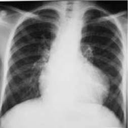
Fig. 148. Tetralogy of Fallot on a chest X-ray.
Tetralogy of Fallot accounts for 2/3 of all cyanotic congenital anomalies. It is characterized by pulmonary stenosis, a high ventricular septal defect, rightward displacement (dextroposition) of the aortic ROOT originating from both ventricles, and secondary right ventricular hypertrophy. Due to pulmonary stenosis and aortic dextroposition, a portion of venous blood flows from the right ventricle into the systemic Circulation, causing inadequate blood oxygenation and cyanosis.
Clinically, it manifests as cyanosis, dyspnea, a characteristic squatting posture in children during hypoxic spells, and a systolic murmur in the 2nd–4th left intercostal spaces along the Sternum caused by blood passing through the narrowed pulmonary outflow tract and the ventricular septal defect.
A direct anteroposterior chest radiograph reveals diminished pulmonary vascular markings, increased lung translucency, and smaller pulmonary roots. The Heart assumes an aortic configuration with a slightly elevated and rounded apex due to right ventricular hypertrophy (Figs. 148, 149). The ascending aorta is dilated and shifted to the right. Fluoroscopy reveals deep pulsations of the ascending aorta and the right ventricle, which predominate over left ventricular pulsations.
During angiocardiography, the contrast medium simultaneously opacifies the pulmonary trunk and the aorta. Ultrasound examination reveals a ventricular septal defect, a dilated aorta positioned over the septal defect, thickening of the right ventricular wall and interventricular septum, and narrowing of the right ventricular outflow tract. Electrocardiographically synchronized cine-Magnetic Resonance imaging produces slices that visualize and measure heart chambers, intracardiac septa, Valves, and great vessels. Doppler sonography and magnetic resonance angiocardiography determine the direction and velocity of blood flow within the heart chambers and great vessels.
To minimize radiation exposure in a child with a Congenital Heart defect, ultrasound (echocardiography) and MRI should be considered the primary Diagnostic Imaging Modalities. These Methods can also be used for postoperative patient follow-up.
The triad of Fallot is characterized by pulmonary stenosis, an atrial septal defect, and secondary right ventricular hypertrophy. Hemodynamic disturbances in isolated pulmonary stenosis also lead to right ventricular wall hypertrophy followed by cavity dilatation, accompanied by right atrial hypertrophy and dilatation. Patients complain of weakness, dizziness, and dyspnea at rest. The pulse is rapid, and a systolic murmur is heard in the 2nd–3rd intercostal spaces along the left sternal border. Radiography shows diminished pulmonary vascular markings, hypertransparent lung fields, and enlargement of the right heart chambers. Ultrasonography, MRI, and angiocardiography verify the Diagnosis.

Fig. 149. Tetralogy of Fallot (diagram).
A - Radiograph:
1 - accentuated cardiac waist;
2 - Left Ventricular Hypertrophy;
3 - pulmonary hypovascularization;
B - Angiocardiogram:
1 - intravascular catheter;
2 - narrowing of the pulmonary trunk;
3 - aorta;
4 - pulmonary hypovascularization.
Patent ductus arteriosus (Botallo's duct) occurs more frequently than other developmental defects. During intrauterine development, this duct connects the aortic arch to the pulmonary trunk. After birth, the duct normally obliterates and closes within the first two years of life; however, in 0.1–0.2% of cases, it remains patent. The shunting of blood from the systemic to the Pulmonary Circulation through the patent ductus arteriosus leads to circulatory insufficiency, clinically manifested in children by weakness, fatigue, dyspnea, tachycardia, palpitations, and delayed physical development. A characteristic systolic-diastolic murmur is auscultated in the left 2nd–3rd intercostal space, and diastolic blood pressure is lowered.
Radiologically, the heart in patent ductus arteriosus has a mitral configuration, and the pulmonary roots are widened, especially on the left side, although their structural definition is preserved. The pulmonary vascular pattern is enhanced either bilaterally (in case of communication with the main pulmonary trunk) or predominantly on the left (in case of connection with the left pulmonary artery). In the lower lung fields, the "amputation" sign of Arteries without branching into smaller divisions is frequently observed, indicating active pulmonary Hypertension.
Angiocardiography reveals retrograde Filling of the pulmonary artery with contrast-enhanced blood from the aorta 5–6 seconds after contrast injection, prolonged retention of the contrast medium in the pulmonary circulation, and a right-to-left shunt from the pulmonary trunk to the aorta in the event of severe pulmonary hypertension. Aortography demonstrates blood shunting from the aorta into the pulmonary trunk.
Ultrasound reveals left ventricular enlargement, an altered aortic-to-left atrial diameter ratio (normally 1:1), and prominent, hyperdynamic pulsations of the pulmonary trunk, aorta, and left ventricle. Contrast echocardiography detects The entry of contrast medium—administered via the umbilical artery in newborns—from the aorta into the pulmonary artery.
Atrial septal defect is one of the most common congenital heart defects, especially when combined with other cardiac anomalies. The size of the defect can vary—from a patent foramen ovale to the complete absence of the septum. A small, patent foramen ovale is not accompanied by hemodynamic disturbances because it is closed by the valve of the foramen ovale during atrial contractions.
Since the pressure in the left atrium is higher than in the right, a large atrial septal defect causes blood to shunt from the left atrium into the right, resulting in volume overload of the right heart chambers. The dimensions of the right atrium, right ventricle, and pulmonary trunk increase. Small atrial septal defects present with no clinical manifestations. Significant defects lead to Heart Failure and shortness of breath. Auscultation reveals a systolic murmur in the II-III intercostal spaces at the left sternal border. Pulse pressure is decreased due to an insufficient volume of blood entering the systemic circulation. The Electrocardiogram shows a right-axis deviation and P-wave deformation.
X-ray Examination reveals widened, fibrotic, and structured lung roots. The pulmonary vasculature is accentuated, exhibiting a sudden pruning of arteries toward the periphery. In the anteroposterior projection, a pronounced bulging of the pulmonary trunk arch is visible, giving the heart a mitral configuration. The cardiac apex is displaced to the left by the enlarged right ventricle. The right atrium arch is enlarged to the right, and the right cardio-vascular angle is shifted upward.
Ultrasound reveals enlargement of the right atrial and ventricular cavities, reduction of the left ventricle, and deep pulsation of both ventricles and the pulmonary trunk. Two-dimensional echocardiography determines the exact Location and dimensions of the atrial septal defect.
The ability to pass a catheter from the right atrium into the left during cardiac catheterization confirms the presence of an atrial septal defect.
Ventricular septal defect, much like atrial septal defect, frequently occurs in combination with other congenital heart anomalies. A small defect in the muscular portion of the septum is not accompanied by hemodynamic disturbances because ventricular contraction during systole closes the defect. Hemodynamic alterations in significant ventricular septal defects involve blood shunting from the left ventricle to the right, leading to pulmonary hyperperfusion and underfilling of the systemic circulation. Clinically, this manifests as delayed physical development in children and Complaints of exertional dyspnea. Objectively, patients exhibit Skin pallor, rightward expansion of cardiac borders, and a harsh systolic murmur at the apex.

Fig. 150. Ventricular septal defect (arrow) on MRI (T1-weighted image).
Radiological examination reveals widened lung roots and an enhanced pulmonary pattern with marked active hypertension. The heart assumes a mitral shape. Along the left contour of the cardiovascular silhouette, the arches of the pulmonary trunk and the left ventricle (displaced by the right ventricle) protrude; the cardiac apex is slightly elevated and rounded. The right borders of the cardiac shadow are enlarged due to the right ventricle. Fluoroscopy demonstrates increased pulsation of both the right and left ventricles.
Angiocardiography shows contrast medium passing from the right ventricle into the left, whereas ventriculography demonstrates contrast flow from the left ventricle into the right. Cardiac probing often allows the catheter to pass from the right ventricle into the left, thereby proving the presence of a ventricular septal defect.
Ultrasound and ultrafast MRI (Fig. 150) enable direct visualization of the defect's localization and dimensions, enlargement of the right ventricular cavity along with increased wall thickness, and a reduction in left ventricular size.
Aortic coarctation is a congenital segmental narrowing of the aorta ranging from 1 cm to 1-2 mm, most commonly localized in the isthmus region. Consequently, blood pressure is elevated in the left ventricle and the arterial branches arising proximal to the narrowing that supply the upper body, whereas pressure is reduced in the arteries originating distal to the narrowing that supply the lower body. This pressure gradient induces the compensatory development of collateral vessels, leading to the dilation of the anterior and posterior intercostal arteries and subcutaneous arterial branches on the anterior chest wall.
Chest radiographs of coarctation patients older than 12 years reveal symmetrical notches (rib notching) on the lower margins of the III-VI Ribs bilaterally (Fig. 151), alongside thickening of the compact bone along the inferior rib margins (observed in 75% of adult coarctation patients). The heart has an aortic configuration, the left ventricle is hypertrophied with its apex rounded and elevated above the Diaphragm, and the contrasted Esophagus is deviated to the right at the cardiac level.

Fig. 151. Coarctation of the aorta.
A - anteroposterior chest radiograph: 1 - prominence of the left Subclavian Artery contour; 2 - effacement of the left contour of the aortic arch; 3 - left ventricular hypertrophy; 4 - inferior rib notching; 5 - esophageal displacement caused by post-stenotic aortic dilatation;
B - aortography in the left lateral projection: 1 - brachiocephalic trunk; 2 - left subclavian artery; 3 - stenosis of the aortic isthmus; 4 - post-stenotic dilatation; 5 - internal thoracic artery.
Fluoroscopically, the pulsation amplitude of the left ventricle and ascending aorta is increased, while that of the descending aorta is diminished.
Aortography reveals dilatation of the ascending aorta. The aortic arch along the left contour of the cardiovascular silhouette is smoothed out at the transition point into the descending aorta, lacking its usual convex contour. In the left oblique projection, narrowing is visualized in the region of the aortic isthmus. In the lateral projection behind the sternum, an enlarged internal thoracic artery is identified, appearing as an elongated shadow with a wavy contour.
Angiocardiography demonstrates enlargement of the left atrial and ventricular cavities, dilatation of the ascending aorta, and stenosis in the aortic isthmus region. Echocardiography shows aortic narrowing, hyperdynamic aortic pulsation, and turbulent blood flow distal to the stenosis. MRI in the sagittal plane clearly demonstrates the site of narrowing and its length.
Dextrocardia is characterized by a mirror-image anatomical position of the heart relative to normal, which is readily detected by all imaging modalities (Fig. 152). Dextrocardia is frequently associated with Bronchiectasis and sinusitis (Kartagener's triad).

Fig. 152. Dextrocardia on a plain chest radiograph. The gastric gas bubble is located on the left side.
Last update: 08/08/2026
Editorial and Educational Adaptation: This material has been compiled based on the primary/original source text. The project team performed an editorial review, corrected technical inaccuracies, structured sections, and adapted the content for an educational format.
What was processed:
- elimination of formatting defects (OCR errors, structural breaks, corrupted characters);
- editorial organization of content;
- standardization of terminology in accordance with academic sources;
- verification of factual statements against the original source text.
All mentions of the author, publication year, and origin of the primary text have been preserved in accordance with the source.