Human Anatomy Part 1 - K. A. Dubenko, A. K. Kolomiysev, Yu. B. Chaykovsky 2002
Special Section
Myology, myologia [the study of muscles] — General Information
Development of Muscles
Striated Muscles develop and differentiate from myotome Cells of the dorsal mesoderm, which is located on either side of the neural tube and notochord (Fig. 121).
By the 3rd week of embryonic development, segmentation occurs in the HEAD region of the dorsal mesoderm, forming sac-like protrusions known as somites. Following The formation of the first pair of head somites, further Cleavage of the dorsal mesoderm proceeds in the caudal direction.
Class="center">
Fig. 116. First-class lever (two-armed lever);
A - fulcrum; B - point of force application; C - point of resistance

Fig. 117. FOOT as a second-class lever;
A - fulcrum; B - point of force application; C - point of resistance

Fig. 118. Forearm as a third-class lever;
A - fulcrum; B - point of force application; C - point of resistance

Fig. 119. Diagram of a "force couple" (after M. F. Ivanytskyi):
AB - resultant force of the biceps brachii; ДЖ - opposing force exerted by the humerus; АБ - effective "component of the biceps brachii"; АГ - second component of the same Muscle, which contributes to the pressure of the forearm against the arm at the elbow joint; Д - Components of the pressure force exerted by the humerus on the forearm; АД - lever arm of the force couple, where one force is АБ and the other is ДЕ
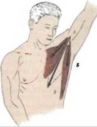
Fig. 120. Parallelogram of forces (after M. F. Ivanytskyi):
АБ - line of pull of the pectoralis major muscle; АГ - line of pull of the latissimus dorsi muscle; AB - diagonal of the parallelogram of forces
By the 4th week of embryonic development, 40 pairs of somites are formed, which include: 5 occipital, 8 cervical, 12 thoracic, 5 lumbar, 5 sacral, and 3 to 4-5 coccygeal pairs. Somites increase in volume and become separated from one another by Connective Tissue septa (myosepta). A small cavity, the myocoel, forms in the center of each somite, dividing it into two plates: medial and lateral. Subsequently, differentiation takes place, and the somite splits into three parts: the myotome, whose cells form striated muscles; the dermatome, which gives rise to the connective tissue layer of the Skin; and the sclerotome, whose cells form the vertebrae and Ribs. The myotomes grow ventrally and divide into ventral and dorsal parts. Later, the dorsal musculature of the trunk, located on the anterior and lateral aspects of the trunk, develops from the dorsal part of the myotomes (see Figs. 121, 122).
In the head region of the embryo, a group of branchiogenic muscles arises from the mesoderm of the pharyngeal (branchial) arches, comprising the Muscles of the head, neck, soft palate, Pharynx, and Larynx. They are innervated by the V, VII, IX, and X cranial nerve pairs. Spinal Nerves (neuromeres), formed at the level of each somite, grow into every myotome (myomere). The nerve divides into anterior and posterior branches corresponding to the division of the myotome into anterior and posterior parts.

Fig. 121. Trunk segment of a primitive vertebrate embryo; semi-schematic diagram (after Braus)

Fig. 122. Cross-section of an adult human body - diagram (after Braus)
As THE POSITION OF a muscle changes, the course of its supplying nerve changes as well. An example is the Diaphragm, which initially originates within the region of the V-VI cervical somites and subsequently shifts to the inferior thoracic aperture. The Innervation of the diaphragm is provided by the phrenic nerve, which is formed from the IV-V cervical spinal nerves and assumes a descending position. Longitudinal splitting of muscle bundles leads to the formation of individual muscles (such as the trapezius and sternocleidomastoid); tangential splitting results in the formation of deep and superficial muscle layers (such as the oblique and transverse muscle layers of the abdominal wall). Certain muscles remain at their original site of development and retain a segmental Structure, forming the so-called local or autochthonous musculature (from Greek *autos* - self, *chthon* - earth). Examples include the intercostal muscles. Muscles that migrate from the trunk to the limbs are termed trunkofugal (from Latin *truncus* - trunk, *fugo* - to flee), whereas those migrating from the limbs to the trunk are termed trunkopetal (from *peta* - to strive toward).
In the Cytology/cytology/16.html">Early stages of embryonic development, connective tissue derived from the remnants of the mesoblast accumulates around the muscles, forming fasciae. This connective tissue is located around the muscles, between muscle layers, and also within the muscles surrounding individual muscle fascicles.
Development of the trunk muscles. As previously noted, striated muscles develop from myotomes. Myotomes are arranged symmetrically as plates on both sides of the notochord (see Fig. 121). They grow in the ventral and dorsal directions and merge in the anterior and posterior regions via a membrane. The ventral and dorsal myotomes are separated by a longitudinal connective tissue septum. Subsequently, the cavity of the myotomes disappears, and their walls fuse to form muscular sheets that retain a segmental structure. These muscular sheets are divided by connective tissue into deep and superficial layers. Short intervertebral muscles are formed from the deep layers of the myotomes in the back region. The superficial layers of the myotomes on the dorsal and anterolateral surfaces of the trunk lose their segmental structure, fuse, and form the primordia for the long and broad Muscles of the Trunk. Fasciae develop around the muscle layers. The largest of these is the thoracolumbar fascia, which separates the trunk muscles from the Muscles of the Pelvic Girdle. In the thoracic region of the trunk, the myotomes grow predominantly ventrally. Connective tissue ribs, which are later replaced by cartilaginous ribs, differentiate within the intermyotomal septa. The PARTS OF THE myotomes directly associated with the ribs split and transform into the external and internal intercostal muscles. The deep layers of the myotomes on the inner surface of the thoracic cavity become the transversus thoracis and subcostal muscles. The Abdominal muscles are formed by the fusion and splitting of several adjacent myotomes. They form three layers: the external oblique, internal oblique, and transversus abdominis muscles. The rectus abdominis muscle forms during the 6th week from the fusion of several central myotomes, as evidenced by the transverse tendinous intersections (intersectiones tendinei) and their sources of innervation.
Development of the head muscles. The myotomes of the First and Second pharyngeal arches serve as the source for the striated muscles of the facial Skull. The mesenchyme bordering the first pharyngeal arch gives rise to the masticatory muscles, mylohyoid muscle, anterior belly of the digastric muscle, tensor veli palatini, and tensor tympani muscle. They are innervated by the motor Branches of the Trigeminal nerve, n. trigeminus (V).
The facial muscles develop from the second pharyngeal arch. They migrate to the head region along with the Facial Nerve, resulting in the Formation of the subcutaneous muscle of the Head and Neck (platysma). The platysma is divided into facial and occipital parts, located anterior and posterior to the primordium of the External ear. The facial part of the platysma differentiates into superficial and deep layers.
In a six-week embryo, the deep layer forms sphincter-like circular muscles around natural orifices. Subsequently, some muscles degenerate, while the orbicularis oris persists, and the buccinator, caninus, nasal, and auricular muscles are formed.
In a seven-week embryo, the superficial layer loses its connection with the platysma of the neck and extends cranially and superficially to the cheek and lower jaw, reaching the temporal region, forehead, Mouth, and eye. Posteriorly, it connects with the occipital part of the platysma, forming the epicranius muscle (occipitofrontalis). This gives rise to the galea aponeurotica, muscles of the eye, Lips, and chin. The posterior divisions of this muscle form the occipital, stylohyoid, and stapedius muscles, as well as the posterior belly of the digastric muscle. The extraocular muscles develop from the head myotomes (preotic) and are innervated by the III, IV, and VI cranial nerve pairs (Fig. 123).
Development of the Neck Muscles. The neck muscles develop from the dorsal parts of the upper four cervical (occipital) myotomes (see Fig. 123). Initially, a common primordium for the trapezius and sternocleidomastoid muscles appears. These muscles subsequently separate but retain a common source of innervation—the Accessory nerve, n. accessorius (XI). More neutrally [medially/laterally in context, keeping anatomical sense: deeper/medial] to these muscles, the primordia of the Tongue and hyoid muscle groups arise, which are innervated by the Hypoglossal nerve, n. hypoglossus (XII). The scalene muscles, Deep Muscles of the head and neck are innervated by the motor branches of the Cervical plexus.
The mesoderm of the pharyngeal arches differentiates into a group of branchiomeric muscles. The mesoderm of the first pharyngeal arch is associated with the motor branches of the trigeminal nerve. The mylohyoid muscles develop from the common primordium for the masticatory muscles medially to the lower edge of the Mandible. Inferiorly, between the Hyoid bone and the chin, the anterior belly of the digastric muscle arises. The stylohyoid muscle, the posterior belly of the digastric muscle, and the platysma develop from the Superficial layer of the second pharyngeal arch, innervated by the facial nerve, n. facialis (VII).

Fig. 123. Location OF THE head and trunk myotomes in the embryo:
I - preotic myotomes; II - occipital myotomes; III - cervical myotomes; IV - thoracic myotomes; V - lumbar myotomes; VI - sacral myotomes; VII - coccygeal myotomes
Development of the limb muscles. At the end of the 4th to the beginning of the 5th week of embryonic development, on the lateral surfaces of the body at the border of the segmented and unsegmented mesoderm, paired
limb buds of the upper extremities appear. During the 6th–7th weeks of intrauterine development, the limbs grow in length. An expansion resembling a paddle forms in the region where the hand and fingers will develop. Initially, the digits lie parallel to each other, and only during the 8th week do the first digits of the upper and lower limbs diverge laterally. In the developing limbs, the mesoderm is located on the ventral and dorsal surfaces of the skeletal axis. Subsequently, the dorsal myotomes give rise to extensors and abductors, while the ventral myotomes serve as the source for flexors and adductors. Nerves from the V–VIII cervical segments and the I thoracic segment grow into the muscle primordium of the upper limb. Based on their function and localization, the muscles are divided into three groups.
The first group consists of autochthonous muscles, which originate from the mesenchymal bud; the shoulder muscles develop first, followed by the forearm and hand muscles. The deltoid muscle develops first, connecting the shoulder girdle with the upper limb, followed sequentially by the scapular muscles. In the forearm, extensors develop earlier than flexors. By the end of the 8th week of intrauterine development, the muscles of the thenar eminence, hypothenar eminence, and the interosseous muscles separate.
The second group comprises axipetal muscles, which include muscles derived from the mesenchyme of the upper limb bud, but one end of these muscles attaches to the BONES OF THE trunk. These include: the pectoralis Major and minor, and latissimus dorsi muscles.
The third group includes axifugal muscles, which develop from a muscle primordium on the trunk, but one end attaches to the bones of the shoulder girdle or humerus. These include: the trapezius and sternocleidomastoid muscles, as well as the omohyoid, subclavius, levator scapulae, serratus anterior, and rhomboideus major and minor muscles.
The Muscles of the Lower Limb develop from the lower limb buds. The mesenchymal primordia are connected with the IV–V lumbar and I–III sacral segments. During growth, a broad proximal part (from which the pelvic girdle develops) and a narrow distal part with a paddle-like tip resembling the fin of an aquatic animal differentiate. During the second month of development, the thigh, leg, and foot are formed, and the digits appear as mounds.
Muscle anomalies. The occurrence of various anatomical variations in Muscle Structure is mainly associated with individual features of Muscle Development in ontogenesis. The main anomalies include: variations in muscle shape, size, topography, attachment sites, as well as the absence or presence of additional parts and accessory muscles. In different individuals, the same muscle may have an oval, fusiform, or elongated shape. A broad muscle can range from triangular to irregular polygonal in shape. In terms of size, facial muscles, and the short Muscles of the hand and foot are the most variable. Variations in Anatomical Structure are most pronounced in complex and broad muscles; they may exhibit varying degrees of subdivision into parts, and the number of heads of origin may vary. Absence of a part of a muscle is found in the deltoid, pectoralis major, and diaphragm, which can lead to diaphragmatic hernias. Individual muscles may feature additional muscle bundles, heads, and tendons. Alterations in TOPOGRAPHIC AND ANATOMICAL relationships with neighboring structures are caused by the sites of origin and insertion, as well as the subdivision of the muscle into parts.
The absence of inconstant muscles (such as the psoas minor, pyramidalis abdominis, peroneus tertius, and coccygeus) is frequently observed. Cases also occur where accessory muscles are present: sternalis, episternal, axillary arch, gastrocnemius tertius, peroneus quartus (or minor), etc. In some muscles, the Separation of parts is not entirely proportional. An example is the complete separation of the sternal and clavicular parts throughout their entire length.
The noted deviations from the norm are of no significant practical importance, but they provide a General Overview of muscle anomalies.
Last update: 08/08/2026
Editorial and Educational Adaptation: This material has been compiled based on the primary/original source text. The project team performed an editorial review, corrected technical inaccuracies, structured sections, and adapted the content for an educational format.
What was processed:
- elimination of formatting defects (OCR errors, structural breaks, corrupted characters);
- editorial organization of content;
- standardization of terminology in accordance with academic sources;
- verification of factual statements against the original source text.
All mentions of the author, publication year, and origin of the primary text have been preserved in accordance with the source.