Medical Radiology - Lazar A.P. 2008
Radiation Examination of the Thoracic Organs
Pleurisy
Pleurisy is a pleural disease that most commonly develops as a secondary process, acting as a complication of Pneumonia, tuberculosis, or other pulmonary, cardiac, and hematological conditions. It is classified into dry and exudative pleurisy.
The Development of dry (fibrinous) pleurisy is driven by productive inflammation of the Pleura. Patients typically present with a cough, fever, and chest pain that intensifies during coughing. Localized dry pleurisy often presents no distinct radiographic findings. Diffuse dry pleurisy is accompanied by thickening of the interlobar and costal pleura (up to 1 cm), decreased lung transparency, and blunting or indistinct contours of the costophrenic angles. Occasionally, ultrasound imaging can detect a small amount of fluid within the pleural cavity.
The presence of pleural fluid is best detected with the patient in the lateral decubitus position. Plain radiography can identify fluid volumes exceeding 0.5 L in the pleural cavity, whereas Ultrasonography can detect volumes greater than 20 mL.
The clinical course of exudative (effusion) pleurisy is characterized by more pronounced clinical manifestations. On standard upright posteroanterior chest radiographs, the accumulation of pleural exudate appears as an opacification of the lateral costophrenic angle or as a triangular shadow in the lateral region of the lower lung field, bounded by a characteristic upper concave meniscus of the fluid level (Fig. 113). If the upper margin of the shadow reaches the level of the 5th rib, the fluid volume is approximately 1 L; at the 4th rib level, it is about 1.5 L; and at the 3rd rib level, approximately 2 L. Larger effusions cause a significant mediastinal shift toward the contralateral (healthy) side.
Pleural adhesions may compartmentalize the pleural cavity into distinct, isolated spaces, resulting in encapsulated (loculated) pleural effusions.
Class="center">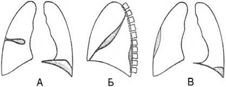
Fig. 114. Encapsulated pleurisy on a radiograph (diagram).
A — interlobar (in the horizontal fissure) and diaphragmatic; B — interlobar (in the oblique fissure) and costovertebral; C — costal and costophrenic.
The shadows corresponding to such effusions do not shift with changes in body position; their contours are distinct and convex, and they tend to be refractory to Treatment and resolve slowly. Depending on their anatomical Location, encapsulated pleurisy is classified into costal, costophrenic, costovertebral, apical, diaphragmatic, mediastinal, and interlobar forms (Fig. 114).
Last update: 08/08/2026
Editorial and Educational Adaptation: This material has been compiled based on the primary/original source text. The project team performed an editorial review, corrected technical inaccuracies, structured sections, and adapted the content for an educational format.
What was processed:
- elimination of formatting defects (OCR errors, structural breaks, corrupted characters);
- editorial organization of content;
- standardization of terminology in accordance with academic sources;
- verification of factual statements against the original source text.
All mentions of the author, publication year, and origin of the primary text have been preserved in accordance with the source.