Critical States in Respiratory Tuberculosis in Adults - N.I. Fomichova 2010
Spontaneous pneumothorax
Spontaneous pneumothorax (SP) is a pathological condition characterized by the accumulation of air in the pleural cavity due to a defect in the visceral Pleura, unrelated to mechanical injury to the lung or chest wall caused by trauma or medical Procedures. The presence of air in the pleural cavity leads to lung compression, a reduction in its respiratory surface area, and The Development of Acute Respiratory Failure. The aggravation of pathophysiological changes is also influenced by the mediastinal shift toward the healthy lung.
Etiology AND Pathogenesis. A healthy pleura is extremely durable; its rupture is only possible under a pressure of 200 mm H2O, which cannot be achieved through coughing or physical exertion. For the pleura to lose its integrity, prior pathological involvement of a congenital or acquired nature is required.
The development of spontaneous pneumothorax is rooted in various bronchopulmonary disorders that differ in etiology, pathogenesis, clinical manifestations, and prognosis. The most common causes of SP include congenital defects of the pulmonary parenchyma accompanied by The formation of bullae and cysts, Pneumonia complications such as Pleural Empyema, lung abscess, Bronchiectasis, and malignant tumors of the lung and pleura. Among all complications associated with respiratory tuberculosis, SP is observed in 1-2% of cases.
Despite The Diversity of etiological factors, the pathogenesis of spontaneous pneumothorax consists of the following components: a breach of the pleural cavity's airtightness (i.e., its connection with the atmosphere), the detachment of the visceral pleura from the parietal pleura, lung compression, and a mediastinal shift toward the healthy side (not a constant component).
In Pulmonary Tuberculosis, SP can occur at any stage of the pathological process. However, it most frequently develops in active progressive tuberculosis As a result of the rupture into the pleural cavity of subpleurally located foci of Caseous pneumonia or tuberculous cavities. In chronic forms, including extensive residual changes following cured tuberculosis in the form of bullous-dystrophic lesions, the rupture of a subpleurally situated bulla or sclerotically altered lung tissue may occur. In such cases, the provoking factor is elevated intrapulmonary pressure, which can result from physical exertion, severe coughing, laughter, high-altitude flying, etc. Consequently, the lung tissue ruptures, and air enters the pleural cavity. Furthermore, tuberculosis can lead to so-called iatrogenic pneumothorax during transthoracic puncture for fluid evacuation from the pleural cavity, lung tissue biopsy, and in the postoperative period. The cause of the latter is the occlusion of small Bronchi by viscous sputum or local bronchospasm, which leads to increased pressure in the distal Airways, overdistension of alveoli in a localized area, and the promotion of thin-walled bullous cavities.
The presence of air in the pleural cavity significantly increases intrapleural pressure (which is normally subatmospheric due to the elastic recoil of the Lungs), resulting in the compression and collapse of the lung tissue, a shift of the Mediastinum to the opposite side, the depression of the diaphragmatic dome, and the compression and kinking of major Blood Vessels in the mediastinum. All these factors lead to impaired Respiration and Circulation.
Classification. Acute, chronic, and recurrent SP are distinguished. In the acute form, clinical manifestations are highly tumultuous, accompanied by severe pain, dyspnea, and coughing. If acute SP does not resolve within 3-4 weeks, it transitions into a chronic form. The recurrent form is characterized by repeated episodes of SP.
Based on its origin, spontaneous pneumothorax is classified as primary (idiopathic) and secondary (symptomatic). Primary SP occurs in virtually healthy individuals against a Background of complete well-being. Its main cause is localized Bullous emphysema of unknown etiology. Secondary SP is a complication of severe pathological processes in the lungs and pleura.
Pneumothorax can be open, closed, or valvular (tension). In open SP, There is a communication between the pleural cavity and the bronchial lumen, and thus with atmospheric air. During inspiration, air enters the pleural cavity, and during expiration, it escapes through the defect in the visceral pleura. Subsequently, the defect becomes sealed by fibrin deposits, forming a closed pneumothorax, whereby the communication between the pleural cavity and atmospheric air ceases. A valvular (tension) pneumothorax with positive pressure in the pleural cavity may develop when an incoming air stream forcefully enters the pleural cavity during inspiration but cannot escape due to a functioning check-valve mechanism. Consequently, the pressure in the pleural cavity progressively rises and exceeds atmospheric pressure. This leads to a complete lung collapse and a significant mediastinal shift to the opposite side.
According to its extent, total (complete) pneumothorax involves an almost complete collapse of the lung; partial pneumothorax involves a lung collapse of 1/3 of its volume; and limited pneumothorax occurs when air is confined to a section of the pleural cavity bounded by adhesions, with the collapse affecting only that part of the lung not fixed to the chest wall.
Depending on the presence of complications, SP can be uncomplicated or complicated (by hemothorax, exudative Pleurisy, mediastinal and subcutaneous emphysema, pleural empyema, acute cardiovascular and respiratory failure).
Clinical manifestations and Diagnosis (Diagram 6). The clinical picture of spontaneous pneumothorax depends on The rate of its development, the degree of lung collapse and mediastinal shift, and the functional state of the respiratory and cardiovascular systems. Thus, in limited SP, clinical symptoms are mild and detected only upon radiography. This is the so-called latent course, and patients are almost asymptomatic. In total pneumothorax, clinical manifestations of acute cardiovascular and respiratory failure come to the fore.
Typically, SP occurs abruptly, although a gradual development is also possible; symptoms are sometimes intermittent, though more often they are persistent and stable.
The primary sign of SP is chest pain on the affected side, localized in the anterior or lateral wall region or the scapular area, occasionally radiating to the shoulder girdle, arm, or neck. The pain is usually sharp, dragging, or compressive in nature, and may be localized more in the epigastric region than in the chest. Pain is typically accompanied by Skin pallor, weakness, cold sweat, rapid thready pulse, and lowered blood pressure. Movement intensifies the pain. Lying on the affected side reduces it. If air inflow into the pleural cavity ceases, the pain becomes less intense. Pain and the reduction of the lung's respiratory surface are accompanied by dyspnea. Air irritation of the pleura induces coughing. The cough is generally non-productive.
Physical examination in limited pneumothorax is of low diagnostic value.
In total pneumothorax, chest Asymmetry is observed, characterized by expansion of the affected hemithorax, cyanosis, rapid breathing, tachycardia, and possibly arrhythmia. Vocal fremitus on the affected side is diminished, and Percussion reveals a tympanic sound with a hyperresonant (box) note. The boundaries of cardiac dullness are shifted toward the healthy side. Auscultation reveals weakened or absent breath sounds on the affected side of the pneumothorax.
A severe and life-threatening condition for the patient is tension pneumothorax, in which The amount of air in the pleural cavity increases with each breath, intrapulmonary pressure rises, and tension pneumothorax ensues, clinically characterized by acute respiratory failure coupled with hemodynamic impairment. Pleural Shock may occasionally occur, and without timely assistance, it can lead to death.
Furthermore, as a result of tension pneumothorax, air can penetrate along the peribronchial and perivascular interstitium into the mediastinum, and from there into the subcutaneous Tissues of the neck, leading to the development of subcutaneous emphysema. Mediastinal emphysema—indicated by hoarseness of the voice—results in the compression of blood vessels and rapid Displacement of the Heart and great vessels, which in turn leads to acute respiratory failure and hemodynamic disturbances.
SP has a typical presentation and generally poses no diagnostic difficulties for the clinician. Occasionally, a sharp surge in the clinical manifestations of pneumothorax is observed, which can lead to the patient's death within minutes.
Class="center">DIAGRAM 6 DIAGNOSTIC ALGORITHM FOR SPONTANEOUS PNEUMOTHORAX
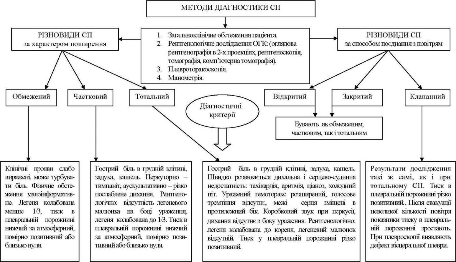
An important diagnostic method for SP is radiography. The presence of gas in the pleural cavity is an absolute sign of pneumothorax. Chest radiographs should be taken in the direct projection during the inspiratory and expiratory phases, as well as a LATERAL VIEW OF the affected side. The main radiological signs of pneumothorax include the absence of a pulmonary vascular pattern on the side of the SP and its enhancement on the healthy side, visualization of the borders of the collapsed lung, a heart shifted toward the healthy side, and a downwardly displaced diaphragmatic dome. Frequently, a small horizontal fluid level is detected beyond the collapsed lung on the affected side of the SP. Fluoroscopy of the affected side reveals the "flapping sail" sign—a sharp contraction of The Heart along the contour directed toward the SP, caused by the loss of the lung's shock-absorbing role.
Manometric examination is used to determine The Nature of SP. Normally, pressure in the pleural cavity is negative. In closed limited SP, it can vary, but is most often subatmospheric or moderately positive. If the pressure fluctuates around zero, it indicates an open SP. In valvular spontaneous pneumothorax, the pressure in the pleural cavity exceeds atmospheric pressure.
To determine the functioning of the bronchopleural/pulmonary-pleural communication, a small amount of gas is aspirated from the pleural cavity, and its pressure is measured a few minutes later. A decrease and stabilization of the pressure readings in the pleural cavity indicate that the visceral pleural defect is closing, whereas an increase in pressure readings indicates the presence of a valve mechanism.
Pleurothoracoscopy is one of the Methods that allows for the diagnosis of the causes of SP. During thoracoscopy, air blebs, pleural adhesions, and other abnormalities are visualized.
Sometimes, particularly in cases of limited SP, a computed tomography (CT) scan of the chest is indicated to perform Differential diagnosis between pneumothorax, cysts, and distended thin-walled bullae.
In spontaneous pneumothorax, various complications occur in 40–45% of cases. The most frequent of these is pneumopleuritis, which develops on the 4th–7th day from the onset of the disease and does not depend on the degree of lung collapse. In cases where a cavity ruptures into the pleural cavity, it becomes infected, subsequently leading to Empyema of the pleura. In 2.2% of patients, SP is complicated by subcutaneous emphysema. A rare but extremely severe complication of SP is hemopneumothorax, which is characterized by a collapse-like state and acute anemia.
EMERGENCY CARE
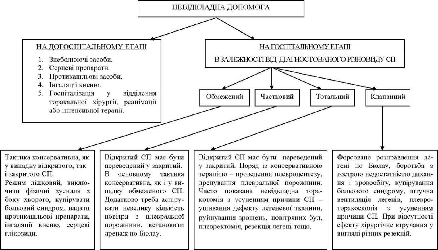
At the pre-hospital stage, the administration of analgesics and cardiac medications, oxygen inhalation, and antitussive drugs is indicated (Diagram 7). All patients with SP are subject to hospitalization and require emergency care. Medical tactics depend on the extent of the lesion and the General condition of the patient. In cases of limited and partial uncomplicated SP, provided that external respiration function is compensated, conservative management is indicated, which aims at the spontaneous resorption of air and excludes physical exertion on the part of the patient. The patient is prescribed bed rest, must remain under constant medical supervision, and receive symptomatic medical Treatment. The patient is placed in a semi-sitting position, provided with humidified oxygen inhalation, and prescribed analgesics, antitussives, and intramuscular injections of 10% sulfocamphocaine 2 ml. Depending on the patient's condition, cardiac Glycosides are administered once or continuously.
In valvular SP, along with conservative therapy, forced lung re-expansion should be performed by placing an intercostal drainage tube. Initially, thoracoscopy should be performed, during which the causes of SP can be eliminated by electrocoagulation of bullae and adhesions, or by suturing bullous-dystrophic areas. If the method of forced lung re-expansion via continuous air aspiration through the drainage tube fails to produce an effect, the patient, in the absence of contraindications, must undergo Surgical treatment in the form of various types of resection (segmental resection, lobectomy, or pulmonectomy) depending on the underlying process that led to the development of spontaneous pneumothorax.
Patients with bilateral SP, pronounced dyspnea (respiratory rate exceeding 45 breaths/min), tachycardia (exceeding 120 beats/min), and a tendency toward lowered blood pressure are transferred to mechanical ventilation. Due to the risk of increasing mediastinal shift, the pleural cavity must be drained prior to initiating mechanical ventilation.
Patients who have contraindications to surgical treatment (severely impaired respiratory and cardiovascular Functions, severe comorbidities) undergo prolonged drainage of the pleural cavity with continuous gentle air aspiration, with periodic replacement of the drainage tube every 15–20 days to reduce the volume of the pleural cavity and limit empyema, along with sanitation of the pleural cavity. Broad-spectrum Antibiotics, detoxification, pathogenetic, and symptomatic agents are indicated.
In tension pneumothorax, emergency pleurocentesis is mandatory to improve gas exchange and reduce venous pressure resistance, which leads to the normalization of Cardiac Output. In life-threatening conditions, the Introduction of a thick needle or trocar into the pleural cavity is possible. In A number of cases, emergency thoracotomy is necessary. If SP is complicated by pneumomediastinum, the patient must be placed in a room with an elevated oxygen concentration. Mediastinal puncture can be performed using a curved needle in the jugular fossa, avoiding the Sternum.
DIAGRAM 7 DIAGRAM OF EMERGENCY CARE PROVISION FOR SPONTANEOUS PNEUMOTHORAX (SP)
CLINICAL CASE EXAMPLE
Patient N., 48 years old, Group II disabled person. She was walking up to the 5th floor. Near her apartment door, she experienced a sharp chest pain, paroxysmal cough, palpitations, weakness, and intensified dyspnea. The physician diagnosed acute cardiopulmonary failure and hospitalized the patient.
At a young age, the patient had suffered from Disseminated pulmonary tuberculosis, recovered, and was discharged from clinical follow-up. Subsequently, marked pulmonary fibrosis and emphysema developed, and respiratory failure progressed.
The patient's condition is severe; her body position in bed is forced — she sits with her lower extremities lowered. Her skin is cold with a bluish tint. Pulse is 112 beats/min, arrhythmic, of weak volume and tension. Blood pressure is 95/60 mm Hg. Heart sounds are muffled. The chest is barrel-shaped. Percussion over the lungs reveals a box Resonance bilaterally, especially over the left lung, where breath sounds are completely absent. Over other lung areas, breath sounds are sharply weakened, with dry rales throughout. The abdomen is soft, tender in the right hypochondrium. The Liver is enlarged by 4 cm and sensitive upon Palpation.
The patient underwent a plain chest X-ray, which revealed: widened intercostal spaces, increased radiolucency of lung fields, deformed roots, and enhanced lung markings bilaterally. Against this background, solitary small, high-intensity foci with clear contours are revealed. On the left, multiple thin-walled ring-like formations are present; from the apex to the Diaphragm, lung markings are not defined, and the contour of a 1/3-collapsed lung is visible. The diaphragmatic domes on both sides are at the level of the VII rib.
Based on the clinical manifestations of the disease, Anamnesis, physical examination data, X-ray Examination, and manometry of the pleural cavity, the diagnosis was established: residual changes of clinically cured bilateral pulmonary tuberculosis with the presence of bullous dystrophy, solitary calcifications, pneumosclerosis, and a partial closed left-sided spontaneous pneumothorax.
The cause of spontaneous pneumothorax in this case was the rupture of a subpleurally located air bulla due to increased intrapulmonary pressure during the ascent to the 5th floor.
The patient received conservative therapy: analgesics, cardiac glycosides, humidified oxygen inhalation, and an intramuscular injection of 2 ml of 10% sulfocamphocaine. Manometry with air aspiration from the pleural cavity was performed using a pneumothorax apparatus, and a Bülau chest tube was installed.
The applied conservative treatment methods were of low efficacy; therefore, the patient underwent pleurothoracoscopy with suturing of the defect in the lung tissue. The patient's condition improved significantly, but a month later everything recurred. The patient underwent resection of the bullously altered lobe of the lung and was discharged home in a satisfactory condition 1.5 months later.
Selection/5.html">Control Questions and TASKS WITH ANSWER KEYS
Task No. 1. Patient S., 59 years old. During a coughing fit, he felt pain in the right half of his chest and dyspnea. The emergency physician suspected spontaneous pneumothorax and took the patient to the surgical department. The patient had experienced a similar condition twice before. At that time, X-ray examination revealed no major pathological Changes in the lung tissue. From the anamnesis, it was established that in his youth the patient suffered from miliary pulmonary tuberculosis, recovered from it with the formation of diffuse pneumosclerosis. He was deregistered from clinical follow-up long ago.
Objectively: the patient's condition is severe, with pronounced pulmonary and cardiac failure. Pulse is 110 beats/min, arrhythmic, of weak volume. Blood pressure is 100/70 mm Hg. Heart sounds are muffled. Percussion over the right lung reveals box resonance throughout, breath sounds are not audible, vocal fremitus is absent, and the heart borders are shifted to the opposite side. On the left, vesicular breathing with a harsh tint is heard. The abdomen is soft and non-tender. The liver protrudes 2.5 cm below the costal margin. A direct-projection plain chest X-ray was performed, which determines: the right hemithorax is widened, its radiolucency is increased, and lung markings are absent throughout. The right diaphragm is at the level of the VII rib. In the ROOT region on the right, the edge of the collapsed lung is determined, and the cardiac shadow is shifted to the left. Enhanced lung markings are visualized throughout the left lung.
The emergency physician's diagnosis was confirmed. When measuring the pressure in the pleural cavity, it turned out to be positive. After evacuating a small amount of air, the pressure readings increased, and the patient's condition rapidly deteriorated again.
Question 1. Determine the type of spontaneous pneumothorax in the patient?
A. Closed, total.
B. Open, total.
C. Limited, tension.
D. Total, tension.
E. Open, partial.
Question 2. What medical care can be provided at the pre-hospital stage to a patient with a total tension pneumothorax?
A. Administer analgesics, prescribe antitussives and sedatives, provide Oxygen therapy, sulfocamphocaine 10% - 2 ml intramuscularly; convert the pneumothorax into an open one, and hospitalize.
B. Administer analgesics, prescribe antitussives, administer cordiamin 25% - 2 ml subcutaneously, and hospitalize immediately.
C. Apply a tight chest bandage, administer cardiac medications, and prescribe antitussives.
D. Transport the patient to a specialized hospital as soon as possible.
E. Evacuate air from the pleural cavity using a syringe.
Question 3. What is the primary medical care to be provided at the hospital stage for a patient with total pneumothorax?
A. Perform pleurodesis.
B. Set up a continuous oxygen supply system.
C. Perform active evacuation of air from the pleural cavity.
D. Perform thoracoscopy to carry out diatermocoagulation of the perforation site and lyse pleural adhesions.
E. Perform resection of the affected lung lobe.
Question 4. What is the most likely cause of spontaneous pneumothorax in this patient?
A. Pleural lesion.
B. Bronchial lesion.
C. Pathological process in the lungs (undiagnosed pulmonary tuberculosis, lung neoplasms).
D. Localized adhesions in the pleural cavity.
E. Dystrophic changes in the lungs.
Question 5. Could the development of recurrent spontaneous pneumothorax in this patient have been prevented? If so, by which of the following methods?
A. No.
B. Perform artificial obliteration of the pleural cavity (pleurodesis).
C. Avoid physical exertion, psycho-emotional stress, and inflammatory processes in the bronchi and lungs.
D. Perform resection of the pathologically altered lobe of the lung.
E. Perform pleural cavity drainage.
Answers and their rationale.
Answers to the questions: 1 D, 2 A, 3 C, 4 E, 5 B
1. Based on the fact that after air aspiration from the pleural cavity, the patient's condition rapidly deteriorated again and pressure indices increased, The Mechanism of pneumothorax development should be considered valvular (tension). The pneumothorax is total, as air occupies the entire hemithorax, while the lung is collapsed to the root, its shadow merging with the shadow of the mediastinum, which is shifted to the healthy side.
2. Administer analgesics (pleural rupture is very painful and can cause pleural shock); prescribe antitussives (coughing maintains valve function) and sedatives. Due to the fact that the right lung is excluded from the act of respiration, which led to a mediastinal shift in the direction opposite to the pneumothorax and, consequently, to the development of pronounced pulmonary and cardiac failure, life support requires the administration of oxygen and sulfocamphocaine 10%–2 ml intramuscularly. Convert the spontaneous pneumothorax into an open one by piercing the chest wall with a thick needle. Hospitalize the patient in the surgical department.
3. To improve the patient's condition in such cases, it is necessary to reduce the pressure in the pleural cavity. For this purpose, active evacuation of air from the pleural cavity is performed using a pneumothorax apparatus, a Bülau drainage system, or a Water-jet pump system. The patient's condition rapidly improves.
4. Rupture of the pleura, even with a slight increase in intrapulmonary pressure when examination seemingly reveals no changes in the lungs, typically occurs in cases of congenital pulmonary weakness (pneumopathies), the presence of a bronchoalveolar valve due to local bronchospasm—which leads to the rupture of overstretched interalveolar septa—or subpleurally located congenital or acquired air cysts and emphysematous bullae. A contributing factor may be a genetically determined alpha-1 antitrypsin deficiency, which leads to the development of emphysematous bullae. This pathology is usually detected at a young age and tends to be familial. Our patient is an elderly person; therefore, the likely cause of spontaneous pneumothorax in his case is bullous-dystrophic changes in the lungs that developed against the background of residual post-tuberculosis changes.
5. Introduce sclerosing agents into the pleural cavity: hypertonic calcium chloride solution, 40% glucose solution with 5% alcohol solution, or insufflate a tetracycline suspension in order to induce aseptic inflammation of the pleural leaves, which will lead to the obliteration of the pleural cavity and the elimination of one of the factors predisposing to the development of spontaneous pneumothorax.
Problem No. 2. Patient S., 36 years old, was delivered by an ambulance to the surgical department with suspected cholelithiasis. He complains of sudden acute pain in the right hypochondrium and shortness of breath. He has been suffering from hepatocholecystitis for 3 years, received irregular treatment, and had been feeling well recently.
Acute pain in the epigastric region and shortness of breath appeared after he attempted to lift a log weighing about 500 kg and fell. In the surgical department, despite treatment, body Temperature rose to 37.8 0C a day later, cough appeared, and shortness of breath increased. Examination of the respiratory Organs revealed changes in the lungs, prompting the patient's transfer to the tuberculosis hospital.
Objectively: the patient's condition is of moderate severity. Body temperature is 38.2 0C, skin is moist. Pulse is 90 bpm, rhythmic, of satisfactory volume and tension. Heart borders are shifted to the left. Heart sounds are muffled. BP is 120/80 mm Hg. Over the upper sections of the right lung, there is a tympanic percussion note, with markedly weakened breath sounds in the same area. From the 4th rib along the posterior surface down to the diaphragm, there is shortening of the percussion note transitioning into dullness; breath sounds are not auscultated. The abdomen is soft, acutely tender in the right hypochondrium. The liver projects 3 cm below the costal margin.
Blood analysis: RBC 3.8 T/L, Hb 130 g/L, WBC 13 G/L, eos. 2%, band neut. 10%, seg. neut. 58%, Lymph. 28%, mon. 5%, ESR 30 mm/h. Urinalysis shows no deviations from the norm. Mycobacterium tuberculosis was not detected in sputum via direct Cell/15.html">Microscopy in 3 analyses.
X-ray examination (Overview straight and lateral radiographs, tomograms through the pathological process): the right lung is reduced by % of its volume. Laterally from the collapsed lung, the pulmonary pattern is absent, and transparency is high. Below the 4th rib, there is opacification with a horizontal level. A cavity measuring 2 x 1.5 cm is revealed in the collapsed lung. The cardiorespiratory shadow is shifted to the left. On the left near the root, there are solitary focal shadows of medium intensity with blurred contours.
Right-sided spontaneous pneumothorax with a complicated course was diagnosed.
Question 1. What is the likely cause of the development of spontaneous pneumothorax in this patient?
A. Infiltrative Tuberculosis of the right lung in the decay phase.
B. Air cyst.
C. Lung neoplasm with decay.
D. Eosinophilic infiltrate.
E. Lung abscess.
Question 2. What factor triggered the development of spontaneous pneumothorax in the patient?
A. Presence of changes in the pulmonary tissue.
B. Physical overexertion.
C. Acute respiratory infection.
D. Presence of hepatocholecystitis.
E. Injury from a fall.
Question 3. What complication of spontaneous pneumothorax was diagnosed in the patient?
A. Hemopneumothorax.
B. Cardiopulmonary failure.
C. Bronchial fistula.
D. Subcutaneous emphysema.
E. Pneumopleuritis.
Question 4. Determine the type of spontaneous pneumothorax in this patient.
A. Partial, complicated.
B. Localized, complicated.
C. Valvular, secondary, total.
D. Acute secondary, partial, complicated.
E. Open, localized, primary, complicated.
Question 5. What is the most appropriate treatment method for the patient in this clinical case?
A. Aspirate fluid from the pleural cavity and prescribe broad-spectrum antibiotics.
B. Prescribe combined anti-tuberculosis therapy, perform thoracotomy with closure of the defect in the lung tissue.
C. Prescribe combined anti-tuberculosis therapy in combination with broad-spectrum antibiotics.
D. Prescribe combined anti-tuberculosis therapy in combination with broad-spectrum antibiotics, and perform pleural punctures.
E. Forced lung re-expansion via Bülau drainage, administration of combined antimicrobial therapy.
Answers and rationale.
ANSWERS TO QUESTIONS: 1 A, 2 B, 3 E, 4 D, 5 D
1. The development of spontaneous pneumothorax in the patient was caused by Infiltrative pulmonary tuberculosis in the phase of destruction and dissemination. The tuberculous etiology of the underlying disease is supported by its paucisymptomatic course, the presence of lung tissue destruction and seeding foci in the contralateral lung, as well as changes in the hemogram (slight leukocytosis with a left shift, moderately accelerated ESR). The absence of MTB in sputum upon microscopic examination does not rule out tuberculosis, as they may be detected using bacteriological methods.
2. Physical exertion led to an increase in intrapulmonary pressure, resulting in the rupture of the wall of a subpleurally located cavity and the visceral pleura, with subsequent air entry into the pleural cavity.
3. Based on the physical examination and chest X-ray findings, it is evident that the patient has developed pneumopleuritis. Due to the rupture of the cavity wall, its contents enter the pleural cavity along with air. This leads to infection of the cavity, followed by inflammation and exudation. There is no reason to suspect hemopneumothorax (or any of the other complications listed) due to the Clinical presentation, which is atypical for hemothorax, as well as the lack of data from pleural puncture results.
4. Spontaneous pneumothorax is acute, secondary, partial, and complicated, as it developed suddenly for the first time against the background of lung Damage caused by a destructive tuberculous process. The lung is collapsed by more than 1/3 of its volume. The condition was complicated by pleural effusion.
5. The patient requires combined antibacterial therapy—a comprehensive anti-tuberculosis regimen combined with broad-spectrum antibiotics, alongside therapeutic thoracentesis. This management strategy is necessitated by the diagnosis of a destructive form of pulmonary tuberculosis in the dissemination phase, which was complicated by pneumopleuritis due to the rupture of a cavity wall. An active, late-diagnosed tuberculous process calls for the immediate administration of anti-tuberculosis drugs. As a result of the spontaneous pneumothorax, air entry likely introduced not only Mycobacterium tuberculosis (МБТ) but also non-specific microflora into the pleural cavity, which in turn necessitates modern broad-spectrum antibacterial therapy. The presence of air (in this case, partial spontaneous pneumothorax) and fluid in the pleural cavity requires thoracentesis with aspiration of the effusion. Provided that the open pneumothorax is converted into a closed one, this management approach can be quite effective.
Last update: 08/08/2026
Editorial and Educational Adaptation: This material has been compiled based on the primary/original source text. The project team performed an editorial review, corrected technical inaccuracies, structured sections, and adapted the content for an educational format.
What was processed:
- elimination of formatting defects (OCR errors, structural breaks, corrupted characters);
- editorial organization of content;
- standardization of terminology in accordance with academic sources;
- verification of factual statements against the original source text.
All mentions of the author, publication year, and origin of the primary text have been preserved in accordance with the source.