Tuberculosis Study Guide - M.M. Savula 2002
Complications of pulmonary tuberculosis
Spontaneous pneumothorax
Spontaneous pneumothorax is defined as The entry of air into the pleural cavity caused by a breach in the integrity of the visceral Pleura, unrelated to chest wall trauma.
Pathogenesis. In tuberculosis, spontaneous pneumothorax occurs due to the rupture of a cavity wall or emphysematous blebs forming in the area of pneumosclerosis. Triggering factors may include physical exertion or a hacking cough, but this complication often develops at complete rest.
Clinical presentation of spontaneous pneumothorax depends on The rate of its development and the presence of adhesions between the pleural layers. It can be localized or total. If the pleural cavity is restricted by adhesions, the clinical signs of this complication are mild. In the presence of a free pleural cavity, air fills it, compressing the lung and mediastinal Organs, which leads to their displacement and the onset of life-threatening acute cardiopulmonary failure. In a pronounced clinical presentation, acute chest pain, dyspnea, and hacking cough occur, and the Skin becomes covered with cold sweat. Objective Examination reveals cyanosis, tachycardia, tympany on Percussion on the side of air accumulation in the pleural cavity, and diminished breath sounds on Auscultation. Sometimes the patient's condition is severe and accompanied by syncope.
The patient must undergo an X-ray Examination (fluoroscopy, radiography). On the side of the spontaneous pneumothorax, a band of air is visible (a zone of hyperlucency along the lateral chest wall without lung markings) surrounding the collapsed lung (Fig. 32).
Treatment. If spontaneous pneumothorax is suspected, the nurse must immediately notify the doctor, and the paramedic must administer analgesics (analgin 50% - 2 mL intramuscularly) and call an ambulance. The patient is placed in a semi-sitting position. In case of worsening Heart Failure, corglycon 0.06% - 1 mL in 0.9% sodium chloride solution must be administered slowly intravenously; to reduce reflex bronchospasm, aminophylline 2.4% - 5 mL with 5 mL of 0.9% sodium chloride solution intravenously; in case of arterial hypotension, cordiamine 2 mL subcutaneously or mesaton 1% - 0.5 mL intramuscularly.
Class="center">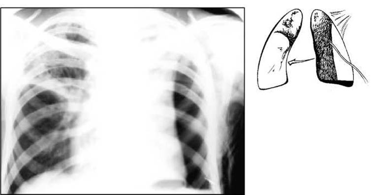
Fig. 32. Chest X-ray. Left-sided spontaneous pneumothorax.
To relieve pain and suppress coughing, the ambulance medical staff administer omnopon or morphine hydrochloride, along with medications aimed at supporting cardiac function, initiate oxygen inhalation, and transport the patient to the nearest surgical department. If the patient's condition does not improve, the doctor performs a pleural puncture over the site of air accumulation and aspirates the air using a large syringe through a rubber adapter tube. After air aspiration, the patient's condition improves rapidly, but in the case of an open or tension pneumothorax, this improvement is only temporary. If the condition deteriorates, the chest wall is punctured with a thick needle, which is secured to the skin with adhesive plaster. The air then escapes through the needle, reducing lung compression. A rubber tube can be attached to the needle, with its free end submerged in a disinfectant solution.
To receive specialized care, patients with spontaneous pneumothorax must be hospitalized in specialized phthisiosurgical or thoracic surgery departments. There, a chest tube is inserted into the pleural cavity using a trocar for continuous air evacuation. A slit rubber finger cot is attached to its outer end and placed in a disinfectant solution (Bülau drainage). During expiration, air escapes from the pleural cavity; during inspiration, the finger cot collapses, thus functioning as a one-way valve. The nurse monitors the function of the drain. If its patency is impaired or subcutaneous emphysema develops (a sensation of "crackling" on Palpation of the soft Tissues near the tube), she notifies the doctor. Daily, the nurse disinfects the skin around the chest tube, changes the sterile dressing, and replaces the disinfectant solution in the container, clamping the tube during this Procedure.
If necessary, the drainage tube is connected to an electric suction pump or a continuous Vacuum system available in specialized departments. Streptomycin and Other Antibiotics are introduced into the pleural cavity by puncturing the rubber tube. Concurrently, the patient is prescribed systemic antituberculosis therapy.
If this treatment method is not sufficiently effective, thoracoscopy is performed to obtain information about pleural changes, adhesions between its layers, and the state of the perforation site, helping to determine further management. In indicated cases, surgery is performed, consisting of suturing the emphysematous blebs or resecting the affected lung area.
If a cavity ruptures and its contents enter the pleural cavity, acute, and subsequently potentially chronic, purulent pleural inflammation (empyema) develops, which is particularly difficult to treat.
Questions
1. What is spontaneous pneumothorax?
2. What are the causes of its occurrence in Pulmonary Tuberculosis?
3. What are the Complaints of a patient who has developed spontaneous pneumothorax?
4. What is detected on percussion and auscultation over the area of pneumothorax?
5. What is the paramedic's management strategy at a feldsher-midwife station (FAP) when spontaneous pneumothorax is suspected?
6. Care provided to the patient prior to hospitalization in a specialized department.
7. What examinations must be performed for a patient with suspected spontaneous pneumothorax?
8. What is Bülau drainage?
Last update: 10/08/2026
Editorial and Educational Adaptation: This material has been compiled based on the primary/original source text. The project team performed an editorial review, corrected technical inaccuracies, structured sections, and adapted the content for an educational format.
What was processed:
- elimination of formatting defects (OCR errors, structural breaks, corrupted characters);
- editorial organization of content;
- standardization of terminology in accordance with academic sources;
- verification of factual statements against the original source text.
All mentions of the author, publication year, and origin of the primary text have been preserved in accordance with the source.