Critical States in Pulmonary Tuberculosis in Adults - N.I. Fomichova 2010
Intrapleural Hemorrhage (Hemothorax)
Intrapleural hemorrhages are characterized by the accumulation of Blood in the pleural cavity.
Etiology AND Pathogenesis. Bleeding into the pleural cavity occurs As a result of compromised vascular integrity in the Lungs, Mediastinum, and chest wall. In Pulmonary Tuberculosis, this is associated with destructive lesions in the lung tissue and Pleura, coagulopathy caused by disseminated intravascular coagulation (DIC) syndrome with secondary Fibrinolysis in patients with Miliary tuberculosis and Caseous Pneumonia, rupture of pleural adhesions during therapeutic and Spontaneous pneumothorax, inadequate hemostasis during surgical intervention and in the postoperative period due to hemostatic disorders, failure of sutures on the lung and pleural vessels, breaches in subclavian vein catheterization technique, or prolonged indwelling of a tubular drain in the pleural cavity.
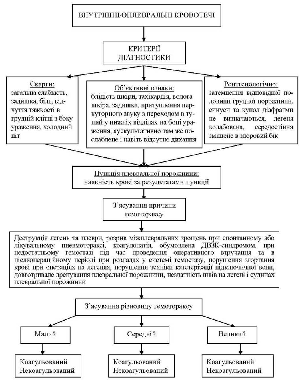
Depending on the volume of blood effused into the pleural cavity, hemothorax is classified as small, moderate, or large (Diagram 4).
In small hemothorax, the blood effused into the pleural cavity fills the costodiaphragmatic sinus up to the level of the diaphragmatic dome, which corresponds to the 7th rib. In moderate hemothorax, blood fills half the volume of the pleural cavity and reaches the level of the 3rd rib. In large hemothorax, blood fills more than half of the pleural cavity and extends above the 3rd rib.
Additionally, hemothorax is classified as coagulated or non-coagulated.
Clinical manifestations (Diagram 4). The Clinical presentation of hemothorax depends on the volume and rate of bleeding into the pleural cavity, as well as on the form and phase of the tuberculous process. Small hemothorax lacks a distinct clinical picture, whereas moderate and large hemothoraces present with signs of blood loss, hypovolemia, respiratory and cardiac failure, and compression of the lung and mediastinal Organs. Patients complain of generalized weakness, dyspnea, pain, and a feeling of heaviness in the affected hemithorax. Symptoms of acute blood loss and hemorrhagic Shock progressively develop: Skin pallor, cold sweat, tachycardia (pulse rate of 80–100 bpm, low volume), and hypotension (blood pressure below 100 mmHg). The respiratory rate may reach 20 breaths per minute or more.
Objective findings. Dullness to Percussion, transitioning to flatness in the lower lung fields on the affected side. Auscultation reveals markedly diminished breath sounds in the same area, which are absent in the lower zones.
Chest radiography demonstrates a collapsed lung and opacification within the pleural cavity. A definitive sign of hemothorax is the aspiration of blood during pleural puncture.
While the aforementioned signs are common to all types of hemothorax, each has its own characteristic features depending on the underlying cause. Consequently, Treatment strategy must be individualized.
Class="center">DIAGRAM 4. DIAGNOSTIC ALGORITHM FOR HEMOTHORAX IN PATIENTS WITH TUBERCULOSIS
Critical conditions in adult pulmonary tuberculosis
EMERGENCY CARE
Management of intrapleural bleeding depends on The rate of Hemorrhage, the underlying cause, and the type of hemothorax (Diagram 5).
DIAGRAM 5. EMERGENCY MANAGEMENT ALGORITHM FOR HEMOTHORAX

In cases of small hemothorax, evacuating the blood from the pleural cavity via thoracentesis is generally sufficient. Broad-spectrum Antibiotics are indicated to prevent empyema. For coagulated hemothorax, streptokinase is used for blood clot lysis. To achieve this, one vial of streptokinase is dissolved in 20 ml of isotonic sodium chloride solution and administered into the pleural cavity of a patient whose bleeding has ceased and in whom there is no risk of recurrence. After 6 hours, a repeat pleural puncture is performed to remove the lysated blood. On days 2–3, the puncture is repeated to remove any residual cavity contents. Depending on the clinical picture, streptokinase administration may be repeated.
For moderate non-coagulated hemothorax, the pleural cavity is drained via the Bülau technique following puncture. Patients with moderate coagulated and large hemothoraces require surgical intervention. The goal of surgery is to stop the bleeding through coagulation, suture ligation of bleeding vessels, and removal of the accumulated blood. Following surgery, the pleural cavity is drained in both upper and lower zones. Concurrently with hemostasis, circulating blood volume (CBV) is restored. This is achieved through rapid intravenous infusion of crystalloid solutions (normal saline, Ringer's solution, hypertonic [7.5%] sodium chloride solution) and colloid solutions (albumin, hydroxyethyl starch, modified gelatin, dextrans).
In severe hemorrhagic shock, preference should be given to low-volume hypertonic resuscitation (a mixture of hypertonic sodium chloride solution and hydroxyethyl starch); when large infusion volumes are required, modified gelatin solutions (Gelofusine), which have virtually no effect on hemostasis, are preferred.
Prior to achieving hemostasis, the infusion rate should maintain blood pressure at no higher than critical levels (80–90 mmHg). Once bleeding is controlled, the infusion rate is determined based on clinical symptoms and measurements of blood pressure and central venous pressure (CVP). Subsequently, intravenous solutions are switched to a drip infusion. Blood transfusion is considered absolute when the Hemoglobin level drops below 60–70 g/L. If the hemoglobin level exceeds 90–100 g/L, restoration of CBV deficits is performed without blood transfusion; the indication for blood transfusion is restricted to profuse bleeding that cannot be promptly controlled. If the hemoglobin level ranges between 70–90 g/L, the physician determines The Need for blood transfusion on an individual basis, guided by parameters such as blood pressure, Heart rate, and blood lactate levels. Nevertheless, it is imperative to replenish the patient's plasma volume deficit and prevent hemoglobin levels from dropping below 80 g/L.
Intrapulmonary and intrapleural hemorrhages associated with destructive forms of pulmonary and pleural tuberculosis pose a severe life threat to the patient. These hemorrhages are typically caused by erosion of pulmonary vessel branches without coagulation disorders. Hemostatic therapy is largely ineffective. In such cases, prompt resection of the affected lung is indicated, along with volume replacement using whole blood and anti-shock agents.
During therapeutic and spontaneous pneumothorax, intrapleural hemorrhages are caused by the rupture of lung tissue at the sites of pleural adhesion attachments. Such bleeding is generally not massive and does not pose diagnostic difficulties. X-ray Examination reveals hydropneumothorax, while pleural puncture yields blood and air. Management of these patients involves Surgical Methods.
Bleeding into the pleural cavity resulting in hemothorax occurs in 5–10% of cases following various thoracic surgeries. The underlying cause of this complication is mechanical hemostasis failure when securing hemostasis from the vessels of dissected Tissues and divided adhesions. Postoperatively, these patients exhibit blood drainage via the tubular drain, which is an inevitable consequence of lung surgery. However, if the blood volume within the first 4 hours exceeds 600 ml with a hemoglobin level above 40 g/L, intrapleural hemorrhage should be suspected. In coagulated hemothorax with minimal blood output through the drain, pleural bleeding should be suspected if tachycardia progressive worsens against the backdrop of falling blood pressure. Such patients require urgent re-thoracotomy to stop the bleeding and evacuate blood clots.
Massive hemorrhages into the pleural cavity resulting from hemostatic disorders may occur following lung surgeries. In such patients, information regarding the fibrinolytic activity of the blood must be obtained immediately. If a detailed assessment of fibrinolytic activity is not feasible, the whole blood clot lysis time test is useful. For this purpose, 4–5 ml of venous blood is placed in a test tube, and its clotting time is recorded. The normal clotting time is 7–8 minutes. A shortened clotting time indicates hypercoagulability, whereas a prolonged time indicates hypocoagulability. In the absence of fibrinolysis, a firm blood clot forms within 3 hours, adhering to the test tube wall, with serum retraction and a small amount of free erythrocytes settling at the bottom. In the presence of fibrinolysis, the clot is friable, floats freely in the test tube, and A large number of erythrocytes settle in the sediment. Thoracotomy is contraindicated in such patients; instead, they require transfusions of Blood and Its components (plasma, fibrinogen), administration of epsilon-aminocaproic acid to inhibit fibrinolysis, infusions of hypertonic sodium chloride and hydroxyethyl starch mixtures, or Gelofusine to restore CBV.
In some instances, pleural hemorrhages may occur due to breaches in subclavian vein catheterization technique. The cause is usually repeated attempts at vein catheterization resulting in vascular wall injury, as well as pressure necrosis caused by the catheter. If this complication arises, the catheter must be promptly removed from the vein, and the intrapleural bleeding may cease spontaneously. Blood accumulated in the pleural cavity is evacuated via puncture and aspiration. In rare cases, immediate surgical intervention is indicated.
Hemothorax may occur due to the prolonged presence of a tubular catheter in the pleural cavity, for example, in cases of tuberculous empyema. Such bleeding is typically caused by pressure erosions of the intercostal Arteries created by the drainage tube. Diagnosing this condition is straightforward. Along with gas from the pleural cavity, blood is discharged through the drainage, and a blood clot forms within the pleural cavity. In such cases, the drainage tube must be removed, the opening closed, and pleural drainage established at a different site.
CASE STUDY
Patient N., 49 years old, has been suffering from pulmonary tuberculosis for 3 years. He has repeatedly and for extended periods undergone inpatient, sanatorium, and outpatient treatment. Fibrocavernous tuberculosis of the upper lobe of the right lung has developed. The patient was admitted to the clinic to address the feasibility of Surgical treatment. His general condition is satisfactory. Symptoms of intoxication are mild. Pulse is 68 bpm, BP is 120/80 mm Hg, and respiratory rate is 18 breaths/min. Percussion reveals a shortened percussion note over the PROJECTION OF THE upper lobe of the right lung; breathing is harsh with a bronchial shade. No rales are auscultated. Heart sounds are clear. The abdomen is soft and non-tender. The Liver is at the edge of the costal arch. Examination of the pulmonary and cardiovascular systems revealed no deviations from the norm. Blood test: Hb 115 g/L, RBC 3.10 /L, WBC 8.10 /L, e. 4%, b. 5%, seg. 67%, l. 20%, m. 4%, ESR 12 mm/h. Mycobacterium tuberculosis was detected in the sputum via direct smear Cell/15.html">Microscopy. Coagulogram: recalcification time 95 s, plasma heparin tolerance 7 min, prothrombin activity 95%, fibrinase 96.3 s, fibrinogen 3 g/L, prothrombin time 23 s, fibrinolytic activity 200 min. Urinalysis is within normal limits. Blood biochemistry tests are within normal limits.
X-ray examination of the lungs revealed: the upper lobe on the right is reduced in volume. A cavity up to 3.5 cm in diameter with thick walls is identified beneath the clavicle.
Given the presence of a unilateral destructive process in the lungs with persistent bacterioshedding, the absence of functional Impairment of the respiratory and cardiovascular systems, and the absence of pathological changes in other organs and the coagulation system, a resection of the upper lobe of the right lung was recommended to the patient, to which he consented.
The surgery was performed under general anesthesia, without blood transfusion, with the support of plasma-expander solutions. The operation lasted 2.5 hours and was uneventful. By the end of the surgery, the patient regained consciousness from anesthesia. Hemodynamic parameters were within normal limits. Pulse was 68 bpm, BP was 115/65 mm Hg. Eight hours postoperatively, the patient's condition sharply deteriorated: dyspnea increased, pain and a feeling of heaviness appeared in the right hemithorax, and general weakness progressed. Skin pallor, tachycardia, and hypotension developed (pulse 96 bpm, BP 95/60 mm Hg). Massive intrapleural hemorrhage was diagnosed. The patient received intravenous infusions of conserved blood followed by intra-arterial jet blood transfusion, as well as colloidal and crystalloid solutions. However, the patient's condition continuously deteriorated. The state of blood fibrinolytic activity was assessed using an express method. The blood clot formed in the test tube after 3 hours was loose and floated freely, indicating enhanced fibrinolysis. The patient was prescribed E-aminocaproic acid 5% - 100 ml, fibrinogen 250 ml intravenously via drip, and hemophobin 5 ml intramuscularly. Despite this, the patient's condition steadily worsened, and 16 hours postoperatively, the patient died amidst symptoms of cardiovascular failure. Autopsy revealed up to 2.5 liters of liquid, non-clotting blood in the pleural cavity. Hemostasis from the surgical site was complete. No failure of postoperative sutures was found. The cause of the fatal intrapleural hemorrhage was a coagulation disorder (enhanced fibrinolysis) in the early postoperative period.
Selection/5.html">Control Questions and TASKS WITH ANSWER KEYS
Task. Patient K., 30 years old, with no prior history of serious illness. She has been undergoing inpatient treatment for two weeks for newly diagnosed pulmonary tuberculosis (infiltrative) of the upper lobe of the right lung, Dest+, MBT+ M+ K0, Resist0, Hist0, Cat. 1.
Throughout this period, the patient's condition was relatively satisfactory. Intoxication symptoms were moderately pronounced, with the primary complaint being a persistent, severe cough with sputum production. Medium-tâched rales were auscultated in the right lung above and below the clavicle against the Background of harsh breathing. The percussion note was shortened. On the chest X-ray taken upon admission, the upper lobe of the right lung showed intensive opacification, and a cavity 3-4 cm in diameter with an irregular inner contour and an infiltrated outer contour was detected subpleurally In the second intercostal space.
The patient tolerated the prescribed etiotropic and pathogenetic treatment well.
During the night, the patient's condition sharply deteriorated. Almost suddenly, she developed general weakness, dyspnea, a feeling of heaviness in the right hemithorax, and cold sweat.
Objective Examination: the patient's condition is severe, skin and mucous membranes are pale, skin is moist to the Touch, pulse is 96 bpm of low volume, respiratory rate is 24 breaths/min, BP is 80/55 mm Hg. Chest percussion on the right from the 3rd rib to the Diaphragm reveals dullness of the percussion note transitioning to flatness in the lower lung regions. Mediastinal organs are shifted to the left. Auscultation reveals sharply diminished breath sounds over the right lung, absent in the lower regions. The physician performed a follow-up examination, established the correct Diagnosis, and provided necessary care.
Question 1. What conditions require Differential diagnosis in this case?
A. Pulmonary infarction.
B. Spontaneous pneumothorax.
C. Myocardial infarction.
D. Exudative Pleurisy.
E. Intrapleural hemorrhage.
Question 2. What additional examinations, besides physical assessment, should be performed to determine the cause of the patient's deterioration?
A. Chest X-ray.
B. Electrocardiography.
C. Pleural puncture.
D. Blood tests: Hb, hematocrit, Lee-White coagulation time, fibrinolytic activity, bleeding time.
E. All of the above.
Question 3. In your opinion, What is the most likely cause of intrapleural Hemorrhage in the patient?
A. Rupture of interpleural adhesions.
B. Presence of a subpleurally located destructive lesion in the lung tissue.
C. Decreased blood clotting.
D. Increased fibrinolytic blood activity.
E. Breakdown of caseous foci on the visceral pleura.
Question 4. What primary medical care should be provided to a female patient diagnosed with moderate uncoagulated hemothorax?
A. Bülau tube thoracostomy, restoration of circulating blood volume (CBV).
B. Evacuation of blood from the pleural cavity via thoracentesis, restoration of CBV.
C. Thoracoscopy with coagulation of the bleeding source, restoration of CBV.
D. Resection of the right upper lobe, restoration of CBV.
E. Administration of hemostatic agents, evacuation of blood from the pleural cavity via thoracentesis, restoration of CBV.
Answers and their rationale.
Answers to the questions: 1 E, 2 E, 3 B, 4 A
1. Differential diagnosis must primarily be performed with intrapleural hemorrhage. The basis for making this preliminary diagnosis in the patient consisted of objective clinical signs indicating the presence of fluid in the pleural cavity: on the right side, percussion reveals dullness extending from the 3rd rib to the diaphragm, transitioning to flatness in the lower parts; the mediastinal organs are shifted to the healthy side; auscultation reveals that breath sounds over the right lung are sharply diminished and absent in the lower sections. Hemothorax is specifically indicated by the sharp deterioration of the patient's condition during treatment, accompanied by signs of hemorrhagic collapse—sudden onset of pronounced general weakness, cold sweat, hypotension, tachycardia, dyspnea, and pallor of the skin and mucous membranes. Neither spontaneous pneumothorax, myocardial or pulmonary infarction, nor exudative pleurisy present with such clinical manifestations.
2. To determine the cause of the patient's deterioration, the following immediate steps must be taken: first, chest X-ray in 2 projections (clinical signs of fluid in the pleural cavity); electrocardiography (signs of collapse, which may be accompanied by acute Heart Failure); pleural puncture, which will confirm the presence of fluid and determine its nature; blood tests will, firstly, detect changes associated with hemorrhage (drop in hemoglobin, abnormal hematocrit values), and secondly, identify the cause of bleeding (blood clotting disorders, etc.).
3. The cause of the intrapleural hemorrhage in this clinical case was the rupture of a subpleurally located cavity. This is proven by the results of the chest X-ray, which revealed a progressive tuberculosis process in the upper lobe of the patient's right lung accompanied by The breakdown of caseous-necrotic masses (a cavity with irregular inner contours), which, in turn, led to the destruction of Blood Vessels in the lung tissue and the adhered pleura.
4. In destructive forms of respiratory tuberculosis, pulmonary hemorrhages are typically caused by the erosion of Branches of the pulmonary vessels without impairment of blood clotting. Therefore, hemostatic therapy has low efficacy. Finding the source of bleeding and coagulating it with such extensive pathomorphological Changes in the lung tissue and pleura is unlikely. Evacuation of blood from the pleural cavity via puncture does not allow for monitoring the bleeding status and may lead to the patient fainting. Therefore, the most appropriate management is Bülau tube thoracostomy with concurrent restoration of CBV. If these measures prove ineffective and bleeding continues, resection of the affected lung lobe should be performed.
Last update: 08/08/2026
Editorial and Educational Adaptation: This material has been compiled based on the primary/original source text. The project team performed an editorial review, corrected technical inaccuracies, structured sections, and adapted the content for an educational format.
What was processed:
- elimination of formatting defects (OCR errors, structural breaks, corrupted characters);
- editorial organization of content;
- standardization of terminology in accordance with academic sources;
- verification of factual statements against the original source text.
All mentions of the author, publication year, and origin of the primary text have been preserved in accordance with the source.