Critical Conditions in Pulmonary Tuberculosis in Adults - N.I. Fomichova 2010
Pulmonary Hemorrhage and Hemoptysis
Hemoptysis and Pulmonary Hemorrhage result from bronchial and pulmonary damage of various etiologies, differing in the volume of Blood discharged.
Hemoptysis is defined as the expectoration of sputum mixed with blood, ranging from streaks to diffuse coloration, as well as distinct spits of blood. Pulmonary hemorrhage refers to the discharge of a significant amount of pure blood through the respiratory tract. Pulmonary hemorrhage and hemoptysis occur in 2.5–25.0% of patients with respiratory tuberculosis. Practically all forms of Pulmonary Tuberculosis can lead to this complication, though it is most frequently observed in Fibrocavernous tuberculosis, cirrhotic tuberculosis, and Caseous Pneumonia.
Depending on the volume of blood discharged, pulmonary hemorrhages are classified as mild (up to 100 mL), moderate (up to 500 mL), and massive (exceeding 500 mL). Based on The rate of blood loss, pulmonary hemorrhages are divided into 3 grades: Grade I – 20 mL/h (1.2% of the normal circulating blood volume, NCBV), Grade II – 50 mL/h (from 5% to 15% of NCBV), and Grade III – 200 mL/h (more than 15% of NCBV). In addition, fulminant (fatal) pulmonary hemorrhages are distinguished. With Grade I, external blood loss does not exceed 5% of NCBV and presents as streaks or admixtures of bright red blood in the sputum (hemoptysis) without hemodynamic disturbances or changes in red blood Cell counts on the hemogram. Grade II blood loss is of moderate intensity and may be accompanied by a 10–20% drop in blood pressure and Hemoglobin levels. Grade III blood loss is a massive hemorrhage characterized by blood gushing out in mouthfuls or in a "fountain" from the Mouth AND Nose, accompanied by a sharp drop in blood pressure, hemoglobin level, hematocrit, and erythrocyte count by 20% or more. Signs of collapse may occur: Skin pallor, dizziness, lightheadedness, sweating, mucosal cyanosis, tachycardia, decreased body Temperature, and occasionally vomiting, nausea, and convulsions.
Pathogenesis. For the proper and timely Treatment of pulmonary hemorrhage in patients with respiratory tuberculosis, it is essential to determine which pathogenetic and functional changes underlie this complication. The Mechanism of development of pulmonary hemorrhage and hemoptysis depends precisely on The Nature of pathomorphological Changes in the Lungs, that is, on the form and phase of the tuberculosis process.
Thus, at the onset of the disease, when the inflammatory process is dominant, capillary and small vessel permeability in the lungs increases under its influence, as well as due to the action of toxins on the vascular wall and tissue allergy with endothelial hypersensitization. On the other hand, cellular-colloidal blood structures are disrupted (disseminated intravascular coagulation). These changes lead to diapedetic hemorrhages, which clinically most often manifest as hemoptysis.
In "fresh" destructive processes in the lungs, erosion of the wall of a large vessel plays a leading role in the pathogenesis of hemorrhage. With progressive pulmonary destruction, capillaries and small vessels form pericavernously, and their walls are subjected to toxic and hyperallergic effects. Small bronchial Arteries around the cavity become severely deformed due to Connective Tissue proliferation.
In chronic forms of pulmonary tuberculosis, the primary cause of pulmonary hemorrhage is the mechanical rupture of aneurysmatically and varicosities-altered walls of large vessels resulting from pulmonary Hypertension.
In the pathogenesis of pulmonary hemorrhages in patients with pulmonary tuberculosis, pathomorphological changes in other body systems also play a significant role by contributing to functional hemostatic alterations. During pulmonary hemorrhages, The primary function of the hemostatic system is, on the one hand, to maintain blood in a fluid state within the Circulation and, on the other hand, to stop bleeding by forming a thrombus at the site of the damaged vessel. The blood clotting time in tuberculosis patients remains virtually unchanged at 3–7 minutes. The blood fibrinogen level in tuberculosis is significantly elevated, and this elevation is closely correlated with The Nature and duration of the process. In newly diagnosed, timely detected respiratory tuberculosis, the blood fibrinogen level is moderately elevated, whereas in widespread destructive processes, its amount increases substantially—reaching up to 12 g/L against a normal range of 2–4 g/L. This represents a defense mechanism of the body aimed at walling off the inflammatory zone from undamaged lung areas. Consequently, clot formation in tuberculosis is unimpaired, with no abnormalities in the 1st and 2nd phases of coagulation. However, the quality of the clot in tuberculosis is altered: it is fragile, loose, and rapidly undergoes lysis, creating conditions for life-threatening hemorrhages and their relapses. This occurs due to reduced activity of the plasma fibrin-stabilizing factor, especially in patients with widespread, chronic destructive forms of tuberculosis. Active tuberculosis also features deviations in heparin METABOLISM—the heparinase activity of the blood is diminished. Furthermore, a fibrinogen-heparin complex, which requires a specific amount of heparin, is detected in the patients' blood. The most substantial abnormalities in tuberculosis are observed in the fibrinolytic system. The fibrinolytic activity of Blood Plasma and sputum in tuberculosis patients is increased by 2 to 10 times, particularly during an episode of pulmonary hemorrhage, which hinders the consolidation of the fibrin clot and creates conditions for recurrent bleeding. Such changes are caused by the fact that the blood of tuberculosis patients is rich in plasminogen and active plasmin while nearly lacking their rapid and slow-acting antagonists—antiplasmins. A pronounced fibrinolytic effect is also exerted by the fibrinogen-heparin complex. In addition, The breakdown of lung tissue Proteins involves massive destruction of cellular structures with the release of substances possessing enhanced lytic activity. These substances exert their effects both systemically and locally (within the tuberculous inflammation zone). Evidence of this is the increased fibrinolytic activity of sputum and lung tissue Fibrinolysis in the affected area, especially pericavernously. Another mechanism of increased fibrinolysis in tuberculosis also exists: it has been proven that M. tuberculosis release a substance that acts as a plasminogen activator and is capable of fibrin lysis.
Thus, during pulmonary hemorrhages in tuberculosis patients, clots form rapidly, but they are highly fragile because The activity of substances acting as fibrinolysis inhibitors is sharply suppressed in both blood and Tissues. All these changes are particularly evident in both "fresh" disseminated forms of tuberculosis and chronic destructive processes in the active phase.
Thus, the significant role of fibrinolysis in the pathogenesis of pulmonary hemorrhages in tuberculosis patients has been established, driven by high lytic activity of plasma and sputum, the release of activators from disintegrating lung tissue and M. tuberculosis, as well as pulmonary hypertension and increased vascular wall permeability caused by intoxication and allergy.
In cases of hemoptysis and pulmonary hemorrhage, it is very difficult to predict their duration and outcomes, and there is also no certainty regarding The Development of relapses. Therefore, patients even with minor hemoptysis must be immediately hospitalized in a specialized ward.
Clinical manifestations. In mild and moderate hemorrhages, the patient initially experiences a scratching sensation in the throat, chest tightness and pain, and a feeling of heat in the chest, followed by a cough with a bloody odor, a salty taste in the mouth, and dyspnea; bright red frothy blood that shows no tendency to clot is expelled with the cough. The clinical picture of massive pulmonary hemorrhages involves the development of collapse and anemia: skin pallor, dizziness, palpitations, adynamia, and a drop in blood pressure appear. The cause of death in patients with massive hemorrhages is asphyxia resulting from bronchospasm and occlusion of the Airways by blood clots.
Diagnostics (Figure 1). When blood is discharged from the Oral Cavity, it is crucial to promptly identify the source of the hemorrhage. This may be the Nasal cavity, oral cavity, Stomach, Esophagus, Trachea, Bronchi, or lungs. Among all the Organs whose pathology can be complicated by bleeding, the greatest threat to human life is posed by hemorrhages originating from the lungs, esophagus, and gastrointestinal tract, which require emergency and sometimes resuscitative care. Examination of the patient should begin with an inspection of the Oral Cavity and nasopharynx, which helps rule out pseudoblooding caused by gingivitis, stomatitis, tonsillar abscess, Osler-Rendu syndrome, hemorrhagic diathesis, or epistaxis in hypertensive disease. Esophageal variceal bleeding occurs in Liver cirrhosis with portal hypertension, accompanied by icterus of the skin and sclera, teleangiectasias, palmar erythema, dilation and distension of chest and abdominal wall Veins, and hepatosplenomegaly. Gastrointestinal bleeding is caused by chronic fistulas, tumors, etc. In recent years, the incidence of acute fistulas caused by the intake of ulcerogenic drugs (acetylsalicylic acid, butadione, indomethacin, corticosteroids), as well as stress and Hypoxia (myocardial infarction, Cor Pulmonale), has increased. In gastrointestinal bleeding, blood is expelled during vomiting, appears dark, and resembles "coffee grounds". The vomitus has an acidic odor, contains food mixtures, and is later followed by loose, tarry stools.
In pulmonary hemorrhages, blood is brought up with a cough, is bright and frothy, and lacks food admixtures. When a pulmonary hemorrhage occurs, the source of the hemorrhage must be identified first and foremost. For this purpose, standard chest X-ray examinations are sufficient in the majority of cases. Major difficulties in diagnosing the bleeding source arise in bilateral lung lesions, in which patients sometimes note a sensation of heat on the side of the bleeding. Auscultation of such an area may reveal an increase in rales during the hemorrhage. Overview radiographs may show Atelectasis, a horizontal fluid level, or a dense spherical formation within a cavity. In complex cases, tracheobronchoscopy is indicated—preferably under anesthesia—during which not only the bleeding site (lung lobe, segment) is identified, but hemostasis is simultaneously performed.
When facilities are available to diagnose the exact cause and source of pulmonary hemorrhage in complex diagnostic cases, chest computed tomography and bronchial arteriography can be performed. Within 1–2 hours from the onset of bleeding, a red blood cell analysis must be performed to determine hemoglobin levels, hematocrit, and bleeding time. If possible, a thromboelastogram and coagulogram should be obtained, along with measurements of plasma fibrin-stabilizing factor (XIII), prothrombin and Thrombin time, plasma and sputum fibrinolytic activity, and blood platelet counts. In life-threatening hemorrhages, rapid diagnostic Assessment of the Blood Coagulation SYSTEM is sufficient:
1. Determine the blood clotting time using the Lee-White method (normally, a clot forms within 5–11 minutes).
2. Monitor the character of clot formation in a test tube over 10 minutes (impaired clot retraction after more than 12–20 minutes indicates a decreased fibrinogen level and increased fibrinolytic activity).
3. Determine the spontaneous clot lysis time (a twofold reduction in clot volume within 15–20 minutes indicates high blood fibrinolytic activity).
4. Determine the fibrinogen content using the thrombin clotting test (a clot forming within 5–11 seconds corresponds to 2–4 g/L of blood fibrinogen).
5. Determine the blood platelet count (normally no less than 50x10%).
Class="center">FIGURE 1 DIAGNOSTIC ALGORITHM FOR PULMONARY HEMORRHAGE AND HEMOPTYSIS

To clarify the pathogenesis of pulmonary hemorrhage, an ECG examination (to detect signs of Chronic cor pulmonale), echocardiography, and spirography should be performed.
Emergency Care
To date, there is no standardized protocol for emergency care in pulmonary hemorrhage among patients with respiratory tuberculosis. For each patient with hemorrhagic syndrome, the emergency management plan depends on the degree of blood loss, the nature and extent of the tuberculosis process, and the availability of modern endoscopic and Surgical Methods for stopping pulmonary hemorrhage.
Pre-hospital care (Scheme 2).
1. Position the patient in a semi-sitting posture to facilitate sputum expectoration and prevent blood from flowing down into the Lower Respiratory Tract.
2. In case of hemoptysis, have the patient drink a saline solution (1 tablespoon of salt per 1 Glass of Water) — 1 tablespoon every 30 minutes.
3. In case of hemorrhage, apply tourniquets to the limbs to redistribute blood volume and reduce its inflow to the lungs (do not compress the arteries!).
4. Administer sedatives (valerian tincture, bromine preparations, motherwort), as bleeding causes psychological stress and fear of death. This triggers the release of vasoconstrictors into the blood, which enhance vascular spasm and promote further bleeding.
5. Immediately call an ambulance.
Pre-hospital medical care (Scheme 2). It aims to stop hemoptysis or hemorrhage and restore circulating blood volume (CBV) during profuse pulmonary hemorrhage. An essential objective is to restore airway patency. To achieve this, the patient is placed in a semi-sitting position with the HEAD turned to the side, and the oral cavity is cleared using a suction device. Sedatives are administered orally, and tourniquets are applied to the extremities.
For hemostatic purposes, calcium chloride, aminophylline, atropine sulfate, sulfocamphocaine, menadione sodium bisulfite (vikasol), etamsylate or dicynone, and routine or ascorutyn (orally) are administered. Relying solely on coagulants for hemostasis fails to account for the physiology of blood clotting, the specific pharmacological actions of coagulants, and the pathogenesis of hemorrhages in pulmonary tuberculosis. It should be emphasized that the administration of fibrinolysis inhibitors in tuberculosis is always appropriate, as elevated fibrinolysis is observed in all patients—especially at the peak of bleeding—while the pool of endogenous inhibitors (antiplasmins) is extremely small. The choice of drug and its route of administration must be determined by the physician, taking into account the degree of blood loss, the patient's condition, and their medical history.
SCHEME 2 EMERGENCY CARE PROTOCOL FOR PULMONARY HEMORRHAGES

It is particularly advisable to use trasylol for grade II–III hemorrhages because it can be administered by rapid intravenous injection, thereby accelerating intervention. Furthermore, trasylol inhibits both enzymatic and non-enzymatic fibrinolysis, which is significantly activated in patients with respiratory tuberculosis. To reduce vascular wall permeability and achieve a desensitizing effect, intravenous administration of calcium chloride, pipolfen, or diphenhydramine is recommended, along with 3–5 ml of a 5% ascorbic acid solution intramuscularly. It is also advisable to use a 0.025% adroxon solution, 1 ml intramuscularly or subcutaneously. The latter does not increase blood pressure or affect Cardiac Activity, but it promotes the spasm of small vessels and capillaries, making it useful for stopping parenchymal and capillary bleeding. If CBV restoration is necessary, 0.9% sodium chloride solution can be used initially, followed by polyglucukin and other plasma expanders in the event of a critical drop in blood pressure.
In any case, the patient must be immediately hospitalized in a specialized department (phthisiology, pulmonology, internal medicine, thoracic surgery, or general surgery), depending on their general condition, degree of blood loss, Anamnesis, etc. If necessary, infusion therapy is continued en route.
Specialized care (Scheme 3).
Before initiating specialized care—and often during its delivery—it is necessary to evaluate the nature and extent of the tuberculosis process, the blood coagulation profile, hemogram, hemodynamics of the pulmonary and systemic circulations, and to identify the source and primary pathogenetic factor of the pulmonary hemorrhage, as well as the intensity and degree of blood loss.
In cases of hemoptysis, the patient is prescribed bed rest and aminocaproic acid (5 g orally 3–4 times a day for 3–5 days). 10% calcium chloride (1 tablespoon 3–4 times daily after meals), ascorbic acid, and desensitizing agents may also be prescribed.
The main therapeutic measures for pulmonary hemorrhage in tuberculosis patients should aim at:
1) reducing pressure in the lesser circulation;
2) enhancing blood coagulability and inhibiting fibrinolysis;
3) reducing vascular wall permeability;
4) restoring CBV in cases of profuse hemorrhage.
In patients with category I blood loss, blood loss generally corresponds to grade I–II. The primary pathogenetic factors of hemorrhage in these patients are increased vascular wall permeability, decreased blood clotting, and increased fibrinolytic activity. Therefore, the patient should primarily be prescribed:
1. Calcium chloride 10% — 10 ml intravenously.
2. Ascorbic acid 5% — 5 ml intramuscularly.
3. Prednisolone 5 mg 3 times daily.
4. Suprastin 2% 1 ml intramuscularly or 0.5 mg 3 times daily.
5. E-aminocaproic acid 10% - 10 ml intravenously twice a day.
6. Dicynone 12.5% - 2 ml subcutaneously.
7. Thrombin inhalations (dissolve 250 mg of thrombin in 5 ml of saline solution).
8. Intensive anti-tuberculosis therapy.
9. Broad-spectrum Antibiotics to prevent aspiration pneumonia.
10. Slow-acting hypotensive drugs can be prescribed: aminophylline 2.4% - 10 ml intravenously.
In patients of category II, alongside grade I-II blood loss, grade III hemorrhages are frequently observed. The pathogenetic factors leading to bleeding are primarily the ulceration of vessel walls resulting from the progression of the tuberculosis process. In addition, these patients exhibit all the pathogenetic factors present in category I patients. In cases of grade I-II blood loss in category II individuals, medical care should be initiated similarly to that for category I patients. In cases of recurrent hemorrhages or their progression to grade III, hypotensive therapy must be intensified. For this purpose, ganglionic blockers are used (1.5% gangleron solution 1-2 ml subcutaneously, 5% pentamine solution 1-2 ml subcutaneously or intravenously, benzhexonium 0.1 g 3-6 times a day orally). When administering ganglionic blockers, blood pressure should be measured every 5 minutes. If systolic pressure drops to 100 mm Hg or by 60 mm Hg in patients with hypertension, ganglionic blockers must be discontinued. For grade II bleeding, 2.4% aminophylline solution 10.0 ml intravenously, 0.01% clonidine solution 1.0 ml subcutaneously, 0.1% atropine sulfate solution 1.0 ml subcutaneously, or 2% papaverine solution 2.0 ml subcutaneously can be used as hypotensive agents. The choice of hypotensive medications is determined by the intensity of blood loss. For low- and moderate-intensity hemorrhages, it is advisable to prescribe slow-acting hypotensive agents for 5-7 days. For high-intensity hemorrhages, fast-acting drugs should be used in the first 1-3 days until bleeding stops. For this category of patients, proteolysis and fibrinolysis inhibitors should be used for hemostatic purposes: contrycal (trasylol) 10,000 - 30,000 IU intravenously, gordox 10,000-30,000 IU intravenously, hydrocortisone 12.0-25 ml intravenously, amben 50-100 mg intravenously.
In cases where bleeding does not stop or recurs, collapse therapy methods should be employed (artificial pneumothorax, pneumoperitoneum).
DIAGRAM 3 SCHEME FOR PROVIDING SPECIALIZED EMERGENCY CARE IN PULMONARY HEMORRHAGE
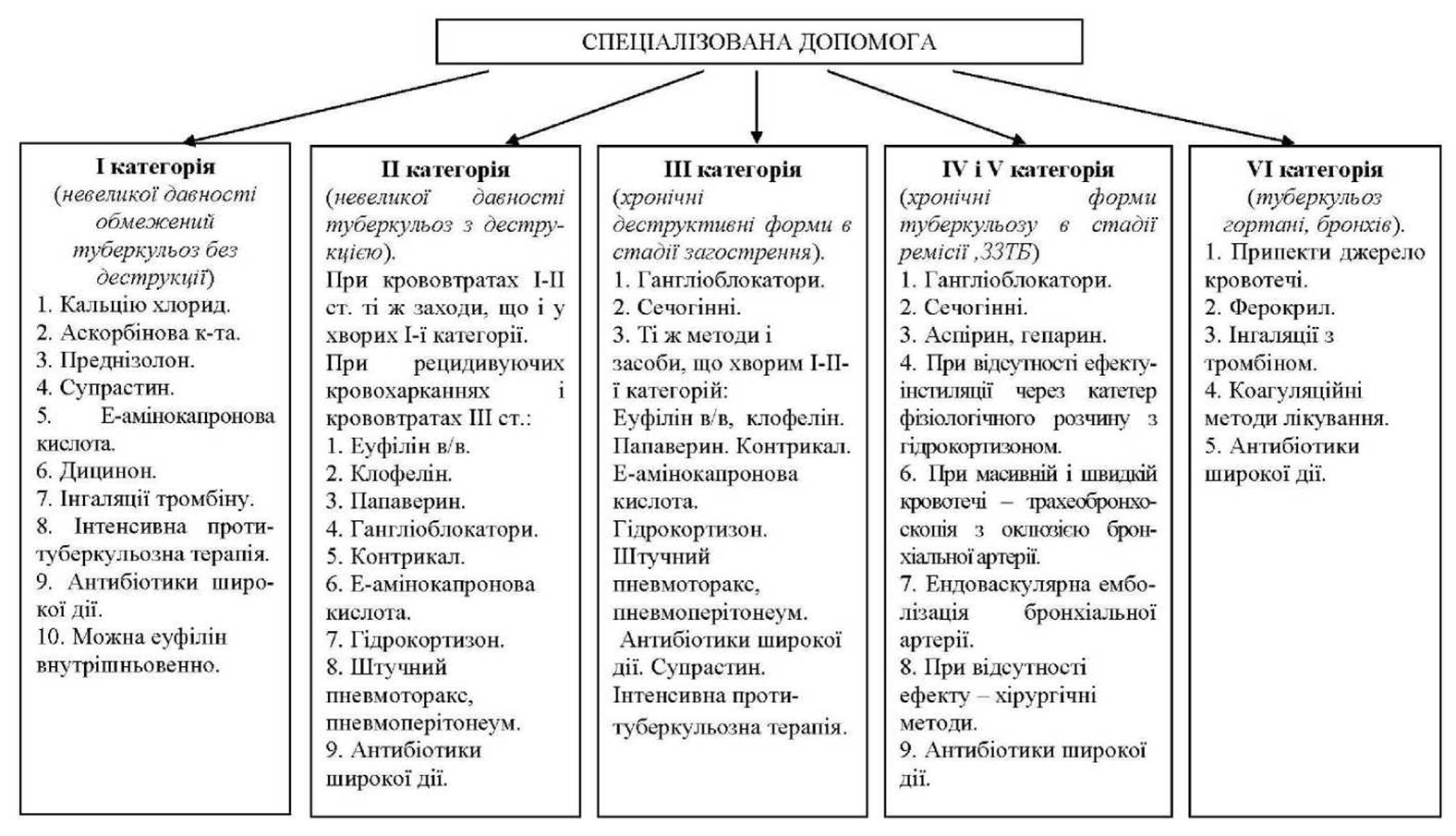
In patients with category III respiratory tuberculosis, the primary pathogenetic factor in the development of pulmonary hemorrhage is the rupture of the wall of a large vessel modified by aneurysmal or varicose nodes As a result of pulmonary hypertension. Since these patients, alongside old fibrous changes, have "fresh," recent lesions in the form of foci, infiltration, and necrotic changes, these can also serve as a source of hemorrhages with their inherent pathogenetic factors. When providing emergency care to such patients, one should first start with hypotensive therapy using fast-acting drugs (ganglionic blockers) with parallel administration of Diuretics (furosemide 40 mg in the morning on an empty stomach). Given that this category of patients has recent focal-infiltrative and necrotic processes that can serve as a source of hemorrhages, the same measures should be applied to them as to individuals in categories I and II.
Patients belonging to categories IV and V share common pathogenetic factors in the development of pulmonary hemorrhages. Pulmonary hypertension can lead to the rupture of sclerosed vessels. Furthermore, narrowing of the Pulmonary Circulation vessels, slowing of blood flow, and increased platelet adhesiveness lead, in turn, to intravascular blood coagulation and, as a consequence, prolonged recurrent hemoptysis. When providing care to patients in these categories, it is necessary first of all to reduce pressure in the pulmonary circulation by prescribing hypotensive and diuretic agents. In the presence of prolonged recurrent hemoptysis, medications that reduce blood coagulation are indicated (aspirin, heparin 10,000-15,000 IU intravenously).
If conservative therapy is ineffective, tracheobronchoscopy is indicated. Hemostatic solutions are instilled via a catheter or fiberbronchoscope with simultaneous aspiration: cold saline solution 40-60 ml; 5% E-aminocaproic acid solution 40-50 ml; 1% ferocril solution 10-15 ml; saline solution 100 ml + hydrocortisone 1 ml (25 mg) + 3-4 ml of 10% calcium chloride solution. In cases of massive and rapid bleeding, tracheobronchoscopy is performed with occlusion of the segmental, lobar, or main bronchus using a sterile foam sponge, which can remain in the bronchial wall for 1-3 days. Good results in pulmonary hemorrhage are achieved by endovascular embolization of the bronchial artery that caused the bleeding. Catheterization is performed via the femoral artery-aorta in an X-ray room. To diagnose the eroded bronchial artery, a contrast agent is first injected, followed by its occlusion with blood clots, spongostan, or teflon velour.
In cases of treatment failure, radical or palliative surgeries are indicated: segmentectomy, lobectomy, pneumonectomy, thoracoplasty, ligation of the pulmonary artery, and others. The most effective measures after successful hemostatic therapy for preventing the recurrence of pulmonary hemorrhage are surgical operations addressing the underlying pulmonary process. They are performed after a detailed examination of the patient and provided that adequate preoperative preparation is completed, which reduces the risk of postoperative complications.
In category VI patients, hemorrhages are most often caused by caseous-destructive processes in the Larynx, inflammatory-fistulous changes in the Trachea and Bronchi in active progressive respiratory tuberculosis, as well as changes in the mucous membrane and vessels (thinning, sclerosis, varicosities) in a healed process. In these cases, coagulation treatment methods can be used. When a source of hemorrhage is detected in the larynx, tracheal mucosa, or a large bronchus, it should be cauterized with a concentrated solution of trichloroacetic acid, 20% silver nitrate solution, 3% hydrogen peroxide solution, or 5% E-aminocaproic acid solution.
Clinical Case Example
Patient A., 42 years old, has been suffering from pulmonary tuberculosis for 9 years. Cirrhosis of the right lung developed in the course of treatment. The patient was hospitalized in a regional anti-tuberculosis dispensary due to persistent hemoptysis and periodic pulmonary hemorrhages. For 6 months, he was on strict bed rest. Intensive anti-tuberculosis therapy was administered in combination with hemostatics: Vikasol 1% - 1 ml intramuscularly, Dicynone 12.5% - 2 ml intramuscularly, calcium chloride 10% - 10 ml intravenously, E-aminocaproic acid - 10 g per day orally, hemophobin 3% - 1 tablespoon 3 times a day, inhalations of thrombin 250 mg dissolved in 5 ml of saline solution. Due to the persistence of hemoptysis, the patient was transferred to the clinic for Surgical treatment. Objective status: the patient's condition is moderately severe, he lies motionless on his back, afraid to turn on his side or breathe deeply. Skin is pale. Muscles of the lower extremities are flaccid. Pulse is 100 beats per minute, rhythmic. Heart sounds are muffled, accent of the second sound over the pulmonary artery. Blood pressure - 110/60 mm Hg. Respiratory rate - 24/min. In the lungs on the right throughout, bronchial breathing is heard along with numerous moist rales of various calibers; on the left in the subscapular region, moist rales are heard. Abdomen is soft. The liver protrudes 3 cm below the costal margin. Sputum analysis: leukocytes - 3-4 in the field of view, erythrocytes - 40-60 in the field of view, epithelial Cells - 1-2 in the field of view. Mycobacterium tuberculosis was not detected. X-ray Examination diagnosed total cirrhosis of the right lung. Blood test: RBC 4.3 T/l, Hb 100 g/l, color index 0.9, WBC 10.5 G/l, e. 4%, b. 6%, s. 69%, l. 16%, m. 5%, ESR - 15 mm/h. Coagulogram: recalcification time - 100 sec, plasma heparin tolerance - 8 min, fibrinogen - 6.9 g/l, prothrombin time - 19.4 sec, fibrinolytic activity - 96 min, Lee-White clotting time - 8 min, platelet count - 52. Spirometric study: VC/predicted VC - 50%, MVV/predicted MVV - 49%, FEV1 - 43%. ECG: decreased voltage of waves, deviation of the electrical axis of The Heart to the right, P2 and P3 pulmonary waves, dominant S waves in standard leads, R in V1 = 11 mm, R/S in V1 = 1.4, S in V1 = 1.6 mm, RV1+SV5=11.5 mm, incomplete right bundle branch block. Echocardiography with dopplerography - pulmonary artery pressure is 70 mm Hg. As a result of the examination, the patient was diagnosed with: major residual changes of Tuberculosis of the right lung (total cirrhosis), Destr-, MBT-, respiratory failure stage II, chronic cor pulmonale stage II, pulmonary hemorrhage, lower left lobe aspiration pneumonia. Based on the Nature of the pulmonary process and the pathogenesis of the aforementioned complications, the patient should be classified into category V. This indicates that the main cause of his pulmonary hemorrhages and hemoptysis was elevated pressure in the pulmonary circulation with the development of chronic cor pulmonale. These factors led to the rupture of sclerosed vessel walls that had lost their elasticity. In addition, the patient had increased fibrinolytic activity of the blood, as evidenced by its parameters (96 min compared to the norm of 180-300 min), as well as an increased amount of fibrinogen (normal 2-4 g/l, patient's level - 6.9 g/l). It should be kept in mind that in patients with chronic pulmonary insufficiency, There is a slowing of blood flow velocity in the Vessels of the Pulmonary Circulation and an increased platelet adhesive capacity, which additionally creates conditions for thrombosis and the development of thromboembolic recurrent hemoptysis. Therefore, the administration of drugs that increase blood clotting only worsened the patient's condition. Since the Organism is an integral self-regulating system, the increase in blood clotting triggered the activation of the anticoagulation system, which led to recurrent hemorrhages in the patient.
Based on the pathogenesis of pulmonary Hemorrhage in the patient, it is first necessary to "unload" the pulmonary circulation and reduce respiratory and cardiac insufficiency. To achieve this, it is necessary to improve bronchial patency, reduce the volume of circulating blood in the lungs, and improve pulmonary blood flow. The patient was prescribed ganglionic blockers, diuretics, and heparin. To reduce the fibrinolytic activity of the blood, high doses of E-aminocaproic acid and glucocorticoids were administered. Due to the development of aspiration pneumonia, broad-spectrum antibiotics were prescribed. Since the patient has no signs of active tuberculosis, intensive anti-tuberculosis therapy is not indicated. Given that pulmonary cirrhosis was complicated by hemorrhage with aspiration pneumonia, prophylactic treatment with isoniazid is advisable to prevent the recurrence of tuberculosis.
Two weeks later, the patient's condition significantly improved, hemoptysis ceased, and signs of respiratory and cardiac insufficiency decreased.
Selection/5.html">Control Questions and tasks with standard Answers
Task No. 1. A general practitioner was called to see a 60-year-old neighbor who had developed pulmonary hemorrhage. At the age of 30, he suffered from Infiltrative pulmonary tuberculosis. He recovered and was removed from the dispensary register. He underwent annual fluoroscopic examinations. There was no reactivation of the process. Ten days ago, his body temperature rose to 38.60C, accompanied by weakness, chest pain, and a cough. He was treated with home remedies without effect. Blood appeared in his sputum, followed by a pulmonary hemorrhage. He expelled over 20 ml of blood.
Community-acquired pneumonia was suspected. The general practitioner provided emergency care and called an ambulance. In the presence of the paramedic, the patient started bleeding again, expelling over 50 ml of blood. Blood pressure - 115/80 mm Hg. The patient received emergency care. He was hospitalized in the therapeutic department with a Diagnosis of community-acquired pneumonia, pulmonary hemorrhage.
Objective status: the patient's condition is severe. Body temperature 38.5 0C, skin is pale and clammy. Pulse is 100 beats/min, rhythmic, weak volume. Heart sounds are muffled. Blood pressure - 100/65 mm Hg. Over the right lung, especially in its upper lobe, the Percussion note is shortened. In the same area against the Background of weakened breathing, moist rales are heard throughout. On the chest X-ray, the right lung shows decreased transparency, especially from the first to the fourth rib. The shadow is heterogeneous, with areas of increased lucency in places. The interlobar Pleura is thickened. Below the 4th rib, there are multiple focal shadows of small and medium intensity.
Blood test: RBC 2.8 T/l, Hb 90 g/l, WBC 12.2 G/l, e. 2%, b. 9%, s. 65%, l. 18%, m. 6%, ESR - 18 mm/h.
Mycobacterium tuberculosis was not detected in the sputum by direct smear Microscopy.
Lee-White blood clotting time is 5 min. Spontaneous clot lysis time: its volume halved within 10 min, indicating high blood fibrinolytic activity.
In the hospital, the patient experienced recurrent hemorrhages, despite being administered subcutaneously 1 ml of 5% pentamine, intravenously 10,000 IU of kontrikal, and an intravenous drip containing: 300 ml of normal saline + 5 ml of 10% calcium chloride + 25 mg of hydrocortisone, ε-aminocaproic acid, Vikasol, Dicinone, and sulfocamphocaine. During one of the bleeding episodes, the patient lost consciousness, but regained it after medical assistance. A day later, during a recurrent hemorrhage, the patient passed away.
Question 1. In your opinion, What is the patient's diagnosis based on the examination findings?
A. Community-acquired destructive pneumonia.
B. Tumor of the upper lobe of the right lung.
C. Relapse of infiltrative tuberculosis (lobitis), dissemination phase, Dest+.
D. Pulmonary infarction.
E. Candidomycosis.
Question 2. What additional examination must be performed primarily to clarify the cause of the hemorrhage?
A. Radiological examination (right lateral radiograph, tomograms through the ROOT and the pathological process in the lung).
B. Coagulogram.
C. Bronchoscopy.
D. Assessment of the functional state of the pulmonary and cardiovascular systems.
E. Microbiological sputum examination.
Question 3. Is it possible to provide first aid to a patient with pulmonary hemorrhage if no hemostatic drugs are available? If so, what is the optimal approach?
A. Not possible.
B. Place the patient horizontally, apply tourniquets to the extremities.
C. Administer antitussive medications.
D. Place the patient in a semi-sitting position in bed, apply tourniquets to the extremities, administer sedatives (valerian tincture, bromine preparations, motherwort).
E. Place the patient in a sitting position, lower the lower extremities to the floor, have them drink a hypertonic saline solution (one tablespoon of salt per 1 glass of water).
Question 4. What emergency first aid should the paramedic/emergency physician have provided to the patient?
A. Apply tourniquets to the extremities, administer Vikasol, calcium chloride.
B. Apply tourniquets, administer sulfocamphocaine with atropine, euphylline, calcium chloride, dicinone, aminocaproic acid.
C. Administer euphylline, pentamine, Vitamin C, Vikasol.
D. Administer sulfocamphocaine, calcium chloride, hemophobin.
E. Administer gangleron or pentamin, vikasol, oxygen inhalation.
Question 5. What emergency care should have been provided to the patient who lost consciousness during bleeding?
A. Place the patient in a horizontal position, administer cordiamine subcutaneously.
B. Lower the head of the bed, administer eufillin, apply oxygen inhalation.
C. Wipe the temples with ammonia, administer corglycon intravenously, perform a blood transfusion.
D. Perform external cardiac massage, artificial Respiration, administer sulfocamphokain.
E. Clear the airways of blood clots, initiate oxygen inhalation.
Question 6. What is the primary cause of sudden death in a patient with pulmonary hemorrhage?
A. Blood loss.
B. Aspiration pneumonia.
C. Asphyxia due to airway obstruction by blood clots.
D. Atelectasis.
E. Anemia, hypoproteinemia.
Question 7. What treatment method could have been used to prevent the patient's death?
A. Controlled hypotension.
B. Artificial pneumothorax, pneumoperitoneum.
C. Surgical treatment.
D. Blood transfusion, corticosteroid Hormones.
E. Endovascular bronchial artery occlusion.
Answers and their rationale.
ANSWERS TO QUESTIONS: 1 C, 2 A, 3 D, 4 B, 5 E, 6 C, 7 C
1. Pulmonary tuberculosis of the upper lobe of the right lung (infiltrative, dissemination phase), Destr + MBT- M- K 0 Hist 0 Resist 0 Cat 2. The patient exhibits a typical clinical and radiologic picture of lobitis-type tuberculous infiltrate: involvement of the upper lobe of the right lung with pronounced interlobar pleura reaction, and dissemination foci in the lower lobe. Changes in the hemogram are characteristic of this form of tuberculosis. The absence of Mycobacterium tuberculosis in sputum by direct microscopy in the presence of lung destruction does not rule out tuberculosis.
2. A right lateral radiogram and tomograms through the root and pathological process will allow a more precise determination of its localization and Structure, which, combined with clinical manifestations and hemogram changes, will help establish a more accurate diagnosis.
3. Position the patient semi-upright to facilitate sputum expectoration and prevent blood from flowing into the lower PARTS OF THE lungs. Apply tourniquets to the extremities to redistribute blood and reduce its inflow to the lungs. During pulmonary hemorrhage, the patient experiences psychological stress and a sense of impending death, which leads to the release of vasoconstrictors into the blood that enhance vascular spasm and promote bleeding; therefore, the patient must be calmed and given sedatives.
4. Upon the arrival of the emergency doctor, the severity of the pulmonary hemorrhage can be assessed as grade I-II (blood loss of 20-50 ml), therefore long-acting antihypertensive drugs are prescribed. Apply tourniquets to the extremities, administer eufillin, sulfocamphokain, atropine sulfate, dicynone, calcium chloride, and aminocaproic acid.
5. The main cause of this condition is airway obstruction by blood clots; therefore, they must be removed mechanically or using a suction device, or, if possible, blood clots should be aspirated from the airways via a bronchoscope.
6. Asphyxia resulting from airway obstruction by blood clots.
7. Vital indication surgery (resection, thoracoplasty, pulmonary artery ligation) because such hemorrhages pose a threat to the patient's life.
Problem No. 2. Patient S., 58 years old, has been suffering from pulmonary tuberculosis for over 10 years. She has repeatedly undergone inpatient treatment. The latest hospitalization was caused by the onset of pulmonary hemorrhage. The patient lost over 500 ml of blood within the last hour.
Upon admission, the patient's condition is of moderate severity, with moderately pronounced Intoxication syndrome; the patient is agitated, pale, and troubled by dyspnea and cough. Respiratory rate is 26 breaths per minute. Auscultation reveals numerous diverse rales throughout the lungs. Heart rate is 90 beats per minute, cardiac activity is rhythmic, and heart sounds are muffled. Blood pressure is 115/60 mm Hg. The liver is enlarged by 3 cm, tender upon Palpation.
A chest X-ray of the upper lobe of the right lung reveals a 3x4 cm cavity with thick walls. Pronounced fibrotic changes surround the cavity. The upper right lobe is reduced in volume, and the root is pulled upward. Signs of emphysema are present in the lower sections of the right lung. The mediastinal shadow is shifted to the right. Throughout the left lung, there are multiple medium-intensity foci with blurred contours, in places merging into infiltrates of non-homogeneous structure due to areas of radiolucency.
MBT were detected in the sputum by direct microscopy.
Blood analysis: RBC 3.3 T/L, Hb 100 g/L, CI 0.9; WBC 13.4 G/L, E 3%, Band 8%, Seg 68%, Lymph 16%, Mon 5%, ESR 16 mm/h.
ECG: decreased wave voltage, rightward axis deviation, P and P waves = 3.0 mm, predominant S waves in standard leads; R in V1 = 7 mm, P/S in V1 = 1.4 mm, S in V1 = 1.6 mm, RV1 + SV5 = 11.5 mm, incomplete right bundle branch block.
Based on the studies performed, the patient was diagnosed with: Upper lobe fibrous-cavernous pulmonary tuberculosis of the right lung, Dest+ (infiltration and dissemination phase), MBT+ M+K0, Resist0, Hist0, Cat. 4, pulmonary hemorrhage.
Proceeding from the established diagnosis and examination results, the primary pathogenetic factor in the Development of the complication was identified, and treatment was administered accordingly. The patient's condition improved.
Question 1. What is the severity grade of the pulmonary hemorrhage in this patient?
A. First.
B. Second.
C. Third.
D. Moderate.
E. Impossible to determine.
Question 2. What examination should be prescribed for the patient to determine the source of bleeding?
A. Chest computed tomography.
B. Fiberoptic bronchoscopy.
C. Bronchial artery arteriography.
D. Chest fluoroscopy.
E. Thoracoscopy.
Question 3. In your opinion, what is the leading pathogenetic factor of pulmonary hemorrhage in this patient?
A. Increased vascular wall permeability.
B. Vessel wall erosion due to a destructive process in the lung tissue.
C. Disseminated intravascular coagulation.
D. Vessel wall erosion due to pulmonary hypertension.
E. Functional disorders of hemostasis.
Question 4. What emergency medical care should be provided to the patient first and foremost?
A. Prescribe ganglionic blockers, fibrinolysis inhibitors, and diuretics.
B. Perform artificial pneumothorax.
C. Prescribe coagulants and peripheral vasodilators.
D. Administer oxygen pneumoperitoneum, prescribe prednisolone, and restore BCC.
E. Prescribe thrombin inhalations and administer aminophylline.
Question 5. What type of therapy is mandatory for the patient along with emergency care?
A. Antituberculosis therapy.
B. Broad-spectrum antibiotics.
C. Antituberculosis therapy combined with broad-spectrum antibacterial drugs.
D. Resolving (resorptive) therapy.
E. Nonsteroidal anti-inflammatory drugs.
Answers and their rationale.
Answers to the questions: 1 C, 2 B, 3 D, 4 А, 5 С
1. The patient has a grade III pulmonary hemorrhage because she lost more than 200 ml of blood in a short period of time.
2. First of all, fiberoptic bronchoscopy is indicated for the patient because she has bilateral lung lesions. Under such conditions, an urgent need for surgery may arise; therefore, identifying the reliable source of bleeding is mandatory.
3. In chronic forms of pulmonary tuberculosis, the main cause of pulmonary hemorrhage is the mechanical rupture of aneurysmatic and varicose-altered walls of large vessels as a result of pulmonary hypertension.
4. In chronic destructive forms of pulmonary tuberculosis, emergency care in the event of pulmonary hemorrhage should begin with the administration of ganglionic blockers, fibrinolysis inhibitors, and diuretics. These measures are aimed at correcting the main pathogenetic factors of pulmonary hemorrhage during the progression of chronic respiratory tuberculosis: elevated pressure in the pulmonary artery system, increased fibrinolytic activity of blood and sputum. The latter leads to the rapid destruction of the blood clot under normal clotting processes.
5. The patient urgently requires massive antituberculosis therapy along with broad-spectrum antibiotics. The Need for antituberculosis drugs is due to the fact that the patient has an active chronic tuberculosis process with a progressive course. Since massive pulmonary hemorrhage can lead to aspiration pneumonia, the patient requires broad-spectrum antibiotics.
Last update: 08/08/2026
Editorial and Educational Adaptation: This material has been compiled based on the primary/original source text. The project team performed an editorial review, corrected technical inaccuracies, structured sections, and adapted the content for an educational format.
What was processed:
- elimination of formatting defects (OCR errors, structural breaks, corrupted characters);
- editorial organization of content;
- standardization of terminology in accordance with academic sources;
- verification of factual statements against the original source text.
All mentions of the author, publication year, and origin of the primary text have been preserved in accordance with the source.