Tuberculosis - I.T. Pyatnochka 2005
Primary tuberculosis
Fibrocavernous pulmonary tuberculosis
Fibrocavernous tuberculosis is a chronic destructive process characterized by the presence of an old fibrous cavity, pronounced fibrosis, and foci of bronchogenic dissemination in the lung tissue surrounding the cavity or in other PARTS OF THE Lungs, featuring a prolonged, undulating course with periods of exacerbation and remission, alongside persistent or intermittent bacterial excretion. Socially, patients with fibrocavernous Pulmonary Tuberculosis are classified as disabled, predominantly in disability group II (Fig. 20).
The prevalence of fibrocavernous tuberculosis among newly diagnosed pulmonary tuberculosis patients is 1.2%, and approximately 10% among outpatients registered at tuberculosis dispensaries. Fibrocavernous tuberculosis is the leading cause of death in patients with pulmonary tuberculosis.
Pathomorphology. Fibrocavernous tuberculosis develops as an unfavorable outcome of infiltrative, disseminated, or Focal pulmonary tuberculosis. The extent of pulmonary changes varies, and the process may be unilateral or bilateral, presenting with one or multiple cavities, as well as pneumosclerosis, emphysema, and Bronchiectasis.
The wall of the cavity has a three-layered Structure: the inner layer is pyogenic, the middle is granulation, and the outer is fibrous.
Clinical Features. The clinical manifestations of fibrocavernous tuberculosis are diverse. They are driven by the tuberculosis infection itself, the extent and phase of the process, Changes in the lung parenchyma, and various complications.
There are several clinical Variants of the course of fibrocavernous pulmonary tuberculosis:
1. Limited fibrocavernous pulmonary tuberculosis with a stable course.
2. Limited or advanced fibrocavernous pulmonary tuberculosis with a progressive course.
3. Fibrocavernous pulmonary tuberculosis with complications.
Class="center">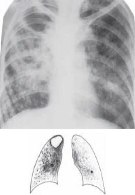
Fig. 20. Fibrocavernous Tuberculosis of the right upper lobe, infiltration and dissemination phase. Overview radiograph
The first variant involves a localized process featuring a cavity and fibrosis, along with a minimal number of foci in the surrounding lung tissue. Symptoms of intoxication and bronchopulmonary syndrome are mild. MBT may be absent in the sputum. Patients remain relatively fit for work. This condition can persist for a long time. However, under unfavorable factors or inadequate medical care, the progression of fibrocavernous pulmonary tuberculosis ensues.
The progressive variant of fibrocavernous pulmonary tuberculosis is characterized by an undulating course. Mild symptoms of intoxication and bronchopulmonary syndrome become more pronounced. During exacerbation, perifocal infiltration, fresh foci, and even new destruction cavities appear around the cavity. MBT are detected in the sputum. Marked Blood test abnormalities and functional impairments are also observed.
Fibrocavernous pulmonary tuberculosis with complications generally follows a course similar to the second variant, but is additionally accompanied by specific and non-specific complications.
Specific complications include tuberculosis of the Larynx, Bronchi, Pleura (Tuberculous Pleurisy), intestines, Kidneys, genitals, and other Organs.
Non-specific complications include Hemoptysis and Pulmonary Hemorrhage, Spontaneous pneumothorax, Chronic Cor Pulmonale, internal organ amyloidosis, Atelectasis, as well as non-specific inflammatory and fungal complications (aspergillosis, candidomycosis).
Diagnosis is based on medical history, radiological findings (presence of a cavity, fibrosis, and foci), Chest deformities, physical examination data, blood test changes, and the presence of MBT in the sputum.
Differential diagnosis is performed to rule out chronic abscess, polycystic lung disease, disintegrating Cancer, and bronchiectasis.
Treatment. Over 95% of patients with fibrocavernous pulmonary tuberculosis have previously received treatment for Other forms of tuberculosis; therefore, the prescription of at least 5 anti-tuberculosis drugs must be strictly individualized based on MBT drug sensitivity, drug tolerance, presence of complications, comorbidities, and patient age. The goals of treatment are to eliminate intoxication, achieve resorption of fresh foci and infiltrates, sanitize cavities, and stop mycobacterial excretion. In addition to Chemotherapy, which promotes cavity healing in 5-10% of patients with fibrocavernous pulmonary tuberculosis, pathogenetic therapies and surgical interventions are utilized. Thus, treatment must be comprehensive—incorporating Antimycobacterial agents, Pathogenetic Therapy, and Surgical Methods—to enhance treatment efficacy, specifically achieving cessation of mycobacterial excretion in 50-60% of patients. The total duration of the main treatment course ranges from 12 to 18 months, whereas for patients with drug-resistant tuberculosis (MBT resistance to at least isoniazid and rifampicin), it typically lasts 14-16 months.
CONTROL QUESTIONS
1. Definition of fibrocavernous pulmonary tuberculosis.
2. Causes leading to The Development of fibrocavernous pulmonary tuberculosis.
3. Clinical and radiological variants of the course of fibrocavernous pulmonary tuberculosis.
4. Complications of fibrocavernous pulmonary tuberculosis.
5. Cytology/practical/136.html">DIFFERENTIAL DIAGNOSIS OF fibrocavernous pulmonary tuberculosis.
6. Main chemotherapy regimens for fibrocavernous pulmonary tuberculosis.
7. Pathogenetic therapy in patients with fibrocavernous pulmonary tuberculosis.
8. Indications and contraindications for Surgical treatment in patients with fibrocavernous pulmonary tuberculosis.
TESTS
1. The frequency of fibrocavernous pulmonary tuberculosis among newly diagnosed pulmonary tuberculosis patients:
A. 1.2 %
B. 6.2 %
C. 8 %
D. 10 %
E. 12 %
2. From which clinical form of tuberculosis does fibrocavernous pulmonary tuberculosis most frequently develop?
A. Tuberculoma
B. Primary tuberculosis complex
C. Infiltrative
D. Focal
E. Cirrhotic
3. The most frequent nonspecific complication of fibrocavernous pulmonary tuberculosis:
A. chronic cor pulmonale
B. laryngeal tuberculosis
C. spontaneous pneumothorax
E. Amyloidosis of internal organs
4. The most frequent specific complication of fibrocavernous pulmonary tuberculosis:
A. laryngeal tuberculosis
B. intestinal tuberculosis
C. Tuberculous pleurisy
5. Patient K., aged 35, complains of a cough with sputum production, weakness, and dyspnea upon minor physical exertion. Three months ago, he returned from a correctional facility. Upon examination, the right hemithorax is narrowed and lags behind in the respiratory act. MBT were detected by bacterioscopy. What is the clinical form of pulmonary tuberculosis?
A. Tuberculoma
B. Focal tuberculosis
C. Tuberculous pleurisy
D. Disseminated pulmonary tuberculosis
E. Fibrocavernous tuberculosis
6. Patient K., aged 36, is newly diagnosed with fibrocavernous pulmonary tuberculosis. MBT are resistant to ethambutol and streptomycin. Prescribe the most optimal combination of antimycobacterial drugs.
A. Rifampicin + isoniazid + kanamycin + pyrazinamide
B. Isoniazid + rifampicin + thioacetazone + viomycin
C. Isoniazid + kanamycin + PASA + ethionamide
D. Kanamycin + ethionamide + rifampicin + phтивазид
E. Isoniazid + cycloserine + protionamide + kanamycin
7. The most frequent immediate cause of death in patients with fibrocavernous pulmonary tuberculosis.
A. Pulmonary atelectasis
B. Chronic cor pulmonale
C. Pulmonary hemorrhage
E. Progression of tuberculosis
8. Pathoanatomical changes in Cirrhotic pulmonary tuberculosis.
A. Pronounced bullous-dystrophic changes
B. Massive proliferation of Dense Connective Tissue in the lungs and pleura with specific active foci in the lungs
C. Encapsulated tuberculous foci
D. Tara cavity with pronounced fibrosis
E. Multiple cavities, fibrosis, and polymorphic focal lesions
9. Fibrous-cavernous pulmonary tuberculosis must be differentiated from which of the following diseases:
A. Eosinophilic infiltrate
D. Lobar Pneumonia
E. Lung cyst
10. Differential diagnosis of fibrous-cavernous tuberculosis is least appropriate with which of the following diseases?
A. Chronic abscess
C. Polycystic lung disease
D. Chronic bronchitis
E. Bronchiectasis
11. Patient Z., aged 45, was admitted to a tuberculosis hospital due to pulmonary hemoptysis. Over the past two years, she has been troubled by cough with sputum production, dyspnea, and low-grade fever up to 37.5 °C. She has not undergone X-ray Examination for five years. On physical examination, the left hemithorax is narrowed and lags behind in the act of breathing. Fine- and medium-moist rales are heard over its upper section. What is the most likely clinical form of pulmonary tuberculosis?
A. Tuberculoma
B. Disseminated
C. Fibrous-cavernous
D. Cirrhotic
12. Differential diagnosis of cirrhotic tuberculosis is performed with:
A. Carcinomatosis
B. Peripheral cancer
C. Aspergilloma
D. Eosinophilic infiltrate
E. pulmonary agenesis
PROBLEMS
1. Patient Z., aged 37, weight 65 kg, has been hospitalized for 5 months for pulmonary tuberculosis (PTB) (04.05.2003) of the right upper lobe (infiltrative), Dest+, MBT+M+K+, Resist+(H), Hist0, Cat2 Cog2 (2003). Treatment included streptomycin 0.5 g, isoniazid 0.3 g, ethambutol 0.8 g. X-ray shows narrowing of the right upper lobe, a ring-shaped shadow 3 cm in diameter beneath the clavicle surrounded by fibrous strands and single focal shadows, MBT (-).
a) Causes of treatment failure.
b) Justify the current diagnosis.
c) Further treatment plan.
2. Patient K., aged 45, carpenter, developed Fibro-cavernous tuberculosis of the right upper lobe 6 years ago. Following a 2-year intensive antimycobacterial therapy, stable cessation of bacterial excretion was achieved; a thin-walled irregular cavity is present in the right upper lobe. There are no signs of activation of the tuberculous process. There are 3 children in the family.
a) Formulate the diagnosis.
b) Further management strategy for the physician.
3. A 50-year-old male patient presents with a destruction cavity with thick, irregular walls and a horizontal fluid level in the upper lobe of the left lung. There are no foci of bronchogenic dissemination; Complaints include pain in the upper left hemithorax, dyspnea, hemoptysis, and body Temperature up to 38 °C. He considers himself ill for 5 months. Blood test: WBC - 10.0×10⁹/L, ESR - 65 mm/h.
a) Formulate a provisional diagnosis.
b) Examination plan.
Last update: 10/08/2026
Editorial and Educational Adaptation: This material has been compiled based on the primary/original source text. The project team performed an editorial review, corrected technical inaccuracies, structured sections, and adapted the content for an educational format.
What was processed:
- elimination of formatting defects (OCR errors, structural breaks, corrupted characters);
- editorial organization of content;
- standardization of terminology in accordance with academic sources;
- verification of factual statements against the original source text.
All mentions of the author, publication year, and origin of the primary text have been preserved in accordance with the source.