Tuberculosis - I.T. Pyatnochka 2005
Primary tuberculosis
Primary tuberculosis complex
A clinical form of Primary tuberculosis characterized by specific inflammation in the Lungs (primary focus), involvement of the intrathoracic Lymph Nodes (lymphadenitis), and lymphangitis (Fig. 13).
Pulmonary localization occurs in 90% of cases, and abdominal localization in 10% of Primary tuberculous complex cases.
Pathogenesis. Following the penetration of MBT into the lungs, the primary lesion (primary focus), ranging in size from a millet seed to a pulmonary lobe, is predominantly localized subpleurally in segments II, III, VIII, and X. From the primary focus, the infection spreads via Lymphatic vessels to the intrathoracic lymph nodes. However, lymphadenitis may also be primary or develop simultaneously with the pulmonary component.
Pathomorphology.
The primary tuberculous complex consists of three components: the primary focus (pneumonitis), lymphadenitis (involvement of the intrathoracic lymph node), and lymphangitis (the 'lymphatic pathway' connecting the primary focus to the lymphadenitis). Specific inflammation may spread to the Pleura and cause Pleurisy.
The Clinical presentation of the primary tuberculous complex depends on the extent of pathomorphological Changes in the lungs and intrathoracic lymph nodes, as well as on various complications. Asymptomatic, mild, Pneumonia-like, and flu-like Variants of the clinical course of the primary tuberculous complex are possible. However, it most commonly develops and proceeds similarly to tuberculous intoxication.
Radiologically, four phases (stages) of the primary tuberculous complex are distinguished: pneumonic (infiltrative), resorption (bipolarity), scarring, and calcification (petrification). In the pre-antibacterial era, calcification processes began after a year and lasted 2–3 years; with modern antimycobacterial therapy, they occur much earlier and are quite rare because resorption and scarring processes predominate.
Class="center">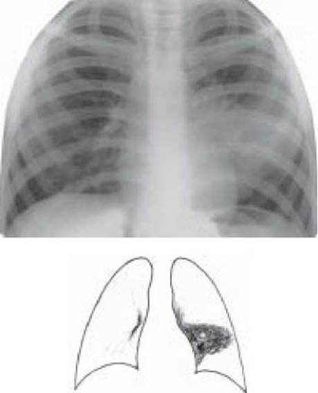
Fig. 13. Primary tuberculous complex (infiltration phase) of the left lung. Plain chest X-ray in the anteroposterior view.
Complications: exudative pleurisy, lymphohematogenous dissemination, tissue breakdown (primary cavity at the site of the pulmonary component), bronchial tuberculosis, segmental or lobar Atelectasis, Caseous pneumonia, and chronic primary tuberculosis developing As a result of severe complications or inadequate Treatment.
Diagnosis is based on Anamnesis (history of contact), tuberculin Skin test conversion ('virage'), a hyperergic Mantoux reaction, symptoms of intoxication and paraspecific reactions, radiological findings (primary focus, lymphangitis, lymphadenitis), hemogram changes (mild leukocytosis with a slight shift to the left, lymphopenia, monocytosis, accelerated ESR), while MBT is rarely detected.
Differential diagnosis is primarily performed with pneumonia, eosinophilic infiltrate, and peripheral or central Cancer.
Treatment begins in a hospital Setting and is carried out using 3 antimycobacterial drugs (isoniazid, rifampicin, pyrazinamide, or ethambutol). After 2 months, once signs of intoxication have disappeared, treatment is continued with 2 drugs (isoniazid with rifampicin or ethambutol) for another 4 months. Concurrently, Vitamins B1, B6, C, desensitizing agents, and symptomatic treatments are administered.
(see also “Tuberculosis of intrathoracic lymph nodes”, ’’Tuberculous intoxication in children”)
1. Pathways of penetration and dissemination of mycobacteria tuberculosis in The Human Body.
2. Definition of primary tuberculosis and its features (characteristics).
3. What is tuberculin reaction conversion ('virage') (possible consequences, preventive measures).
4. Tuberculous intoxication in children (definition, clinical presentation, diagnosis, differential diagnosis, treatment).
5. Which groups of intrathoracic lymph nodes can be affected in tuberculosis?
6. Definition of Tuberculosis of the intrathoracic lymph nodes (pathogenesis, pathomorphology, clinical and radiological variants, clinical presentation, and diagnosis).
7. Main diseases requiring differential diagnosis with tuberculosis of the intrathoracic lymph nodes.
8. Primary tuberculous complex: pathogenesis, pathomorphology, clinical and radiological characteristics, diagnosis, and differential diagnosis.
9. Complications of primary forms of tuberculosis (primary tuberculous complex and tuberculosis of the intrathoracic lymph nodes).
10. Treatment of primary forms of tuberculosis.
11. Methods for the early detection of primary forms of tuberculosis.
TESTS
1. The most informative X-ray method for diagnosing minor forms of tuberculosis of the intrathoracic lymph nodes:
A. targeted radiograph
B. fluorogram
C. tomogram at the level of the tracheal bifurcation
D. chest X-ray
E. bronchogram
2. What is meant by the diagnosis of "tuberculous intoxication"?
A. A symptom complex of functional and objective signs of intoxication resulting from primary infection with Mycobacterium tuberculosis with an undetermined localization.
B. Intoxication syndrome in a minor form of intrathoracic lymph node tuberculosis.
C. Intoxication syndrome in primary Pulmonary Tuberculosis complex.
D. Intoxication syndrome in primary tuberculosis complex of the ileocecal region of the intestine.
E. Low-grade fever, sweating, cough, hoarseness.
3. Paraspecific manifestations in primary tuberculosis:
A. micropolyadenitis, erythema nodosum, phlyctenular keratoconjunctivitis
B. tuberculosis of the skin and Tonsils
C. Amyloidosis of internal Organs, Pleural Empyema
D. Tuberculous pleurisy and pericarditis
E. tuberculous Peritonitis and intestinal tuberculosis
4. What is primary tuberculosis?
A. Newly diagnosed tuberculosis
B. Tuberculosis that develops in individuals newly infected with M. tuberculosis
C. Tuberculosis that develops after a previous primary tuberculosis complex
D. Tuberculosis detected during a preventive examination
E. Tuberculosis caused by bovine mycobacteria
5. Management strategy of a phthisiatrician regarding a 7-year-old child diagnosed with tuberculous intoxication.
A. Follow up at the TB dispensary for 2 years
B. Administer treatment with 3 antimycobacterial drugs for 4–6 months, provided that sanatorium and hygienic regimens are observed
C. Follow up at the children's outpatient clinic until 14 years of age
D. Perform Chemoprophylaxis with isoniazid for 3 months
E. Send the child to a recreation cAMP for health improvement
6. The most characteristic complication of the primary tuberculosis complex.
D. Exudative pleurisy
E. Amyloidosis of Internal Organs
7. To detect the "minor" form of tuberculous bronchoadenitis, it is necessary to perform:
A. plain chest radiography
B. targeted radiography
C. fibrobronchoscopy
D. tomography at the level of the tracheal bifurcation
E. ultrasound.
8. A 6-year-old boy complains of cough, poor appetite, sweating, and low-grade fever up to 37.5 °C. The radiogram shows enlarged bronchopulmonary lymph nodes on the left with indistinct outer contours. The Mantoux test with 2 TU shows a 15 mm infiltrate. Blood test: WBC - 9.0×10⁹/L, ESR - 30 mm/h. The most likely diagnosis.
A. Nonspecific pneumonia
B. Central cancer
C. Sarcoidosis
D. Tuberculosis of intrathoracic lymph nodes
E. Lymphosarcoma
9. During a routine check-up, a 17-year-old boy was found to have bilateral enlargement of bronchopulmonary lymph nodes. His general condition is satisfactory, and he has no Complaints. Physical examination revealed no pathological changes. The Mantoux test with 2 TU is negative. Complete blood count shows no abnormalities. What is the most likely diagnosis?
A. Lymphogranulomatosis
B. Nonspecific adenopathy
C. Sarcoidosis
D. Tuberculosis of intrathoracic lymph nodes
10. What is the most frequent segmental localization of the primary pulmonary affect?
A. Segments I, II, III, IV
B. Segments I, II, IV, VII
C. Segments II, III, VIII, IX
D. Segments I, II, IV, VI
E. Segments I, II, VI, VII
11. A 7-year-old girl suffered from "flu" 2 months ago, followed by the onset of cough, general weakness, decreased appetite, sweating, and body Temperature up to 37.5 °С. Percussion and Auscultation revealed no pathological changes. X-ray shows enlarged tracheobronchial and bronchopulmonary lymph nodes on the left. Blood test: WBC - 9.0× 10⁹/L, ESR - 22 mm/h. Mantoux test with 2 TU shows a 17 mm infiltrate. What is the most likely diagnosis?
A. Sarcoidosis
B. Lymphogranulomatosis
C. Lymphosarcoma
D. Tuberculosis of intrathoracic lymph nodes
E. Central cancer
12. A 5-year-old boy with tuberculosis of the intrathoracic lymph nodes suddenly developed a hacking cough, retrosternal pain, dyspnea, and moderate cyanosis of the lip mucosa. Body temperature is 38.4 °С. Percussion dullness and weakened breath sounds are detected over the upper section of the right lung. What is the most likely complication of intrathoracic lymph node tuberculosis?
A. Exudative pleurisy
B. Spontaneous pneumothorax
C. Atelectasis
D. Tuberculosis of the bronchus
E. Empyema of the pleura
PROBLEMS
1. A 3-year-old child with a tuberculin skin test conversion underwent a chest X-ray, which showed the shadow of the right pulmonary hilum to be widened, elongated, convex, and featuring fuzzy, blurred outlines.
a) What conditions might present with a similar radiological syndrome?
b) What diagnostic tests should be performed to confirm or rule out tuberculosis?
2. In a 7-year-old child, the Mantoux test with 2 TU shows an induration of 19 mm in diameter. The Mantoux test results were negative for the previous 2 years. The child was BCG-vaccinated in the maternity hospital and has a post-vaccination scar. The parents are healthy.
a) Determine The Nature of the tuberculin reaction.
b) Justify your Conclusion.
c) What should be the physician's management strategy regarding the child?
3. Girl S., aged 6, feels well. Her father has pulmonary tuberculosis (active, infectious) (07/22/2003) (disseminated) (infiltration), Destr+, MBT+M-K+, Resist-, HistO, Respiratory Failure II st., Cat 1 Coh 3 (2003).
a) The physician's management strategy regarding the girl.
b) The Scope of diagnostic examination for the child.
c) Your management strategy if she shows no evidence of tuberculosis.
4. A 6-year-old child's mother suffers from open pulmonary tuberculosis. The child was vaccinated in the maternity hospital and has a post-vaccination scar. Symptoms of intoxication are moderately expressed. Radiologically, an infiltrative shadow is present in the 3rd segment on the right, merging with the widened, infiltrated pulmonary hilum. Blood count is within normal limits.
a) What condition should be suspected?
b) What diseases should it be differentiated from?
c) Treatment plan.
5. In a 3-year-old child vaccinated in the maternity hospital, the Mantoux test with 2 TU has increased from 6 to 20 mm over the last 2 years. For over 3 months, the child has had a poor appetite, is cranky, frequently complains of abdominal pain, and occasionally runs a low-grade fever. A chest X-ray reveals no pathological changes.
a) What condition can be suspected?
b) What diseases should it be differentiated from?
c) Plan for further examination and treatment.
Last update: 10/08/2026
Editorial and Educational Adaptation: This material has been compiled based on the primary/original source text. The project team performed an editorial review, corrected technical inaccuracies, structured sections, and adapted the content for an educational format.
What was processed:
- elimination of formatting defects (OCR errors, structural breaks, corrupted characters);
- editorial organization of content;
- standardization of terminology in accordance with academic sources;
- verification of factual statements against the original source text.
All mentions of the author, publication year, and origin of the primary text have been preserved in accordance with the source.