Tuberculosis - I.T. Piatnochka 2005
Primary tuberculosis
Chronic disseminated tuberculosis
This is the most common form of disseminated Pulmonary Tuberculosis, primarily of hematogenous origin, which usually develops either as a primary presentation or against the Background of inadequately managed Subacute disseminated pulmonary tuberculosis. It is characterized by an apicocaudal spread of the disease process. Sequential involvement of various Organs and systems is possible. Chronic disseminated tuberculosis has a wave-like course, during which intoxication symptoms partially subside in periods of remission, but intensify during flare-ups with the appearance of fresh pulmonary and extrapulmonary focal lesions. X-ray findings reveal polymorphic focal shadows against a background of distorted lung markings, fibrosis, and emphysema (Fig. 15). Cavities may develop in one or both Lungs at any stage of the disease. Consequently, over a prolonged course of disseminated tuberculosis—As a result of recurring flare-ups and remissions, repeated waves of dissemination, and The Development of pneumosclerosis and emphysema—Chronic Cor Pulmonale eventually ensues.
Class="center">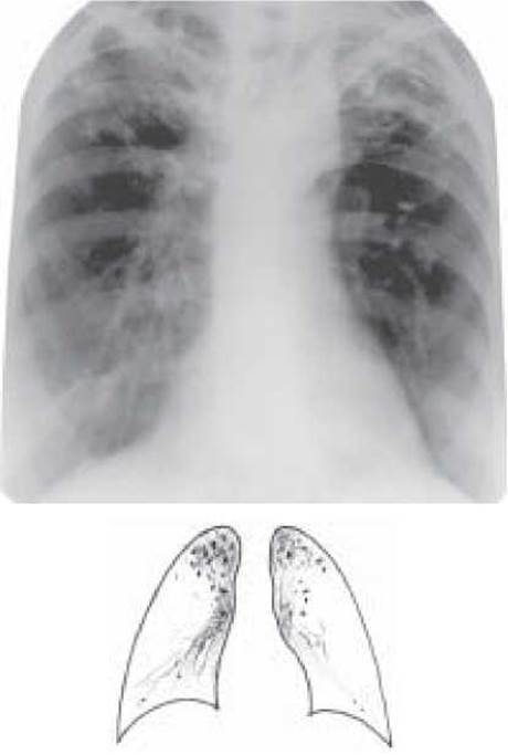
Fig. 15. Disseminated pulmonary tuberculosis (chronic). Overview radiograph
When diagnosing chronic disseminated tuberculosis, key diagnostic factors include a history of contact with a bacterium carrier, past Primary tuberculosis, Pleurisy, Extrapulmonary tuberculosis, and a prolonged, wave-like disease progression. Radiologic findings show polymorphic focal shadows, predominantly in the upper and middle lung fields, against a background of distorted pulmonary markings and fibrosis, often accompanied by irregularly shaped cavities of disintegration. Confirmation of the specific Etiology of the disease relies on the detection of MBT.
Cytology/practical/136.html">Differential Diagnosis OF chronic disseminated tuberculosis is performed against the same conditions considered in the differential diagnosis of subacute disseminated tuberculosis, particularly those with a protracted clinical course.
The mainstay of Treatment is prolonged antimycobacterial therapy, taking into account the drug susceptibility of MBT, patient tolerance, and potential complications (see "Treatment of tuberculosis").
CONTROL QUESTIONS (see "Miliary tuberculosis")
1. Definition of disseminated pulmonary tuberculosis, its Pathogenesis and pathomorphology.
2. Clinical and radiological variants of disseminated pulmonary tuberculosis.
3. Miliary pulmonary tuberculosis, its radiological presentation and clinical forms.
4. Differential diagnosis of miliary pulmonary tuberculosis.
5. Subacute disseminated pulmonary tuberculosis: Clinical Features, radiological presentation, course, and complications.
6. Chronic disseminated pulmonary tuberculosis: clinical features, radiological presentation, course, and complications.
7. Main conditions that must be differentiated from subacute and chronic disseminated pulmonary tuberculosis.
8. Management of patients with miliary and disseminated pulmonary tuberculosis.
TESTS
1. How soon after the onset of the disease can pathological changes be detected on a radiograph in miliary pulmonary tuberculosis:
A. 1-2 days
B. 3-5 days
C. 7-14 days
D. 21-30 days
E. 1-2 months
2. Which clinical course form is atypical for miliary tuberculosis:
A. pulmonary,
B. meningeal,
C. Typhoid,
D. Septic (Landouzy's disease),
E. Renal.
3. A patient D. was diagnosed with miliary pulmonary tuberculosis, MBT (-). Four antimycobacterial drugs were prescribed. Which Pathogenetic Therapy agents are most advisable to use for this patient?
A. Thymus preparations
B. Corticosteroids
C. Nonsteroidal anti-inflammatory drugs
D. Calcium chloride Electrophoresis
E. Tissue preparations (plasmol, aloe extract)
4. A patient K. was diagnosed with miliary pulmonary tuberculosis, infiltration phase, MBT (-). Which combination of antimycobacterial drugs is most optimal for the 1st stage of treatment?
A. Isoniazid + rifampicin + ethambutol
B. Isoniazid + rifampicin + ethionamide
C. Isoniazid + rifampicin + streptomycin + pyrazinamide
D. Isoniazid + streptomycin + pyrazinamide + ethambutol
E. Isoniazid + ethambutol + kanamycin + pyrazinamide
5. Patient L., aged 35. The disease onset was acute, presenting with a body Temperature rise up to 39 °C and a cough with sputum production. He had been receiving Antibiotics for a week with no effect. A few moist fine-bubble rales were heard between the scapulae. A chest X-ray showed foci of varying sizes with blurred contours throughout both lungs. Blood test: WBC - 13.2x10⁹/L, ESR - 45 mm/h. What is the most probable diagnosis?
A. Disseminated pulmonary tuberculosis
C. Infarction pneumonia
D. Pulmonary congestion
6. Fluorographic examination of a 48-year-old man revealed numerous foci of various sizes, low and moderate intensity, with blurred contours in the upper lobes of both lungs. The patient's general well-being is unaffected. Blood test: 8.2x10⁹/L, ESR - 20 mm/h. What form of pulmonary tuberculosis does the patient have?
A. Miliary
B. Disseminated
C. Focal
D. Fibrocavernous
E. Infiltrative
7. A 46-year-old man, released from prison two months ago, developed an insidious onset of illness featuring a cough, dyspnea, and a low-grade fever up to 38 °C. Chest radiography reveals faint focal shadows with hazy margins in the upper lung lobes. What is the most likely diagnosis?
A. Carcinomatosis
B. Disseminated tuberculosis
C. Focal pneumonia
D. Focal tuberculosis
8. A 35-year-old male underground miner with a 10-year occupational history complains of exertional dyspnea and a cough productive of scanty sputum. His body temperature is normal. Lung Auscultation reveals no rales. Complete blood count: WBC 7.8×10⁹/L, ESR 8 mm/h. Mantoux test with 2 TU shows a 10 mm infiltrate. Chest radiograph demonstrates bilateral, dense, well-defined small focal shadows, predominantly in the mid-lateral lung zones. What is the preliminary diagnosis?
A. Microfocal pneumonia
B. Miliary tuberculosis
C. Disseminated tuberculosis
D. Carcinomatosis
9. A 62-year-old male presents with paroxysmal cough, shortness of breath, decreased appetite, and a 10 kg weight loss. He has a history of household contact with a brother suffering from pulmonary tuberculosis and underwent prostate Cancer surgery 6 months ago. Blood work reveals anemia and an ESR of 65 mm/h. Chest X-ray shows multiple well-demarcated focal shadows, 5–6 mm in size, distributed mainly in the middle and lower lung fields. What is your preliminary diagnosis?
A. Disseminated tuberculosis
B. Carcinomatosis
C. Focal tuberculosis
E. Bilateral focal pneumonia
10. Main radiological features of subacute disseminated pulmonary tuberculosis.
A. Total bilateral microfocal involvement
B. Bilateral symmetrical focal lesions, predominantly in the upper and middle lung fields
C. Unilateral focal lesion
D. Bilateral focal lesions confined to the apical segments
E. Focal infiltrative bilateral process in the lower lobes of both lungs
PROBLEMS
1. A 19-year-old woman developed a dry cough, dyspnea, sweating, and a body temperature up to 38.5 °C following an Artificial abortion on March 24, 2004. Respiratory rate was 32 breaths/min. Harsh breath sounds were auscultated over the lungs. A chest X-ray showed low-intensity small focal shadows throughout both lungs. A year ago, the Mantoux test with 2 TU was 19 mm; currently, it is negative. Hemogram: WBC 9×10⁹/L, eosinophils 2%, band neutrophils 4%, segmented neutrophils 74%, lymphocytes 12%, monocytes 8%, ESR 11 mm/h.
a) Formulate a preliminary diagnosis.
b) What diseases should be included in the differential diagnosis?
c) Management.
Answer: a) Disseminated pulmonary tuberculosis (24.03.2004) (miliary), Destr-, MBT-M-K-, Hist0, Respiratory failure grade II, Cat 1 Coh 1 (2004); b) focal pneumonia; c) isoniazid 0.3 g, rifampicin 0.6 g, streptomycin 1.0 g intramuscularly, pyrazinamide 2.0 g daily, desensitizing agents, Vitamins B1, B6.
2. Patient Z., 29 years old, developed a cough with sputum production, dyspnea, generalized weakness, sweating, and a body temperature up to 39 °C three weeks after having the "flu". Two years ago, he was treated for right-sided exudative pleurisy. A chest X-ray revealed medium and large focal shadows with indistinct margins throughout both lungs.
a) Formulate a preliminary diagnosis.
b) Investigation plan for the patient.
c) What diseases require differential diagnosis?
d) Treatment plan.
3. Following a routine fluorographic examination, a 35-year-old woman was found to have foci of varying size and intensity in the upper lobes of both lungs. She had a history of frequent "flu" infections. Her last chest X-ray was 7 years ago, showing no abnormalities. The patient's general condition is satisfactory.
a) Formulate a preliminary diagnosis.
b) Investigation plan for the patient.
c) To which medical institution should the patient be referred?
4. Patient P., 20 years old, experienced an acute onset of illness following a cold: body temperature rose to 39.5 °C, accompanied by generalized weakness, sweating, marked dyspnea, and a dry cough. Dry and moist rales were heard over the lower lung fields. Chest radiography showed dense, medium-intensity foci in the lower lobes against the background of an enhanced pulmonary pattern.
a) Formulate a preliminary diagnosis.
b) To which medical institution should the patient be referred?
c) Investigation and treatment plan.
5. Patient K., 30 years old. Reports being ill for the past 7 months. The onset of the disease was insidious. Currently presents with pronounced general weakness, sweating, cough with sputum production, and dyspnea. Has lost 12 kg of weight. X-ray Examination reveals focal infiltrative shadows with multiple cavity formation throughout both lungs.
a) Formulate a preliminary diagnosis.
b) Outline an investigation plan.
c) Prognosis of the disease.
Last update: 10/08/2026
Editorial and Educational Adaptation: This material has been compiled based on the primary/original source text. The project team performed an editorial review, corrected technical inaccuracies, structured sections, and adapted the content for an educational format.
What was processed:
- elimination of formatting defects (OCR errors, structural breaks, corrupted characters);
- editorial organization of content;
- standardization of terminology in accordance with academic sources;
- verification of factual statements against the original source text.
All mentions of the author, publication year, and origin of the primary text have been preserved in accordance with the source.