Tuberculosis - I.T. Pyatnochka 2005
Primary tuberculosis
Focal pulmonary tuberculosis
A mild form of tuberculosis characterized by the presence of a focus (a specific lesion up to 1 cm in diameter) or a group of foci within no more than two segments of one or both Lungs (Fig. 16). Among newly diagnosed Pulmonary Tuberculosis patients, Focal pulmonary tuberculosis accounts for 18.9%.
Pathogenesis. Focal pulmonary tuberculosis may develop As a result of exogenous superinfection or endogenous dissemination of MBT from latent tuberculosis foci in intrathoracic Lymph Nodes, bones, or Kidneys, most commonly from old encapsulated or calcified lung foci during reactivation. The third pathway of focal tuberculosis development is the involution of other forms, such as infiltrative or disseminated tuberculosis.
Clinical Features. Symptoms of intoxication in focal tuberculosis are mild or absent. However, it should be noted that only conditions with an active, unfinished specific process are classified as focal tuberculosis.
There are two types of pulmonary tuberculosis: soft-focal (in the infiltration phase) and fibro-focal (in the scarring phase).
Soft-focal pulmonary tuberculosis is always a recent and active specific process.
Fibro-focal (chronic) tuberculosis is characterized by dense foci containing calcium, sometimes with caseation, alongside fibrous Changes in the form of cords and areas of hyperpneumatosis. When fibro-focal changes are detected radiologically, a thorough examination of the patient is necessary to determine The activity of the process.
Focal pulmonary tuberculosis is usually detected during routine preventive fluorographic examinations, as clinical symptoms are mild or absent. Only in 1/3 of patients are intoxication symptoms or Complaints related to bronchopulmonary system disorders observed. Intoxication symptoms are more common in soft-focal tuberculosis, whereas respiratory symptoms are more characteristic of the fibro-focal form. Percussion and Auscultation are of little diagnostic value in focal tuberculosis. The hemogram remains unchanged in the vast majority of patients. Mycobacterium excretion is scanty and does not exceed 15% of thoroughly examined patients.
X-ray Examination is the primary and most informative method for detecting focal pulmonary tuberculosis. For this purpose, inspiration and expiration fluorography, targeted radiography, and tomograms at optimal slice levels are used. Radiological signs of focal tuberculosis include: foci limited to no more than two segments; in the soft-focal form — small to medium-intensity foci with blurred margins against the Background of an enhanced vascular pattern in the affected lung area; in the fibro-focal form — high-intensity foci with clear margins against a background of a distorted pulmonary pattern and fibrous cords. Sometimes, in fibro-focal tuberculosis, it is necessary to determine the activity of the process (i.e., when the specific process is not yet complete and progression or regression is still possible).
Class="center">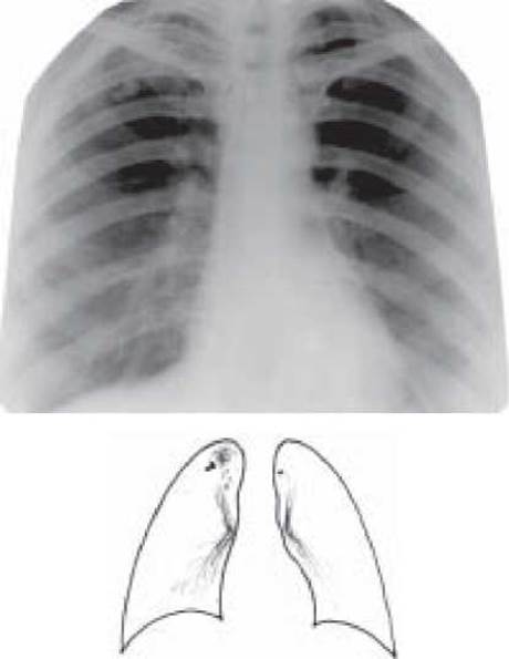
Fig. 16. Focal Tuberculosis of the upper lobe of the right lung. Overview radiograph
The activity of the tuberculosis process is determined based on the following criteria:
1. clinical criteria (presence of bronchopulmonary or intoxication symptoms);
2. radiological criteria (foci of low intensity with blurred margins, numerous or large foci);
3. bacteriological criteria (detection of MBT);
4. hemogram changes (leukocytosis, left shift of the leukocyte formula, lymphopenia, accelerated ESR);
5. tuberculin tests (hyperergic Mantoux test with 2 TU, positive local, focal, and general reactions following subcutaneous tuberculin administration — Koch's test);
6. if previous tests do not provide a definitive answer regarding the activity of pulmonary lesions, a trial Treatment is administered, which involves prescribing 2–3 antituberculosis drugs (isoniazid, ethambutol, and/or streptomycin for no more than two months)
for 3 months. Afterwards, a follow-up X-ray examination is performed; positive radiological dynamics indicate an active tuberculosis process, and treatment is continued. A stable X-ray picture indicates an inactive process, and antimycobacterial therapy is discontinued. However, it should be noted that the most informative tests for tuberculosis activity are radiological, bacteriological, and trial treatment.
Differential Diagnosis. Nonspecific Pneumonia, neoplasms, or metastatic lung lesions can present with clinical and radiological symptoms similar to those of focal pulmonary tuberculosis.
Treatment of patients with active focal pulmonary tuberculosis must begin with three antimycobacterial drugs: isoniazid, rifampicin, and pyrazinamide for 2 months. This is followed by continuation therapy with isoniazid combined with rifampicin for 4 months.
Disease progression may occur in 2–5% of patients with focal pulmonary tuberculosis. In such cases, indications for partial lung resection may arise.
1. Definition of focal pulmonary tuberculosis.
2. Pathogenesis and pathomorphology of focal tuberculosis.
3. Clinical and radiological variants of focal tuberculosis.
4. Clinical features, diagnosis, and Cytology/practical/136.html">DIFFERENTIAL DIAGNOSIS OF focal tuberculosis.
5. Criteria for the activity of focal tuberculosis.
6. The Essence of individual tuberculin Skin testing and trial treatment in determining the activity of the tuberculous process.
7. Treatment of patients with focal pulmonary tuberculosis.
TESTS
1. Which radiological signs reliably indicate the activity of focal tuberculosis?
A. Medium-intensity foci with well-defined outer contours
B. A group of high-intensity foci of varying sizes
C. Low-intensity foci with poorly defined contours
D. Ghon Focus
E. Medium-intensity foci against the background of limited pneumosclerosis
2. Which medications are appropriate for trial treatment to differentially diagnose focal tuberculosis and pneumonia?
A. Streptomycin + sulfalene
B. Streptomycin + isoniazid
C. Doxycycline + cefazolin
D. Doxycycline + rifampicin
E. Doxycycline + streptomycin
3. A 25-year-old patient undergoing a routine fluorographic examination presented with low-intensity focal shadows with poorly defined contours in segments 1–2 of the left lung. The Mantoux test with 2 TU showed a 7 mm infiltrate. Blood test: WBC - 9.9x10⁹/L, ESR - 26 mm/h. What is the most likely diagnosis?
A. Focal tuberculosis
B. Infiltrative tuberculosis
E. Eosinophilic infiltrate
4. The maximum size of shadows in focal pulmonary tuberculosis:
A. 1 mm
B. 1.5 mm
C. 5 mm
D. 10 mm
E. 25 mm
5. The most reliable criterion for the activity of focal tuberculosis is:
B. changes in the hemogram
C. detection of Mycobacterium tuberculosis
D. focal shadow of medium intensity with clear contours
E. positive Mantoux test with 2 TU
6. The maximum number of segments affected in focal pulmonary tuberculosis.
A. 1
B. 2
C. 3
D. 4
E. 6
7. A 45-year-old female patient complains of a cough and a body Temperature rise to 37.5 °C. She fell ill 6 weeks ago after having the "flu". Following a 2-week course of treatment for focal pneumonia, focal shadows in the upper lobe of the right lung remained unchanged. Blood test shows: eosinophilia – 6%. What is the most probable diagnosis?
A. Protracted pneumonia
B. Lung cancer
C. Eosinophilic infiltrate
D. Focal tuberculosis
E. Aspergillosis
8. Patient K., aged 27, was treated in a hospital for infiltrative tuberculosis of the upper lobe of the left lung in the decay phase for 4 months. Abacillarity was achieved, but a round-shaped formation 2 cm in diameter, of medium intensity with clear outer contours, formed at the site of the infiltrate. What treatment method is most appropriate at this stage?
A. Refer the patient for sanatorium-and-spa treatment
B. Continue treatment with antimycobacterial drugs
C. Perform surgical intervention
D. Administer a 1.5–2-month course of hormone therapy
Е. Use traditional folk remedies
9. A 35-year-old patient, who suffered from infiltrative tuberculosis of the right upper lobe 5 years ago, MBT (-). Currently, he complains of a dry cough and sweating. Fine moist rales are heard over the upper part of the right lung after coughing. Over the past 3 years, the radiological picture has been stable (single focal shadows of high and moderate intensity in the right upper lobe). Which dispensary observation group should the patient be assigned to?
A. 5.1
B. 5.2
C. 5.3
D. 5.4
E. 5.5
10. A 43-year-old male smoker complains of a cough with scanty sputum production, low-grade fever, and weight loss. He was hospitalized in the diagnostic department with suspected abscessing pneumonia. The chest X-ray shows a 4x4 cm opacity in the upper lobe of the right lung with a clear outer contour and a crescent-shaped lucency. Blood test: WBC - 9.0х109/L, ESR - 19 mm/h. Sputum analysis: WBC - 8-12 per high-power field. Mantoux test (with 2 TU) - 22 mm infiltrate. What is the most likely diagnosis?
A. Peripheral cancer
B. Lung abscess
C. Tuberculoma
D. Benign tumor
E. Aspergilloma
11. A 38-year-old man complains of pain in The Heart area, low-grade fever, sweating, and weight loss. Pulse 82 bpm, BP 110/75 mmHg, systolic murmur over the cardiac apex. Chest X-ray reveals focal shadows up to 5 mm in diameter in both lung apices. What is the most probable disease?
A. Sarcoidosis
B. Eosinophilic infiltrate
C. Focal pneumonia
D. Focal tuberculosis
E. Peripheral cancer
12. Which tuberculin test has the highest diagnostic value for determining the activity of the tuberculosis process:
A. von Pirquet test
B. Mantoux test
C. Koch test
D. Moro test
E. graduated von Pirquet test
PROBLEMS
1. During a routine fluorographic screening, a 20-year-old young man is found to have medium-intensity focal shadows with blurred contours in the apex of the left lung. His general condition is good. The Mantoux test with 2 TU shows a 19 mm infiltrate.
a) What is your preliminary diagnosis?
b) To which medical facility should the patient be referred?
c) What additional diagnostic examinations should be performed?
Answer: a) focal tuberculosis of the upper lobe of the left lung; b) anti-tuberculosis dispensary; c) chest X-ray and targeted radiograph, tomogram; triple sputum smear Cell/15.html">Microscopy and culture for MTB.
2. A 25-year-old patient's fluorogram reveals a group of low- to medium-intensity shadows, 3-8 mm in diameter, in the upper lobe of the right lung. The patient's condition is satisfactory. The Mantoux test with 2 TU reveals a 16 mm infiltrate.
a) Formulate a preliminary diagnosis.
b) Examination plan.
3. During a fluorographic examination, a 30-year-old man is found to have small and medium low-intensity foci in the apex of the right lung. Over the past month, he has noted decreased appetite, sweating, and a mild cough.
a) What disease can be suspected?
b) To which medical facility should the patient be referred?
c) Examination plan.
Last update: 10/08/2026
Editorial and Educational Adaptation: This material has been compiled based on the primary/original source text. The project team performed an editorial review, corrected technical inaccuracies, structured sections, and adapted the content for an educational format.
What was processed:
- elimination of formatting defects (OCR errors, structural breaks, corrupted characters);
- editorial organization of content;
- standardization of terminology in accordance with academic sources;
- verification of factual statements against the original source text.
All mentions of the author, publication year, and origin of the primary text have been preserved in accordance with the source.