Tuberculosis - I.T. Pyatnochka 2005
Primary tuberculosis
Infiltrative pulmonary tuberculosis
Infiltrative Pulmonary Tuberculosis is a specific exudative-pneumonic process exceeding 1 cm in size, characterized by a tendency toward rapid progression and breakdown (Fig. 17). Currently, it is the most common form of pulmonary tuberculosis, accounting for 54.9% of newly diagnosed cases.
Pathogenesis. Infiltrative pulmonary tuberculosis develops either As a result of perifocal inflammation surrounding older tuberculous foci or due to the progression of soft-focal tuberculosis. The Development of the infiltrate is driven by a hyperergic tissue reaction to A large number of rapidly multiplying virulent MBT. Significant contributing factors include the massiveness of superinfection, concomitant diseases, psychological trauma, and other conditions that impair the body's resistance.
Pathomorphology.
Infiltrative pulmonary tuberculosis may involve one or several lobules, a subsegment, a segment, or an entire lobe.
The focus around which the infiltrate forms represents exudative or productive inflammation prone to caseous necrosis. Under the action of Proteolytic Enzymes, this necrosis leads to liquefaction, breakthrough through the damaged draining bronchus, and The formation of a breakdown cavity accompanied by fresh foci appearing around the infiltrate. Subsequently, under METABOLISM/18.html">The Influence of Treatment, scar changes, focal alterations, Puhl's foci (foci with a caseous center), tuberculomas, indurative areas, and even localized cirrhosis form at the site of the infiltrate, while complete resolution is much rarer. In cases of ineffective treatment, infiltrative pulmonary tuberculosis can progress to fibro-cavitary pulmonary tuberculosis.
Class="center">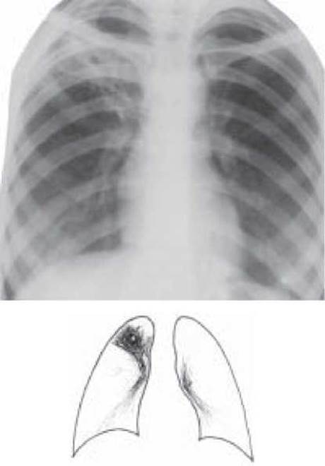
Fig. 17. Infiltrative Tuberculosis of the upper lobe of the right lung, breakdown phase. Overview radiograph
Clinical Features. A distinction is made between an infiltrate and Caseous Pneumonia (the latter is classified as a separate clinical entity). Clinically and radiologically, the following variants of infiltrative pulmonary tuberculosis are distinguished:
1. Cloud-like infiltrate is characterized by a faint, low-intensity, inhomogeneous shadow with indistinct, blurred contours and a tendency toward rapid breakdown.
2. Rounded infiltrate presents as a nearly homogeneous, medium-intensity shadow of a rounded shape with distinct contours (Assmann type).
3. Lobular infiltrate appears as an inhomogeneous shadow representing a conglomerate of fused foci.
4. Lobar infiltrate (lobitis) is an extensive infiltrative process involving an entire lung lobe, frequently inhomogeneous in character and containing one or several breakdown cavities.
5. Perississuritis is an infiltrate located along the interlobar fissure, resulting in a sharp contour on one side and a blurred contour on the opposite side.
The clinical course of infiltrative tuberculosis depends on the morphological Structure, the size of the infiltrate, and the extent of caseation. Most often, infiltrative tuberculosis begins acutely or subacutely, clinically resembling Influenza, an acute respiratory viral infection, or pneumonia. One of the symptoms of infiltrative tuberculosis may be hemoptysis while the patient's general condition remains satisfactory. Sometimes the clinical course is asymptomatic (inapparent), although even in such cases, detailed examination can reveal minor signs of intoxication. Physical findings depend on the size and phase of the process. If the infiltrate exceeds 4 cm in diameter, Percussion dullness, harsh breathing, and moist rales during infiltrate breakdown are observed.
Radiologically, infiltrative tuberculosis is characterized by a shadow exceeding 1 cm in diameter, inhomogeneous in character, of medium or low intensity, connected to the ROOT by a "linear shadow" (trail), predominantly localized in the I, II, or VI segments, featuring a breakdown cavity in the center and surrounded by foci of bronchogenic dissemination either locally or in other areas of the Lungs.
Regarding the hemogram, the vast majority of patients exhibit leukocytosis not exceeding 15.0x10⁹/L, a leftward shift of the leukocyte formula, lymphopenia, and an accelerated ESR. The Mantoo tuberculin Skin test with 2 TU is positive (normergic).
In 40–50% of cases, infiltrative pulmonary tuberculosis is accompanied by Bacteria excretion, which rises to 95% of patients when breakdown occurs.
Cytology/practical/136.html">Differential Diagnosis OF infiltrative pulmonary tuberculosis is performed primarily with nonspecific pneumonia, eosinophilic infiltrate, Lung Cancer, and pulmonary infarction.
Treatment of patients involves continuous antimycobacterial therapy for 6–8 months. For the first 2–3 months, optimal doses of antituberculosis drugs (isoniazid, rifampicin, streptomycin, ethambutol, or pyrazinamide) are prescribed alongside desensitizing, vitamin, and symptomatic therapy. Often, a course of glucocorticoid therapy is advisable in the initial stage of treatment. After 2 months, streptomycin is discontinued, and treatment is continued for another 4 months with isoniazid, rifampicin, and ethambutol (or pyrazinamide) until the breakdown cavities close. In cases where treatment remains ineffective for 3–6 months, surgical intervention—specifically, partial lung resection—is indicated.
Last update: 10/08/2026
Editorial and Educational Adaptation: This material has been compiled based on the primary/original source text. The project team performed an editorial review, corrected technical inaccuracies, structured sections, and adapted the content for an educational format.
What was processed:
- elimination of formatting defects (OCR errors, structural breaks, corrupted characters);
- editorial organization of content;
- standardization of terminology in accordance with academic sources;
- verification of factual statements against the original source text.
All mentions of the author, publication year, and origin of the primary text have been preserved in accordance with the source.