Tuberculosis Study Guide - M. M. Savula 2002
Extrapulmonary Tuberculosis
Urogenital Tuberculosis
Female Genital Tuberculosis
Pathogenesis. Female Genital Tuberculosis occurs when MTB invades the genital Organs via hematogenous or lymphogenous routes, most commonly during the Primary tuberculosis infection. In cases of mesenteric Lymph node and peritoneal involvement, the tuberculous infection can spread to the fallopian tubes by contiguity. In nearly 90 % of cases, genital tuberculosis is localized in the fallopian tubes (bilaterally) or in the endometrium, and rarely in the Ovaries, cervix, and Vagina. Tuberculous tubercles form on the inner lining of the tubes and in the endometrium, sometimes followed by caseous necrosis. Involvement of the inner lining of the tubes (endosalpinx) leads to their adhesion and impaired patency. Exudate and caseous material can accumulate in them, causing the tube to distend and fill with purulent-caseous masses (pyosalpinx). The process can spread to the ovaries and Peritoneum, leading to adhesion formation. Regression of the process is accompanied by encapsulation and calcification in the affected areas.
Clinical presentation and Diagnosis. It most commonly affects young women aged 16 to 30. Subclinical chronic forms are asymptomatic, but they often result in Infertility.
In chronic cases, patients sometimes complain of a low-grade fever, dull pain in the lower abdomen and lumbar region, and occasionally menstrual irregularities.
In subacute and caseous forms, which are rare, local symptoms are accompanied by remittent fever, night sweats, poor appetite, and weight loss.
A routine gynecological examination is performed to establish the diagnosis. On Palpation, when the adnexa are symmetrically enlarged, tenderness is less pronounced in tuberculous lesions compared to non-specific inflammatory processes. A history of contact with a tuberculosis patient, past Pleurisy, or intrathoracic lymph node tuberculosis is of significant diagnostic value. A comprehensive evaluation of the patient is required to detect other potential sites of tuberculosis.
Tuberculin Skin tests are usually positive or strongly positive. To clarify the diagnosis, a subcutaneous tuberculin test (Koch's test) is performed, which provokes an exacerbation of symptoms in cases of active genital tuberculosis. A more accurate diagnosis is possible through histological examination of tissue obtained by needle aspiration of the enlarged adnexa or endometrial curettage. The same specimen or menstrual Blood must be cultured for MTB.
For diagnostic purposes, radiography and hysterosalpingography are performed (Fig. 31). Calcifications of the tubes, Lymph Nodes, and ovaries detected on radiography confirm the diagnosis of tuberculosis. Hysterosalpingography reveals indirect signs of tuberculous involvement: tubal deformity, club-shaped dilation of their ends, rigidity, beaded appearance of the tubes, etc. Differential diagnosis is made with other chronic inflammatory processes and tumors.
In case of diagnostic difficulties, laparoscopy with biopsy of the affected organ is performed.
Treatment. Antibacterial therapy is the mainstay of treatment. In tuberculous endometritis, streptomycin is additionally injected into the myometrium; in Tuberculosis of the adnexa, streptomycin is administered into the posterior fornix after prior aspiration of the exudate. In cases of purulent tubal lesions and failure of antibacterial therapy, surgical removal of the tubes is performed.
Class="center">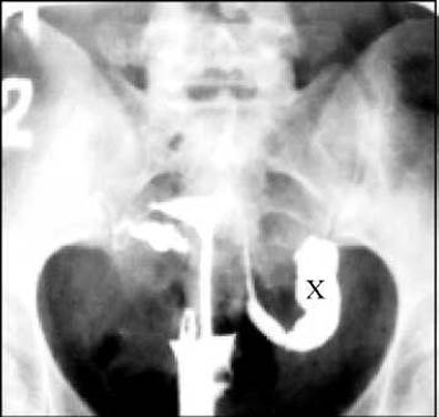
Fig. 31. Salpingogram. Sequelae of tuberculous involvement of the fallopian tubes. The fallopian tubes are deformed, the left one is severely dilated (X).
Last update: 10/08/2026
Editorial and Educational Adaptation: This material has been compiled based on the primary/original source text. The project team performed an editorial review, corrected technical inaccuracies, structured sections, and adapted the content for an educational format.
What was processed:
- elimination of formatting defects (OCR errors, structural breaks, corrupted characters);
- editorial organization of content;
- standardization of terminology in accordance with academic sources;
- verification of factual statements against the original source text.
All mentions of the author, publication year, and origin of the primary text have been preserved in accordance with the source.