Antibiotics (Properties, Administration, Interactions) - M.P. Cherenko 1999
Trauma and Traumatism. Shock. Collapse. Syncope
General concept of INJURY
An injury (from Greek trauma — wound) is an anatomical or functional damage (typically sudden, rarely chronic) to the body, its Tissues, or Organs caused by mechanical, physical, chemical, or psychological factors, accompanied by The Development of local and systemic reactions.
Traumatism refers to injuries that recur under certain circumstances among specific groups of people exposed to identical working, living, and environmental conditions.
Injuries, particularly acute mechanical ones, as one of the most common forms of pathology, show a steady upward trend. This is driven by the increasing mechanization and complexity of industrial production, the saturation of streets and roads with motor vehicles and other transport, and the proliferation of household and office appliances. Furthermore, injuries are becoming more complex and severe, meaning the proportion of Polytrauma is rising. This is evidenced by casualty statistics; notably, injury ranks as the leading cause of death among young men (under 40 years of age). For these reasons, trauma, along with its Prevention and Treatment, has become a paramount social and medical issue of today.
Injuries can be accidental or intentional. Accidental injuries include those caused by external factors, as well as those that frequently accompany physiological processes, such as childbirth (injuries affecting both the infant and the mother). Intentional injuries include surgical Procedures and other harms inflicted for therapeutic purposes. Injuries sustained during warfare are also classified as intentional, alongside those inflicted by homicide and suicide.
A mechanical injury is the most common type, caused by the application of a specific force to the body—that is, the kinetic energy of a moving object or, conversely, the movement of a person's body against The surface of a stationary medium (e.g., falling from a height onto the ground or into Water).
A physical injury is caused either by high or low temperatures (Burns and frostbite), electric current, radiant energy (solar radiation, ionizing radiation), or the Shock wave of air resulting from the explosion of weapons and devices.
Chemical injuries result from the action of chemical agents on human tissues (chemical burns of the Skin and mucous membranes).
A psychological trauma is a nervous shock caused by tragic or adverse life events, insults, or harsh words.
Mechanical trauma most commonly manifests as a wound—a disruption of skin integrity, primarily involving soft tissue damage (with tearing of the subcutaneous tissue, minor vessels, and hemorrhaging). Less frequently, it is accompanied by bone fractures, joint dislocations, and internal organ damage (contusions, ruptures, avulsions, concussions, etc.). The study and treatment of injuries are the focus of such fields as combustiology (The Study of burn treatment) and traumatology (the study of disorders and injuries of The Musculoskeletal System).
Mechanical injury is the most prevalent and complex form of trauma, representing one of the major challenges in surgery.
Classification OF INJURIES
Mechanical injuries are classified according to numerous criteria. Based on the number of sustained injuries, they are categorized into solitary, multiple (polytrauma), and combined injuries.
A solitary injury is damage to a single anatomical region, regardless of the number of affected tissues (e.g., a forearm fracture, a chest contusion with fractures of 2 or 3 Ribs).
A multiple injury involves damage to multiple anatomical regions within a single body system (integumentary, musculoskeletal, digestive, etc.), such as several soft tissue wounds, multiple fractures, or damage to two or more abdominal organs (intestines and Liver, etc.).
An injury is classified as polytrauma when tissues and organs from different body systems are simultaneously damaged. For instance, a combination of soft tissue wounds, bone fractures, and injuries to the abdominal, thoracic, or Brain organs in various combinations is termed polytrauma. It is characteristic of transport accidents (motor vehicle, railway, etc.) and disasters associated with natural calamities (earthquakes, mountain or mine collapses, etc.).
An injury caused by the simultaneous action of several traumatic factors of different nature (mechanical, blast wave, thermal, and penetrating radiation) is termed a combined injury. This is typical of injuries resulting from nuclear missile weapons and accidents at nuclear power plants and facilities.
Depending on The Nature of the traumatic agent, mechanical injuries are subdivided into gunshot and non-gunshot injuries.
An injury accompanied by damage to the body's covering (skin and superficial mucous membranes) is termed open. Injury to tissues and Internal Organs without compromising the body's outer coverings is considered closed (blunt).
If an injury penetrates a body cavity (abdominal, pleural, cranial, or joint, etc.), it is classified as penetrating; otherwise, it is non-penetrating.
Based on the nature of tissue damage, injuries are categorized as follows: skin injuries; injuries to the subcutaneous tissue and Muscles; bone injuries; and injuries to Body Cavities and hollow organs.
A complex injury is characterized by significant damage to multiple tissues and potentially internal organs, major Blood Vessels, or nerves, or severe destruction of a single tissue along with the entire organ (e.g., a comminuted fracture of a long bone).
Injuries can be uncomplicated or complicated. Complicated injuries include those (regardless of their scale) that are compounded by severe general symptoms and reactions (Hemorrhage, Traumatic shock, Crush syndrome, air or fat embolism, various types of pneumothorax, Pleurisy, Peritonitis, concussion-contusion syndrome, etc.).
Clinically, injuries are differentiated into mild, moderate, severe, and extremely severe. The latter may either be incompatible with life or pose an extreme threat to it—so-called critical injury.
Mild injuries include minor damage to soft tissues, skin, subcutaneous tissue, or mucous membranes; moderate injuries include substantial tissue damage and simple, uncomplicated fractures of tubular or small spongy bones.
Severe trauma is defined as complex soft tissue injuries involving damage to major blood vessels or nerves, scalp avulsions, complex closed fractures, and certain internal organ injuries, among others.
Very severe, or critical, traumas include most injuries to the abdominal and thoracic cavities, TRAUMATIC BRAIN INJURIES, pelvic bone fractures, and complex fractures of the lower and upper limbs. Such injuries can be fatal or life-threatening due to hemorrhage, shock, open or tension pneumothorax, cardiac contusion or tamponade, impaired brain function, thromboembolic complications, or secondary infections (peritonitis, pleurisy, anaerobic infection, Sepsis, etc.). Critical injuries also encompass those that, while not immediately life-threatening, result in prolonged disability, such as injuries to the eyes, face, and extremities.
MECHANISMS OF INJURY
The nature of an injury, its clinical course, and severity depend on numerous factors and the Specific characteristics of the traumatic agent. These include the mass and shape of the striking object, the MECHANISM OF ACTION, its velocity, the angle of force application, the contact area and duration of impact, The properties of the body tissues, and environmental conditions. The mechanism by which a traumatic agent affects the body may manifest as compression of the body, an organ, or its tissues; bending of a body part or organ beyond its natural physiological mobility; or the stretching or twisting of a specific organ or body part. A significant number of injuries result from a combination of these mechanisms, as occurs when struck by a moving heavy object or during a fall from a height.
The damaging effect of a mechanical agent depends on its force, kinetic energy (determined by its mass and velocity), shape and surface characteristics (blunt or sharp), contact area, direction of force, as well as the specific Properties of the tissues and the Organism as a whole. Tissue resistance to injury relies on elasticity, extensibility, and mechanical strength, qualities that are fundamentally determined by tissue Structure.
Sharp objects, as well as heavy blunt objects with a small contact area acting with high kinetic energy, typically cause open injuries. These include various types of wounds (incised, lacerated, etc.) that may either penetrate body cavities or remain non-penetrating while still involving internal organ damage. Particularly severe open injuries occur when hands, feet, or Hair are caught and stretched in moving machinery parts (transmissions, flywheels, wheels, etc.). Such traumas are accompanied by traumatic amputation of the distal extremities (sometimes the entire arm) and scalp avulsions.
Blunt objects with a large surface area cause closed injuries, meaning there is no breach of the skin integrity (blunt trauma). For instance, compression of the body between two vehicles or between a vehicle and a wall, particularly during inspiration, leads to chest trauma and traumatic asphyxia. This results in Circulatory Disorders in the upper chest and HEAD (venous congestion accompanied by rupture of small vessels in the skin, mucous membranes of the Upper Respiratory Tract, and conjunctiva, as well as cerebral Hypoxia) and impaired respiratory function (a sharp drop in pulmonary pressure with the rupture of numerous alveoli). The heavier the blunt object and the higher its velocity, the more severe the injury. Tissues with low resistance to deformation, poor elasticity, and minimal mechanical strength are the first to be injured. These include the subcutaneous tissue, small-caliber blood vessels, muscles, and parenchymal or hollow organs. Internal organs with high fluid content easily rupture upon impact or compression. For this reason, blunt impacts to the torso (with a stone, board, fist, etc.) frequently cause closed injuries to internal organs such as the Spleen, liver, Kidneys, or distended small intestines and Urinary Bladder. The vulnerability of these organs is exacerbated by the inhibition of protective Reflexes (absence of abdominal wall Muscle contraction), which commonly occurs in states of intoxication or poisoning.
Blunt injuries occur when the body falls onto a hard surface or moves with massive acceleration, as experienced in aeronautics and astronautics. A body moving at high velocity sustains injury during both positive rectilinear and radial acceleration, and particularly during negative (decelerating) acceleration, which occurs the moment The Human Body collides with an immovable obstacle. Thus, in falls from a height, as well as in high-speed head-on vehicle collisions (or a vehicle colliding with an obstacle such as a wall), injuries manifest not only at the site of primary impact but also at a distance. For example, in a fall onto the feet or buttocks, alongside lower limb fractures, a fracture of the Base of the Skull or spinal Column may be observed; a contusion of the brain can occur on the side opposite to the cranial impact; and a cervical spine fracture may result from the driver's or passenger's head whipping backward upon impact. Traumas associated with high-force acceleration are termed acceleration injuries (from Latin *celer* — swift). They occur due to the sudden deceleration of the body or a part of it upon impact, while other structures—such as the cranial or abdominal contents, or body parts superior to the point of impact—continue moving forward. In aeronautics and astronautics, where multi-directional accelerations (rectilinear-transversal, radial-craniocaudal) generate massive gravitational overloads, conditions conducive to these macro- and microstructural injuries (concussions, circulatory disturbances, microhemorrhages, etc.) arise very frequently.
Skin, bones, and tendons possess a natural capacity to resist deformation and mechanical force. However, significant force applied via bending or twisting mechanisms frequently causes injuries to long tubular bones, such as fractures sustained from perpendicular impacts to the legs of a standing person, or fractures in skiers and skaters during abrupt turns.
Injuries caused by tissue stretching, particularly of tendons, are commonly observed in tennis players, soccer players, and dancers. The resistance of bones to trauma diminishes with age; it is particularly compromised in the elderly due to Osteoporosis and a reduction in the organic content of the bones. In children, bone resilience is much higher, making fractures less frequent. When fractures do occur in children, they often take the form of a "greenstick" fracture, without tearing the periosteum and consequently without fragment displacement. An even greater predisposition to fractures is observed in patients suffering from hyperparathyroidism, osteodystrophy, and specific bone diseases.
The angle of force application also significantly influences the Nature of the injury.
If a traumatic agent strikes the body at a right angle, compression and bending forces cause damage directly at the site of impact. If the force is minor, it results in a simple bruise, whereas high force may fracture bones or injure an internal organ. An agent acting at an acute angle to the body may produce a contused or avulsed wound either at the point of contact or at a certain distance from it (such as a dislocation, fracture, or, less commonly, Other types of injuries).
Chronic trauma (such as corns, abrasions, and bursitis of the knee and elbow joints) results from prolonged, low-intensity pressure applied to tissues (skin and underlying structures).
The condition of the tissues and the organism as a whole also significantly influences the nature of the injury. Dystrophic, inelastic tissues in individuals with atherosclerosis are much more prone to damage than healthy tissues. The increased bone fragility in the elderly was mentioned above. The spleen and liver are also extremely vulnerable when enlarged or hyperplastic due to various conditions (such as malaria, portal Hypertension, or hemolytic anemia) or affected by dystrophic changes. They frequently rupture—especially the spleen—even under minor mechanical impact applied to their anatomical Location or elsewhere; sometimes, even physiological acts (such as coughing, straining, or turning in bed) can trigger this pathology.
A significantly reduced tissue resistance, particularly of the skin, is observed in patients undergoing prolonged or continuous corticosteroid therapy with hydrocortisone or prednisolone (for Bronchial Asthma, Rheumatoid Polyarthritis, and other so-called collagenoses), those suffering from endogenous hypercorticism (Itsenko–Cushing's Disease and syndrome), as well as patients with hypovitaminosis, especially Vitamin C Deficiency (scurvy). In hypercorticism, decreased tissue resistance is caused by protein breakdown, whereas ascorbic acid deficiency disrupts and blocks Collagen synthesis.
Alongside pathological or physiological organ changes that facilitate injury, one must consider the natural vulnerability of specific organs and body regions. For instance, the heightened sensitivity of certain organs leads to complex reflex disorders in the body when they are injured. Areas particularly sensitive to trauma include the neck—especially the Larynx and carotid sinus—the Testes, the epigastric region, the Pleura, and the rectum. This is due to their rich innervation.
The external environment has a substantial impact on The Nature and frequency of injuries. For example, during the autumn and winter periods, adverse weather conditions lead to a sharp increase in musculoskeletal injuries (fractures, dislocations, contusions, etc.). Injuries are more frequently complicated by systemic reactions due to cold exposure and similar factors. However, the overall incidence of trauma is influenced far more by poor road conditions and excessive traffic, particularly motor vehicles, than by meteorological conditions. The steady rise in the number of victims with severe multiple injuries and polytrauma is primarily associated with motor vehicle transport. Nevertheless, many such cases also result from other transport accidents (railway, aviation) and natural disasters (earthquakes, typhoons, etc.). Polytraumas are often caused by a combination of mechanical and physical factors (such as burns and frostbite) resulting from burning vehicles, collapsing buildings, and adverse environmental conditions.
TRAUMA Diagnostics AND PRE-HOSPITAL CARE FOR VICTIMS
The diagnostics of acute trauma require promptness and, above all, high precision in identifying life-threatening injuries. At the pre-hospital stage, this is carried out using physical and simple instrumental Methods, whereas at the hospital stage, both physical and various auxiliary (instrumental and laboratory) methods are employed.
Due to the sudden onset of injuries, their diagnostics are characterized by urgency (owing to potential life-threatening complications) and a very tight timeframe for establishing a Diagnosis. Furthermore, diagnostics often have to be performed while the patient is unconscious, which precludes taking a medical history and necessitates relying solely on objective signs of trauma. Therefore, a physician must possess sound medical and general surgical training and be able to conduct examinations rapidly, following a rational scheme.
Tactically, the examination follows THE PRINCIPLE OF "from the general to the specific," regardless of the scope or severity of the injury. Both the course of the injury and the patient's prognosis often depend on diagnostic accuracy and the quality of primary (pre-hospital) medical care provided. This principle remains equally valid for the hospital stage. Naturally, mild, minor injuries are relatively straightforward to diagnose at all stages. However, determining the severity of an injury is no easy task and requires a comprehensive examination of the victim. Even a patient who walks into a trauma center or outpatient clinic independently after an injury and appears to have a minor wound or closed injury without significant systemic disturbances may actually be facing a life-threatening condition. Such injuries include stab and incised wounds of the abdominal and thoracic cavities, as well as head contusions, which can lead to the slow development of internal bleeding into the abdominal, pleural, pericardial, or cranial cavities due to vascular damage in the organs located within those spaces.
Class="center">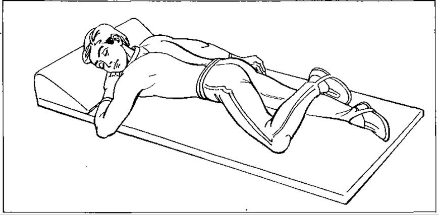
Fig. 30. Patient positioning in an unconscious state
Victims with injuries who independently visit a clinic or trauma center are generally treated on an outpatient basis in surgical rooms after a thorough examination by physicians and the provision of primary medical care. Only a small fraction of injured individuals are referred to surgical inpatient departments based on clinical indications.
For trauma victims of varying severity who are unable to move independently, primary medical care is provided by specialized ambulance teams. In such cases, during the initial pre-hospital stage of diagnosis, attention is focused primarily on assessing the patient's general condition: checking for the presence or absence of heartbeat, respiration, external bleeding, limb paralysis, or major bone fractures. First and foremost, especially in the absence of consciousness, the patency of the airways is checked, and any foreign bodies found are removed. If respiration and heartbeat are absent, cardiopulmonary resuscitation (Artificial ventilation and closed chest massage) is initiated. In cases of external bleeding, temporary hemostasis is performed using a tourniquet, pressure bandage, digital pressure, or other appropriate methods.
In cases of major limb fractures, they are immobilized using standard transport splints (without removing clothing), and wounds are covered with sterile bandages. Unconscious casualties are placed on their side in the "three-quarter" recovery position (Fig. 30) to prevent potential asphyxiation from vomit.
Victims showing signs of or at risk for traumatic shock receive prompt anti-shock therapy (pain relief, warming, cardiac medications, etc.). Infusion therapy is initiated already in the ambulance and continued en route to the hospital. Patients are urgently transported to surgical or specialized trauma units for polytrauma management. Even at the pre-hospital stage, patients with open injuries receive tetanus immunization and broad-spectrum Antibiotics or other antibacterial agents.
To improve (speed up and enhance) trauma diagnosis and prognosis, as well as to ensure the rational use of therapeutic resources and a qualitative assessment of emergency care for trauma victims, several countries—most notably the USA—have developed prognostic systems. These are based not on a physician's subjective Assessment of the injury, but on its objective anatomical or functional characteristics. Among these schemes, the most well-known are the AIS (Abbreviated Injury Scale) and its derivative, the ISS (Injury Severity Score).
These anatomical schemes are highly cumbersome (the list of injury score codes alone spans several pages) and therefore, despite providing reliable criteria for predicting injury outcomes, are practically rarely used.
Recently, a simpler functional scheme proposed by N. Champion et al. (1981), known as the TS (Trauma Score) system, has gained wider acceptance. It evaluates 5 main functional parameters: A — respiratory rate per min; B — respiratory effort (normal or using accessory muscles); C — systolic blood pressure; D — capillary refill time after releasing pressure on the forearm; E — Glasgow Coma Scale (CNS status). The scoring of these parameters is subjective. This scheme yields prognostic results comparable to the AIS and ISS, but it is more portable and therefore more widely used in emergency medical facilities.
TRAUMATISM
Alongside various types of isolated injuries, surgery also deals with group injuries that occur recurrently among individuals of specific professions or groups operating under similar conditions. Such injuries are grouped under METABOLISM/2.html">THE CONCEPT OF "traumatism." The term "traumatism" encompasses both major, severe injuries and minor lesions or microtraumas. The latter are numerous and significant not so much in their own right, but as a source of infectious complications leading to temporary disability.
Traumatism is divided into occupational and non-occupational. The latter accounts for about 20%, meaning non-occupational traumatism heavily predominates in terms of sheer numbers. Occupational traumatism is further subdivided into industrial and agricultural. Industrial traumatism includes injuries not only in manufacturing plants, factories, and workshops, but also among transport workers, construction workers, and others.
Non-occupational traumatism includes the following categories: transport (rail, road, aviation), street, domestic, sports, and childhood injuries. Childhood traumatism is singled out into a separate group purely for biological reasons—specifically due to children's inadequate perception of environmental hazards resulting from the immaturity of their Central Nervous system, lack of life experience, and heightened curiosity and mobility. The causes of non-occupational traumatism in children and adults are otherwise identical.
In addition to the aforementioned types of traumatism, intentional injuries inflicted by individuals with the intent of suicide (suicidal) or self-mutilation are also distinguished. Wartime injuries—that is, injuries sustained during war—are likewise classified as intentional.
Trauma is a leading cause of death among individuals under 44 years of age. It ranks third in overall mortality causes.
Mechanical trauma holds the largest share in overall traumatism and forms its foundation. Its rates are rising worldwide. This is driven by the mechanization of industry and daily life, electrification and gasification, the significant growth of motor transport and aviation, intensive high-rise construction, the development of major technical complexes (underground pipelines, canals, television towers, high-voltage power lines, etc.), mass participation in sports, and increased consumption of alcohol, medications, and narcotic or toxic substances by the population. A key feature of mechanical injuries is their multiplicity, which is why they are currently referred to as polytrauma.
According to WHO statistics, motor vehicle accidents alone claim the lives of over 250,000 people worldwide every year, while the number of injured exceeds 2 million (with other reports citing 8 million). These figures are far from accurate (generally underreported), as there is still no universally accepted definition of trauma-related mortality. While Belgium defines trauma mortality as death at the scene of the injury, the US includes deaths occurring within a year post-injury. In England, road traffic accidents kill more people annually than all other types of trauma combined. Most regrettably, these and other injuries are the leading cause of death among young people (aged 20–40).
The nature of an injury depends on the circumstances in which it was sustained, its cause, and the specific CHARACTERISTICS OF THE damaged tissue and the organism as a whole.
Road traffic traumatism encompasses all injuries caused by vehicles (provided the victims are not performing occupational duties), regardless of whether the victim was inside the vehicle (driver, passenger) or outside (pedestrian, cyclist). The majority of such injuries are the fault of pedestrians. Many traffic accidents occur As a result of driving under The Influence of alcohol.
A street injury is an incident that occurs on a street, in a courtyard, forest, field, etc., regardless of the cause (including those caused by vehicles). Most street injuries result from the victim falling. This happens particularly frequently during the autumn-winter period and in the evening hours.
These injuries are dominated by fractures of the upper and lower limbs, with bruises, sprains, and wounds occurring less frequently. Street traumatism is caused by poor traffic Organization, substandard road surfaces and lighting, and poor maintenance—particularly during icy conditions.
Domestic traumatism comprises injuries occurring in home environments (apartment, yard, garage). A large number of these are related to housekeeping and food preparation. Contusions, wounds, and burns predominate, with the hand being the most frequently affected site. Many injuries arise during domestic arguments and fights, often fueled by alcohol. Domestic injuries occur more frequently in men, particularly younger ones.
Sports traumatism refers to injuries sustained while engaging in sports on fields and in gymnasiums. This type of injury accounts for about 2–3% of all trauma-related damage. It is typical for soccer players, boxers, gymnasts, hockey players, and motorcyclists. Musculoskeletal injuries (contusions, fractures, sprains, dislocations) predominate, along with injuries to the head and trunk.
Childhood traumatism includes all types of non-occupational injuries (road traffic, domestic, school, street, sports, etc.) sustained by children. Domestic accidents involving children occur most frequently under the age of 3. The majority of injuries in children under 13 occur during school age while playing and participating in sports (50%), at home (30%), and in road traffic accidents (where mortality reaches 80%).
According to WHO data, the overall mortality rate of children from accidents is 37.2 per 100,000 boys and 22.8 per 100,000 girls.
Occupational traumatism is associated with the performance of job duties in industry or agriculture.
The primary causes of occupational traumatism include poor working conditions, imperfect or faulty tools of production or their improper operation, and the employee's personal state. Beneath these general causes lie a multitude of specific circumstances related to the organization of workplace labor, equipment, workforce composition, and worker qualifications. Violations of safety regulations, outdated and faulty machinery, poor Sanitary and hygienic working conditions (cluttered workspaces, air pollution, inadequate lighting), insufficient worker qualifications, fatigue, and numerous design flaws in machinery and tools all contribute to traumatism.
The most common industrial injuries are wounds, contusions, burns, and Electrical injuries. Due to mechanization and electrification, The structure of agricultural traumatism today differs little from industrial traumatism. Wounds, contusions, and closed injuries—predominantly of the limbs (over 80%)—also predominate, while internal injuries are much rarer. Up to 70% of injuries are classified as mild.
The majority of injuries are non-occupational. Of greatest concern is road traffic traumatism, which is figuratively referred to as a "war on the roads." Millions of people fall victim to this type of injury every year, with a quarter of a million dying at the scene. Taking into account imperfect record-keeping, injury statistics, and the very definition of trauma-related death, it is safe to say that the actual toll of this traumatism is far greater than officially reported.
INJURY PREVENTION
To combat trauma effectively, it is essential first to create favorable socio-economic and environmental conditions for individuals and to implement targeted preventive measures encompassing organizational, technical, sanitary, hygienic, medical, educational, and ethical aspects.
Occupational injuries are the most amenable to regulation and control. The primary conditions for their prevention include the meticulous organization of safety protocols in the workplace and strict enforcement by all personnel.
Road traffic accidents require significant attention from both the state and its regulatory bodies, as well as vehicle designers, drivers, and pedestrians. Road infrastructure improvements, the manufacturing of highly stable and resilient vehicles equipped with safe steering mechanisms, the mandatory use of seat belts, strict adherence to traffic regulations, proper vehicle maintenance, and advanced driver training are all vital components of road accident prevention. The Introduction of seat belts alone has contributed to a dramatic reduction in fatalities at accident scenes.
Domestic injuries can be mitigated by improving living conditions, developing municipal services, increasing the availability and accessibility of preschool childcare facilities, raising public awareness regarding the safe operation of household appliances (particularly electric and gas stoves), and combating alcoholism and substance abuse.
In street injury prevention, the primary focus is on urban infrastructure maintenance (timely pavement repairs, sanding sidewalks and walkways during icy conditions, adequate nighttime street lighting, traffic management, and maintaining the functionality of transport facilities), alongside the provision of safe playgrounds and adequate supervision of children.
Sports injury prevention relies on improving the organization of training sessions and competitions, enhancing athletic equipment, maintaining discipline, optimizing the performance of coaches and sports team leaders, and monitoring compliance with sanitary and hygienic standards during athletic activities (avoiding overexertion). It also provides for well-organized medical supervision of athletes and proper medical screening for specific sports disciplines.
Childhood injury is an area of concern for everyone—the state, the ministries of internal affairs, education, and healthcare, preschool institutions, schools, sports organizations, drivers of all vehicle types, and, above all, the family.
The most critical preventive measures include adult supervision of children's behavior, keeping hazardous objects out of their reach—such as knives, sharp sticks and knitting needles, matches, chemicals, and medications—as well as prohibiting play in streets and roadways, swimming without adult supervision, and swimming in unverified locations. To prevent injuries in infants, caretakers must avoid lifting them overhead or placing them on chairs from which they could fall.
On the front line of injury prevention, The Role of medical science and its practitioners is to scientifically substantiate The impact of socio-economic and environmental factors on the prevention and treatment of trauma. Recommendations from hygienists, physiologists, and, above all, general practitioners, surgeons, and traumatologists play a particularly vital role here.
The second tier of injury prevention involves the collaborative efforts of enterprise managers, institution and organization leaders, community collectives, and parents working alongside physicians to develop and enforce safety protocols, analyze incidents, draw concrete Conclusions from preventive measures, and drive continuous improvement.
The third line of defense—the primary medical line—is executed with the support and assistance of law enforcement agencies (police, traffic police, firefighters, etc.). It provides for emergency first aid administration to injured individuals, alongside their subsequent treatment and rehabilitation.
Advisory roles in this domain are fulfilled by the Ministry of Health, the National Academy of Medical Sciences, and medical research and educational universities, academies, and institutes. Active participants In the second tier of prevention include personnel from hospitals, sanitary-epidemiological stations, research laboratories, academic departments, enterprise medical units and first-aid posts, as well as school and district physicians.
TREATMENT OF INJURED PATIENTS
First aid for trauma is one of the most crucial links in the patient treatment continuum. The quality and timeliness of first aid frequently dictate not only the subsequent course of the illness and treatment efficacy, but also the ultimate survival of the patient. This is precisely why global healthcare places such immense emphasis on first-aid training. Given the critical role of time in the development of trauma complications—some of which can be fatal within minutes, such as asphyxia from neck, facial, or thoracic trauma, or severe, lethal hemorrhagic shock resulting from injuries to major superficial vessels—first-aid procedures (especially external hemorrhage control and artificial pulmonary ventilation) must be mastered not only by medical professionals, but also by bystanders, particularly police officers, traffic police, and firefighters.
The primary assessment of an injured person involves evaluating vital organ Functions and bodily systems: respiration, Circulation, and the central nervous system (CNS). In the USA and other Western countries, this initial survey protocol is known by the acronym ABCD: A (Airway), B (Breathing), C (Circulation), and D (Disability/neurological status).
When rendering first aid, one must first verify the patency of the patient's airway, ensuring there are no obstructions in the upper respiratory tract or oropharynx (such as soil, blood, vomit, displaced Tongue or soft tissues, or jaw fragments), while simultaneously checking for the presence or absence of breathing, pulse, and consciousness. Concurrently with the physical examination, the responder must check for severe external hemorrhage. Any abnormalities identified during the primary survey must be addressed immediately.
If an unconscious patient is lying supine, they should be placed in the recovery position (on their side) to prevent vomit-induced asphyxia. If they are lying prone face-down, the head must be turned to the side.
If the patient's oropharynx is obstructed with soil or other matter, these substances must be carefully cleared from the Mouth to secure an unobstructed airway before placing the patient on their side.
If the patient's airway is clear but pulse and respiration are absent, artificial pulmonary ventilation must be initiated immediately, coupled with closed-chest cardiac massage (cardiopulmonary resuscitation). The most effective method of artificial ventilation is mouth-to-mouth or mouth-to-Nose breathing via a specialized nasopharyngeal or oropharyngeal airway tube. If a tube is inserted orally, the nose is pinched shut with the fingers, and vice versa if a nasal tube is used. The rescuer blows air through the tube into the patient's Lungs at a rhythm of 15–20 breaths per minute. In the absence of such a tube, a double-folded piece of gauze can be placed over the patient's mouth, and air blown directly into their mouth while pinching their nose shut. Cardiac massage is performed simultaneously with artificial ventilation. This must be carried out while the patient is positioned supine on a firm surface (ground, asphalt, table, backboard, etc.).
If the patient exhibits a heartbeat or a pulse in peripheral or major carotid vessels, but lacks spontaneous respiration, artificial ventilation alone is sufficient and should be maintained until spontaneous breathing resumes. In the absence of a heartbeat, artificial pulmonary ventilation is continued for 15 minutes.
A patient with external arterial bleeding (characterized by spurting bright red blood) requires the application of a tourniquet to the limb proximal to the wound (in the upper third) or firm circular constriction (until bleeding ceases) using a rubber tubing or alternative improvised material (belt, bandage, etc.), followed by slight elevation of the limb.
An unconscious or immobile trauma patient, alongside other severely injured individuals, must be protected from cold, heat, and rain, and warmed if necessary, pending the arrival of an ambulance or other transport.
Patients with mild injuries (who remained conscious), devoid of severe cardiovascular or respiratory failure, as well as those with applied tourniquets or fractures of the FOOT or hand bones, may, weather permitting, be moved indoors for warmth prior to ambulance arrival. In our Setting, emergency care is primarily delivered by specialized paramedic ambulance teams typically staffed by a physician, or more rarely, a paramedic. These crews are fully equipped for urgent first aid, resuscitation (artificial ventilation and cardiac massage), major vessel hemorrhage control, transport immobilization for long bone fractures of the extremities, and the management of traumatic shock (analgesics, cardiac medications, hydrocortisone, anti-shock fluids, etc.). Severely traumatized patients presenting in shock or at high risk of developing it are urgently transported to the nearest surgical department of a district, municipal, or regional hospital, where both critical care management and comprehensive diagnostics are performed.
Trauma victims who show no critical life-threatening abnormalities during the primary assessment should undergo certain secondary assessment procedures ('second-line' diagnostics) during the initial first-aid phase to determine the exact scale and nature of their injuries.
When providing first aid, examining the patient is of paramount importance as it determines the further course of medical procedures. An Anamnesis is gathered regarding the time and mechanism of the injury, symptoms are clarified, the pre-injury status is assessed, and the functioning of the main bodily systems—such as the circulatory, respiratory, nervous, abdominal, and skeletal systems—is examined.
If the casualty is unconscious, the Origin of the injury is ascertained from witnesses. The primary role in such situations belongs to the Objective Examination of the patient, which begins with assessing the condition of The Heart and Respiratory system. The examination is conducted in whatever position the patient is found (supine or otherwise).
Measures for restoring cardiovascular and respiratory functions were discussed above. It is crucial to assess the condition of the Skeleton, particularly the spine. In doing so, one must take into account The Mechanism of the injury (falling on the feet or buttocks, hyperextension of the head during a head-on vehicle collision on the road, or frontal impacts into barriers, poles, etc.). Attention is paid to the point of force application, THE POSITION OF the body, head, and lower limbs, and the presence or absence of impaired mobility and sensation.
Integrity disruptions of the bones in the upper and lower extremities are evaluated based on the posture of the torso and limbs, as well as the presence of deformities and pathological mobility.
Craniocerebral trauma is determined by the localization of head injury signs and its mechanism, the presence of impaired consciousness, limb mobility and sensation, and bleeding from the ears. External bleeding is detected during the inspection of the injury site, while tissue hemorrhage—when the skin remains intact—is identified by Swelling along the projection of major blood vessels and the state of peripheral arterial pulsation (distal to the injury site). Chest (and lung) injuries are identified by the presence of trauma signs on the chest wall, its shape, the presence or absence of open pneumothorax, subcutaneous emphysema, and localized pain. Lung rupture is accompanied by cervical emphysema, tension pneumothorax, and severe respiratory disorders (hypoxemia, cyanosis, tachypnea, often accompanied by hemothorax).
Abdominal and visceral trauma occurs very frequently, making a physical examination of the abdomen mandatory. Injury to intra-abdominal organs manifests as symptoms of peritonitis (pain and abdominal wall muscle rigidity upon Palpation) or hemorrhage (abdominal pain and hemodynamic instability, signs of hemorrhagic shock), with both syndromes (peritonitis and intra-abdominal hemorrhage) frequently observed simultaneously.
The general signs of internal bleeding include pallor, tachycardia, hypotension, and dizziness, especially when the patient attempts to stand up. Hemorrhage into the Abdominal cavity most commonly results from injuries to the spleen, liver, or mesentery. In such cases, local signs include peritoneal irritation with a soft abdominal wall (if hollow organs remain intact) and a dullness sensation in the flanks during Percussion (if blood loss amounts to 1 liter or more).
If There is a suspicion of injury to hollow abdominal organs (intestines, Stomach), the patient must not be given anything to drink.
Urine spilling into the abdominal cavity due to a bladder rupture does not irritate the Peritoneum as severely as intestinal contents; therefore, peritonitis symptoms are much less pronounced than in injuries to The Stomach and intestines. One should be warned against the mistake of evaluating bladder integrity based on the presence of urine draining through an inserted urinary catheter. Often, in cases of extensive bladder ruptures and the accumulation of a significant amount of urine in the abdominal cavity, urine also flows out through the catheter.
Diagnosis of renal trauma is based on patient Complaints, physical examination data (inspection and palpation) of the anterior and posterior abdomen, and urinalysis results. However, complete avulsion of the Kidney (rupture of its vascular pedicle) is not accompanied by A change in urine color, because urine flows not into the Urinary Tract, but into the perirenal adipose tissue. The accumulation of blood and urine here generally leads to a visible bulge in the lumbar region and abdominal flank. In severe cases, further in-depth diagnostics utilizing instrumental and laboratory methods for renal trauma are performed within surgical and traumatological inpatient departments.
For minor soft tissue injuries (wounds, contusions, sprains, or even fractures of small hand or foot bones) or dislocations, medical care is provided in trauma clinics and outpatient departments. Only wounds of the abdomen, neck, and head—especially stab and stab-slash wounds—require surgical exploration in an inpatient setting.
Modern Instrumental Diagnostic Methods applied include: X-ray, ultrasound, endoscopy, paracentesis and laparoscopy, lumbar puncture, thoracentesis, computed tomography, and Magnetic Resonance imaging.
Microtrauma accounts for a significant proportion of the traumatism structure. Among microtraumas, superficial small wounds, abrasions, scratches, and superficial punctures predominate. Treatment for patients with microtraumas is carried out at enterprise medical posts and outpatient clinics. However, both patients themselves and their family members must know how to administer first aid. These wounds and injuries—especially scratches and punctures—should be immediately treated with an alcoholic iodine solution, followed by the application of an adhesive composition such as BF-2 glue or Novikov's liquid, etc.
Large wounds are covered with a dressing. One should not attempt to stop bleeding from puncture wounds; on the contrary, in the absence of bleeding, the wound should be gently compressed from both sides with the fingers to encourage blood drainage. Timely treatment of minor wounds, punctures, or scratches prevents the development of common purulent infections.
Casualties with open injuries, including microtraumas (especially puncture wounds), must receive tetanus immunization if they have not been immunized in the recent past (within the last 5 years) According to the standard schedule.
Injuries, particularly severe ones, can be complicated either immediately after they occur or somewhat later. Among immediate general complications, hemorrhagic and traumatic shock (which may be combined with hemorrhage), crush syndrome, air embolism, traumatic phlebothrombosis, and massive hematomas are most frequently observed. Somewhat later, particularly in fractures of long tubular bones, fat embolism and infectious complications—predominantly purulent in nature, though acute specific and anaerobic clostridial infections also occasionally occur—are observed.
To prevent injury complications, measures identical to those used for traumatism prophylaxis are implemented. A major role in this regard is played by the quality of first-aid organization for casualties and their qualified subsequent treatment.
SHOCK
Shock is a severe disruption of vital bodily functions resulting from ACUTE CIRCULATORY FAILURE (with a drastic reduction in Blood supply and tissue hypoperfusion) under the influence of exogenous or endogenous stimuli. It represents one of the most dangerous general nonspecific Responses of the organism to certain factors, primarily of traumatic origin. Although this reaction was recognized by physicians of antiquity, notably Hippocrates, it owes its modern name to the French physician H.F. Le Dran. In 1737, he published the book Treatise on Reflections upon Gunshot Wounds, in which he termed this reaction "secousse" (meaning shock, jolt, or impact) and described its Clinical presentation. The book was soon translated into English, and the term "secousse" was replaced by the corresponding English word "shock." Although other names were subsequently proposed for this reaction, the English term "shock" has remained.
For a long time, shock was considered exclusively a reaction to trauma. Therefore, practically until the mid-20th century, shock was understood to mean traumatic shock. For these reasons, the latter has been the most thoroughly studied both clinically and experimentally.
With the advancement of science, The Emergence of new industrial technologies, novel methods of treatment and disease prevention (blood transfusions, blood components and products, antibiotic therapy, vaccination, etc.), and the study of a broader range of pathological conditions, it became evident that shock as a general bodily response develops not only in response to trauma, but frequently arises from other pathological states and stimuli (intestinal obstruction, acute pancreatitis, myocardial infarction, anaphylaxis, incompatible blood transfusion, etc.). The application of the term "shock" to general bodily reactions of origins other than mechanical trauma led, for a time, to a certain blurring of the concept itself. The study of shock over recent decades at a higher methodological level has helped clarify the pathophysiological essence of this complex phenomenon and removed doubts regarding its existence as a typical response to A wide variety of stimuli. Progress in studying the etiological and pathophysiological foundations of shock has facilitated the extension of this term to an even greater number of general bodily reactions in response to various harmful influences—specifically those previously grouped under the concept of "collapse" (hemorrhage, cardiovascular insufficiency, etc.). Western scientists have long equated The concepts of "shock" and "collapse." Nevertheless, some researchers deem it necessary to retain both concepts to designate certain acute hemodynamic disturbances of limited genesis.
Among the numerous researchers of the shock problem, mention must be made of De La Tourette, C. Bernard, N.I. Pirogov, W. Cannon, Goltz, MacCallum, Henderson, Quénu, Lillehei, O.O. Bogomolets, N.N. Burdenko, N.I. Ishchenko, and others. These scientists proposed original hypotheses regarding THE ORIGIN OF traumatic shock: various neurogenic variants, cardiovascular, toxemic, acapnial theory, etc. Naturally, none of these theories encompasses the complete Pathogenesis of such a complex phenomenon as traumatic shock, but all of them have contributed to The formation of our modern understanding of this pathological state.
Clinical and experimental studies of shock conducted in recent decades provide grounds to view it as a typical bodily reaction to various nociceptive stimuli. Although initiated via neuro-reflex pathways—specifically through the activation of the sympathoadrenal system—the central place in shock is occupied by cardiovascular dysfunction, resulting in decreased tissue perfusion, cellular hypoxia, and damage to internal organs, particularly the kidneys, lungs, liver, heart, and brain.
The pathophysiological basis of shock—namely, the restriction (deficit) of tissue blood perfusion—is primarily caused by three factors: a reduction in circulating blood volume; inadequate Cardiac Output and pumping function; and the loss of The Vascular System's ability to autonomously regulate vascular tone and blood distribution. In accordance with these mechanisms, three groups of shock are distinguished.
1. Hypovolemic shock: a type of shock fundamentally caused by a significant reduction in circulating blood volume (plasma). This group includes traumatic (including postoperative and burn), hemorrhagic (resulting from bleeding), and hydro- or ion-depletion shock (caused by intestinal obstruction, acute pancreatitis, diarrhea, etc.).
2. Cardiogenic shock is associated with myocardial infarction, myocardial and valvular trauma, and other myocardial injuries, as well as factors that severely impair cardiac function (e.g., cardiac tamponade resulting from a heart wound and blood accumulation in the Pericardium, which restricts ventricular relaxation; Pulmonary Embolism of the main trunk, or aortic dissection, etc.). Shock resulting from extracardiac causes is classified by some authors as obstructive.
3. Shock caused by the loss of autonomic vascular control (severe impairment of vascular tone and blood redistribution), or vasogenic shock. This group includes: septic shock, which complicates the clinical course of sepsis and Other forms of infection (in this type of shock, cardiac function is actually enhanced in the initial phase, and circulating blood volume (CBV) is little changed, but microcirculation is severely impaired due to the opening of arteriovenous shunts caused by intoxication); shock resulting from CNS trauma, specifically Spinal Cord injury and the abolition of sympathetic afferentation; shock due to peridural anesthesia; and anaphylactic and hemolytic shock, among others.
Although types of shock with diverse etiologies ultimately share similar core pathophysiological features, they exhibit somewhat different pathogenesis, clinical courses, and management and prevention strategies.
Traumatic shock—which also includes postoperative and burn shock—along with other forms of hypovolemic shock, is of primary importance to surgeons given its high proportion among shock states caused by surgical pathologies (internal hemorrhage, peritonitis, etc.).
The high prevalence of mechanical trauma, particularly motor vehicle accidents, gunshot wounds, and natural disasters characterized by multiple organ injuries, leads to a high frequency of severe forms of traumatic shock. The latter occurs in 5–10% of trauma patients and in 30–50% of victims with severe injuries. Mortality ranges from 10 to 40%.
Most frequently, traumatic shock results from severe polytrauma or isolated abdominal organ injuries—specifically the liver, spleen, Pancreas, or mesentery—abdominal wall wounds with Evisceration, open fractures of the Femur or lower leg, pelvic bone fractures with pelvic organ involvement, deep burns, and less commonly, complex prolonged surgeries with inadequate analgesia. Alongside trauma itself, contributing factors play a major role in the development of traumatic shock: hemorrhage (external or internal), hypothermia or overheating, and the patient's unsatisfactory baseline condition prior to trauma (dehydration and hypoproteinemia, hypovitaminosis; endocrine dysfunction, particularly of the adrenal cortex, thyroid, or pancreas—Diabetes Mellitus; tumor growth and other diseases; childhood or old age, fear and depressed emotional state, obesity, exhaustion, etc.). Factors that reduce the risk of shock include a good physical condition, pharmacological depression of The Nervous System (a narcotic state, including a mild degree of intoxication), or chemical blockade (denervation) of the receptor zone of the injured area (e.g., via local anesthesia), etc.
Clinically, a distinction is made between primary shock, which occurs immediately after trauma, and secondary shock, which develops predominantly under the influence of compounding external or negative internal factors (Temperature, painful stimuli, etc.).
There is also the so-called recurrent shock, which occurs in a patient who has been revived from a shock state with unstable circulatory compensation, triggered by traumatic (painful) therapeutic procedures, including surgery.
Traumatic shock is a dynamic reaction whose development (pathophysiological changes) can be conventionally divided into three stages: neuroendocrine (reflex), cardiovascular or hemodynamic, and metabolic. The First stage is triggered by severe CNS irritation—primarily a barrage of pain impulses from the injury site—as well as signals from vascular baroreceptors due to hemorrhage or plasma loss (in burns). These stimuli activate the sympathoadrenal system, leading to a sharp surge in the secretion of catecholamines: adrenaline (by 50–100 times) and noradrenaline (by 10–40 times). This response serves as a protective, compensatory mechanism aimed at maintaining an adequate ratio between the (reduced) blood volume and the capacity of the vascular bed. Catecholamines cause constriction of peripheral vessels (skin, extremities), renal vessels (cortex), and the Veins of The Liver and spleen, thereby maintaining the Blood supply to vital organs (heart and brain) at a satisfactory level for some time. This redistribution of blood is termed the centralization of Blood Circulation and marks the transition into the cardiovascular stage.
Along with the activation of the sympathoadrenal system (mobilization of catecholamines), reflex stimulation via hypothalamic centers also triggers the secretion of various Hormones, notably adrenocorticotropic hormone (ACTH), antidiuretic hormone (vasopressin), Somatostatin, cortisol, aldosterone, and Glucagon. The increased secretion of these hormones is similarly aimed at protecting the organism from trauma and its effects. With timely therapy—specifically hemorrhage control and pain management—the aforementioned compensatory mechanisms, primarily the centralization of blood circulation, can avert the deepening of shock and promote its reversal with a gradual reduction of both neuroendocrine and cardiovascular disorders.
If the traumatic impact and its associated hemorrhage, dehydration, pain, and other adverse factors persist, the centralization of blood circulation—initially protective—turns detrimental, as it severely compromises tissue perfusion. Due to the spasm of peripheral vessels, particularly the microvasculature of the skin, extremities, and renal cortex, the Cells in these tissues suffer from hypoxia caused by decreased blood perfusion.
Hypoxia initiates metabolic disturbances within cells and triggers cellular destruction. Deprived of an adequate oxygen supply, cells shift to anaerobic Glycolysis, leading to a sharp drop in energy (ATP) production in the Cell/35.html">Mitochondria. This, in turn, causes the cessation of cellular protein and enzyme synthesis, and impairs the sodium-potassium pump (sodium rapidly enters The Cell, while potassium exits). Lysosomal membranes break down, releasing proteases and esterases into the Cytosol, leading to cell death. An increasing level of Proteolytic Enzymes in the blood stimulates kinin formation from kininogens, accompanied by a sharp rise in vascular and cell membrane permeability. Tissue hypoxia leads to acidosis, with the accumulation of acidic products in the blood—particularly lactic acid (lactate) instead of Pyruvate, which is normally formed during the aerobic oxidation of glucose. Acidosis paralyzes precapillary sphincters that are normally closed. Consequently, a large volume of blood floods the capillary bed, and its flow slows down dramatically. Capillary Wall permeability rises sharply due to increased hydrostatic pressure and the action of kinins, causing the fluid portion of the blood to escape from the vessels into the tissues. Such blood pooling in the capillary bed ("capillary hemorrhage") and fluid extravasation result in a drastic reduction in circulating blood volume (CBV) and an escalation of hemodynamic disturbances (worsening hypotension and tachycardia).
Impaired capillary blood flow leads to the opening of arteriovenous anastomoses (shunts). This does little to restore venous return, yet further deepens tissue hypoxia. The sluggish BLOOD FLOW IN the microvasculature, coupled with increased blood viscosity and hypercoagulability due to various factors (fluid loss, elevated levels of fibrinogen, Prostaglandins, thromboxane, etc.), leads to the Development of the so-called sludge syndrome—platelet and erythrocyte aggregation along with disseminated intravascular coagulation (DIC). The latter is accompanied by a further drop in tissue perfusion, worsening cellular hypoxia and acidosis, and culminating in cell necrosis. Disseminated intravascular coagulation (DIC syndrome) leads to a deficiency of clotting factors resulting from their excessive consumption during thrombus formation. Hemostatic disorders shift in the opposite direction—reduced clotting capacity and increased bleeding tendencies. The latter manifest as petechial hemorrhages in the skin and mucous membranes. Circulatory and metabolic disturbances, along with cell necrosis, lead to The production of toxic products, notably Biogenic Amines and toxic Peptides (kinins). Kinins belong to myodepressant substances that exert a detrimental effect on the heart (impairing its contractile properties). Hemodynamic disorders causing reduced tissue perfusion, together with associated hypoxia and acidosis, adversely affect internal organs.
Liver injury is accompanied by hyperlactatemia and impairment of its protein-synthesizing and other functions. The kidneys initially undergo ischemia and hypotension, leading to decreased urine filtration, followed by hypoxia and acidosis, which cause tubular epithelial destruction, oliguria, anuria, and disturbances in sodium bicarbonate regulation (potentially leading to metabolic alkalosis).
In the lungs, shock may manifest either as pulmonary edema ("wet lungs")—a consequence of heart failure and increased vascular permeability induced by histamine and kinins—or Atelectasis ("dry lungs") with bronchiolar occlusion by proteinaceous debris and increased pulmonary vascular resistance. These changes frequently drive the Development of Respiratory failure and Pneumonia, which are the most common causes of mortality in patients.
Cardiac function and output decline as a result of reduced myocardial blood supply (inadequate coronary filling), increased workload due to elevated peripheral vascular resistance, hypoxia and acidosis (impaired myocardial energy production), and direct myocardial damage by toxic metabolites.
Cerebral circulatory disturbances and brain hypoxia, occurring only when systemic arterial pressure drops significantly (below 10.64 kPa), lead to agitation and, in some cases, coma. Other internal organs, particularly the digestive tract, are also affected. In the stomach (and occasionally the intestines), the mucous membrane becomes edematous, and erosions and ulcers appear (as a result of dystrophic-necrotic processes), sometimes complicated by acute hemorrhage. These are the so-called stress ulcers.
Autopsy findings in patients who died from shock are generally nonspecific. Pallor of the skin and internal organs is observed, with the exception of the lungs, which exhibit congestion and edema (in prolonged shock). The veins contain little fluid blood. Capillaries and venules are dilated and congested. Petechial hemorrhages are present in the serous and mucous membranes of the abdominal cavity. Body cavities show a marked increase in fluid volume. Pneumonia is invariably found upon autopsy in patients who die of shock 48 hours or more after onset.
Clinical Features OF SHOCK
Shock is characterized by numerous dysfunctions across Organ Systems, primarily the nervous, cardiovascular, respiratory, and excretory systems. In the initial (threatening) stage—which N.I. Pirogov termed erectile shock and which is rarely observed due to its brief duration—patients are excessively agitated, shout, and move rapidly. Their actions lack logical coherence, and they fail to respond to requests.
Facial skin is hyperemic, cyanotic, and slightly edematous. Their behavior resembles that of an intoxicated person. The pulse is accelerated but of good volume, arterial pressure is elevated, and breathing is rapid. This stage is observed primarily in patients with thermal burns or electrical injuries, whereas in those with mechanical trauma it is either absent or extremely transient. If medical aid is delayed, this state quickly transitions into its opposite—emotional and physical exhaustion characteristic of the fully developed phase or stage of shock, known as torpid shock (Lat. torpidus — sluggish, numb). This constitutes shock in the generally accepted sense of the term.
A classic description of torpid shock was provided by N.I. Pirogov: "With a torn-off arm or leg, such a patient lies motionless and frozen at the dressing station. He does not cry, shout, complain, participate, or demand anything. The body is cold, the face is pale as a corpse. The gaze is fixed and stares into the distance. The pulse is thready, barely palpable under the fingers. He either does not answer questions at all or whispers to himself; breathing is also barely perceptible. The wound and surrounding skin are insensitive, but if a major nerve exposed in the wound is irritated in any way, the patient reveals signs of sensation through a slight facial muscle contraction. In this frozen state, there are neither convulsions nor unconsciousness."
Thus, the primary manifestations in a patient in shock are CNS depression with preserved consciousness, alongside profound circulatory and respiratory disorders. Further Study of the shock problem has enabled the objectification (quantification) of various organ system dysfunctions in this condition, thereby expanding its clinical profile.
Constant signs of shock include suppressed mental and motor activity, a sense of terror, thirst, a drop in arterial pressure, tachycardia accompanied by a weak pulse, collapse of superficial veins, lowered body temperature, rapid shallow breathing, ashen-gray skin, cold clammy sweat on the forehead and temples, and decreased urine output (oliguria progressing to anuria). Central venous pressure, cardiac output, stroke volume, circulating blood volume (CBV), and arterial and venous oxygen partial pressure are reduced. Conversely, peripheral vascular resistance is elevated. Blood adrenaline and noradrenaline levels are sharply increased, accompanied by the spasm of small vessels in peripheral tissues, particularly the skin. Changes in the Circulatory system serve as the criterion for assessing shock severity. According to most classifications, shock is divided into 4 degrees:
I (mild) — arterial pressure drops to 100 mm Hg (13.3 kPa), pulse accelerates to 100 bpm, and central venous pressure remains close to normal (4–10 cm H2O);
II (moderate) — blood pressure 90–80 mm Hg (12–10.7 kPa), pulse rate 100–120 bpm, central venous pressure — 2–6 cm H2O;
III (severe) — blood pressure 80–70 mm Hg (10.7–9.1 kPa) or even lower, pulse rate 140–160 bpm, central venous pressure — 0–2 cm H2O;
IV (extremely severe, agonal) — blood pressure below 50 mm Hg (6.6 kPa) or unmeasurable, peripheral pulses disappear and can only be palpated over the carotid artery, central venous pressure is zero.
There are numerous scoring systems and predictive models for the course and outcomes of shock, based either solely on anatomical injury severity scores or accounting for functional disorders and age. All of them are cumbersome, subjective, and of limited practical value. The highest prognostic risk is associated with multiple injuries to parenchymal organs, particularly the liver and spleen; trauma to one of these organs combined with intestinal or pancreatic injury; pelvic trauma associated with bone fractures; multiple rib fractures combined with abdominal organ injuries; traumatic amputation of the thigh and both lower legs, etc.
To assess the severity of shock, it is advisable to determine the Allgöwer-Burri index, which is The ratio of the pulse rate to the systolic blood pressure. Under normal conditions, this index is 0.5 (60–70: 120–140). In grade I (mild) shock, it is 1, and in more severe forms, it reaches 1.5–2 or higher. In grade I shock (where the Allgöwer-Burri index is 1), the condition is compensated, and the changes remain reversible. In all other grades, it is decompensated.
Naturally, such a complex phenomenon as shock cannot be characterized solely by hemodynamic parameters. Therefore, other clinical signs and symptoms must also be considered, including the patient's general condition, the degree of central nervous system impairment (level of depression, reflex responses), respiratory function, body temperature, hourly urine output, skin color, and microcirculation. In severe and extremely severe shock, central nervous system disorders are pronounced. In grade IV shock, the patient is typically unconscious. Some authors consider this state to be the threshold where shock transitions into collapse. Urine output changes in direct proportion to the severity of shock. Urine is collected hourly via an indwelling bladder catheter (until shock manifestations resolve). In mild and moderate shock, urine output drops below 20–30 mL/h. In grades III–IV, it decreases to 10–0 mL/h. Body temperature drops as shock progresses, which is assessed by measuring the gradient between rectal temperature and the temperature of the big toe. In grades II–III, this difference reaches 3–5 °C. Clinically, microcirculation is evaluated based on skin temperature, pallor, and the capillary refill time after applying pressure with a finger to the skin (normally 2–3 s). In severe forms of shock, capillary refill is severely delayed and sluggish (10–20 s).
SHOCK DIAGNOSIS
Initial diagnosis of shock is performed using relatively simple diagnostic tests that provide a General Overview of the patient's condition and serve as a basis for immediate therapeutic interventions. First and foremost, this includes a physical examination evaluating the patient's general status, brain function, respiration, body temperature, skin color, pulse, blood pressure, ECG, Hemoglobin levels, central venous pressure, and hourly urine output. Central venous pressure is measured via a catheter introduced into the SUPERIOR VENA CAVA (by puncturing the subclavian or Internal jugular vein using the Seldinger technique and securing it to the skin), while hourly urine output is measured via an indwelling bladder catheter (normal hourly urine output is 40–60 mL). During the initial phase of shock treatment, central venous pressure is assessed every half hour, with 6–10 cm H2O considered normal. These assessments may suffice for mild, moderate, or even brief uncomplicated shock (grade III). However, subsequent management of severe shock in major surgical centers involves a comprehensive diagnostic panel to monitor various bodily functions. Specifically, cardiac output (CO) and circulating blood volume (CBV) are determined, along with hematocrit, and monitoring is established for pulmonary, hepatic, and renal functions, acid-base balance, coagulation status, plasma electrolyte and protein levels, blood glucose, and microcirculation. These investigations are crucial not only for assessing the patient's condition and shock severity but also for justifying the quantitative and qualitative aspects of therapy, primarily fluid resuscitation. Cardiac output (CO) is predominantly measured using non-Invasive Methods, such as rheocardiography or nomograms, though invasive techniques are sometimes required. Resting CO normally ranges from 3 to 6 L/min, averaging 5 L/min, and can increase to 12 L/min or more under physical stress. Currently, the cardiac index—defined as the ratio of CO to body surface area (averaging 1.7 m2)—is more commonly used, normally ranging from 3 to 4 L/min/m2.
CBV is most accurately determined either by the dye dilution method (10 minutes after intravenous administration of T-1824, measuring its concentration colorimetrically) or by administering radioactive albumin labeled with 131I following prior blockade of The Thyroid Gland with iodine preparations.
It should be noted that CBV is a highly variable parameter. Normal CBV is considered to be 2.4 L/m2 in men and 2.8 L/m2 in women. The measured CBV is compared against predicted normal values, which are calculated based on ideal body weight using the Lorentz formula and reference tables.
In clinical practice, CBV is frequently estimated using simplified formulas based on hematocrit levels or both hematocrit and hemoglobin (effective O2 transport is achieved at an Hb transport rate of at least 400 mL/min), or by analyzing systemic Oxygen transport or blood hemoglobin concentration (averaging 100 g/L).
Monitoring pulmonary function in shock involves blood gas analysis (partial pressure of oxygen in arterial blood), radiographic imaging (fluid accumulation in the lungs appears as patchy infiltrates on X-rays), and other methods. With normal renal function, urine output is at least 30 mL/h. Oliguria and high urinary sodium levels indicate impaired capillary blood flow and tubular renal damage.
There are no specific tests for evaluating liver function. Instead, clinicians measure liver enzymes (GPT, LDH, GGT) and lactate levels, which rise as a result of hypoxia, and assess blood clotting factors. Initially, coagulation monitoring may be limited to a basic panel of tests. Specifically, this includes platelet count, Quick's prothrombin time, and partial thromboplastin time. Microcirculation is evaluated by examining the skin, paying particular attention to its color and temperature (measured at the big toe).
Metabolic Disorders in the terminal stage depend on cellular oxygen and energy consumption. A critical consequence of cellular hypoxia is the accumulation of H+ ions and lactate. Functional metabolic impairment is indicated by a critical drop in oxygen consumption and the onset of acidosis accompanied by hypoxia-induced hyperlactatemia.
Acid-base balance is determined through arterial blood gas analysis. Normally, the standard sodium bicarbonate level ranges from 22—25 mmol/L, with a buffer base deficit between -3 and +3. A deficit exceeding 5 mmol/L indicates metabolic acidosis. Biochemical tests are designed to measure electrolytes, glucose, total protein, and other key parameters.
TREATMENT AND PREVENTION OF SHOCK
A patient's prognosis largely depends on the promptness and quality of medical care provided, starting at the pre-hospital stage. The primary interventions must include removing the source of trauma, securing a patent airway, temporarily halting external hemorrhage, immobilizing bone fractures for safe transport, and providing protection against adverse environmental conditions. During the cold season, the victim should be moved to a warm room. In the summer, the patient should also be kept warm and given warm fluids (tea, coffee) unless injury to the digestive tract is suspected. Analgesics should be administered intravenously without delay—opioids should be avoided if possible (and are strictly contraindicated in cases of suspected abdominal organ injury). In stage II—IV shock, infusion therapy is initiated in the ambulance, administering anti-shock agents (such as rheopolyglucukin, polyglucukin, rheoghluman) or Ringer-Locke solutions, glucose, and sodium lactate, along with supportive medications (cardiac drugs, hydrocortisone, ascorbic acid). The patient is then transported to the nearest surgical department. If unconscious, the patient must be placed in the recovery position (on their side) to prevent aspiration in the event of vomiting.
Upon arrival at the surgical department, after measuring heart rate and blood pressure, infusion therapy is either initiated or continued. This therapy relies primarily on plasma volume expanders and is aimed at increasing circulating blood volume (CBV) and improving tissue perfusion.
The initial volume of therapy is determined by clinical parameters, particularly hemodynamics. Solutions are initially administered primarily into the median cubital vein (at least 1 L within the first 15 minutes). Simultaneously, a central venous catheter is inserted (typically via subclavian vein puncture) to measure central venous pressure, and a urinary bladder catheter is placed to monitor hourly diuresis. Physical, laboratory, and instrumental examinations (the "second-round" investigations) are performed to detect other injuries, particularly internal organ damage—especially abdominal—and internal bleeding. Treatment is then adjusted based on these findings.
Experience shows that the total volume of infusion-Transfusion Therapy depends on the severity of shock and averages: 0.5 to 1 L for stage I; 1.5 to 2 L for stage II; 2.5 to 3.5 L for stage III; and 4 to 5 L for stage IV. If blood loss exceeds 20% of the CBV (or if the hematocrit drops below 30% and hemoglobin falls to 120 g/L), blood transfusion is indicated, comprising one-third of the total infused volume. Blood is typically not transfused in stage I shock. In cases of internal hemorrhage, particularly intra-abdominal bleeding, immediate surgery (to stop the source of bleeding) combined with concurrent blood transfusion is performed to combat traumatic-hemorrhagic shock. If intra-abdominal bleeding results from trauma to the liver, spleen, or intestinal mesentery without a breach in bowel integrity, the blood from the abdominal cavity can be collected, filtered, and reinfused (provided the free plasma hemoglobin level does not exceed 0.5% and no more than 8 hours have passed since the injury).
Surgery is also performed in cases of visceral injury (stomach, intestines, etc.) to prevent purulent peritonitis.
In addition to crystalloids, anti-shock solutions, and blood, the infusion regimen includes protein preparations (plasma, protein, albumin, etc.) and hypertonic glucose solutions (10–20% or higher) supplemented with 12–20 IU of Insulin. Glucose solutions not only serve as an energy source but also correct electrolyte imbalances by helping restore sodium-potassium pump function.
The volume of infused fluids is monitored using pulse rate, blood pressure, superficial vein filling, skin color, central venous pressure, and urine output. Urine output should be at least 50 mL per hour. A lower urine output combined with a high relative urine density indicates The Need for continued fluid administration. Low urine output accompanied by low relative urine density and high sodium levels points to ACUTE RENAL FAILURE.
Central venous pressure should be brought to 6–10–12 cm $H_2O$. Once it normalizes, infusion therapy is slowed down. The volume of blood to be administered is determined by hematocrit levels, taking into account cardiac function, the patient's age, and the condition of their coronary vessels. A hematocrit below 30 in patients with coronary insufficiency requires blood transfusions until this value reaches 30–35. In healthy young individuals with a hematocrit of 26–30, blood transfusion may be omitted entirely, whereas a hematocrit below 25 requires correction to at least 25. In traumatic shock without significant blood loss and with a hematocrit above 30, management is typically limited to administering anti-shock solutions (polyglucukin, rheopolyglucukin, hemodez), saline, protein solutions, and glucose (preferably hypertonic). In recent years, artificial "blood" based on oxygen-carrying fluorocarbons, as well as modified solutions and natural hemoglobin, has been used to treat shock. These are highly promising anti-shock agents that offer the distinct advantage over donor blood of being universally compatible with all blood types.
Oxygen therapy is an essential component of treatment, administered by delivering humidified oxygen via a nasal catheter or face mask. In cases of respiratory distress (with a respiratory rate exceeding 30 breaths per minute), patients must be transitioned to mechanical ventilation via endotracheal intubation (if mechanical ventilation is required for more than 5 days, a tracheostomy should be performed).
Oxygen therapy should maintain the partial pressure of oxygen in arterial blood at 70–90 mm Hg (9.2–12 kPa). In cases of pulmonary edema, antibiotics, cardiac Glycosides, and Diuretics such as furosemide are administered.
Acidosis is managed by correcting serum potassium levels and administering bicarbonates. If the bicarbonate level is 15 mmol/L, bicarbonate (70–100 mmol/L of an 8.4% solution) must be infused.
Potassium levels during shock can be significantly elevated, which adversely affects cardiac function. Therefore, it is necessary to limit potassium administration (via blood and penicillin group drugs) and to administer a hypertonic glucose solution with insulin (up to 20 IU) and calcium gluconate (20–40 mL of a 10% solution per 500 mL of isotonic glucose solution). To normalize microcirculation—alongside infusion therapy, particularly the administration of blood-flow-improving solutions (such as polyvinylpyrrolidone and rheopolyglucukin)—alpha-adrenergic blockers are used to relieve peripheral vascular spasm (especially in the skin), along with high doses of hydrocortisone.
Anticoagulants, primarily heparin, also improve blood flow in capillaries and venules, preventing and arresting the development of disseminated intravascular coagulation. In cases of coagulopathy and Fibrinolysis, along with heparin, aminocaproic acid, fibrinogen, and other clotting factors are administered.
Cardiotonic agents, particularly cardiac glycosides (such as strophanthidin, corglycon, digoxin, and lanatoside C), are used to treat cardiac disorders. In the event of renal failure manifested by either anuria or escalating potassium intoxication and increased blood urea levels, peritoneal dialysis or hemodialysis is indicated, depending on the localization and nature of the trauma. A high-calorie, easily digestible diet that can partially serve as an energy source, combined with good nursing care, constitutes a vital component of shock management measures.
Although traumatic shock is accompanied by greater or lesser blood loss (external, intracavitary, or interstitial hemorrhage), its pathogenesis is significantly more complex than that of purely hemorrhagic shock. Specifically, tissue loss and organism intoxication by necrosis products play a major role.
Therefore, in combating traumatic shock, an important role is assigned (following the patient's resuscitation from the shock state and hemodynamic stabilization) to surgical control of the injury site with continuous hemostasis of external bleeding, maximal removal of traumatized tissue (up to the amputation of crushed limb segments), hematomas, grafting of major blood Vessels and nerves, fracture immobilization, cavity drainage, and, whenever possible, primary or secondary wound closure. Surgical intervention must be performed under anesthesia. In the postoperative period, pain management must also be maintained for an extended period (3–5 days).
Postoperative traumatic shock does not differ in its pathogenesis from conventional traumatic shock, as it is associated with the same causes: trauma, hemorrhage, and inadequate anesthesia. Consequently, the therapy is generally the same as that for shock following accidental injuries.
Traumatic shock of burn genesis belongs to the hypovolumic group, developing primarily due to massive plasma loss (the liquid part of the blood), whereas erythrocyte loss is practically negligible. Therefore, burn shock is characterized by a high hematocrit and hemoglobin level. In addition, the loss of a significant skin area—a sensory and temperature-regulation organ—disrupts The regulatory mechanisms of the body's vital functions. This occurs due to a large zone of pain stimulation, escalating fluid loss, and intoxication. Consequently, in the treatment of burn shock, analgesics, replacement infusion therapy, and measures aimed at limiting heat and plasma loss are of exceptional importance.
Other types of hypovolumic shock, notably hemorrhagic and water-electrolyte depletion shock resulting from acute surgical abdominal conditions (perforated gastric ulcer, intestinal obstruction, acute pancreatic necrosis, etc.), require pathogenetic management strategies (pain control, circulating blood-fluid balance, and tissue perfusion improvement) combined with simultaneous or sequential surgical intervention to eliminate the cause of circulatory failure (stopping bleeding, treating peritonitis, and draining the abdominal cavity).
CARDIOGENIC SHOCK
Cardiogenic shock associated with direct myocardial damage (such as myocardial infarction) primarily requires measures to enhance the contractile (pumping) function of the heart: administration of cardiotonic drugs (cardiac glycosides), oxygen therapy, analgesics, antiarrhythmic agents, restriction of venous return to the heart, reduction of peripheral resistance, and occasionally intra-aortic balloon counterpulsation. Infusion therapy is restricted to the slow administration of small quantities of glucose solutions, rheopolyglucukin, hemodes, or vinylpyrrolidone, which simultaneously serve as carriers for the aforementioned cardiotonic agents.
Cardiogenic shock caused by extracardiac factors, such as cardiac tamponade or pulmonary embolism (obstructive cardiogenic shock), is treated primarily by eliminating the causes of circulatory disorder and impaired myocardial contractility—namely, relieving cardiac tamponade, suturing a heart wound, aspirating pericardial exudate, or restoring patency to the pulmonary artery trunk.
SHOCK ASSOCIATED WITH THE LOSS OF AUTONOMIC VASCULAR REGULATION (VASOGENIC SHOCK)
Among the types of shock under consideration, the so-called septic or bacteremic shock falls within the purview of surgeons. Although this shock, like hypovolumic shock, is also characterized by a decreased circulating blood volume (CBV), it is primarily caused by a reduction in vascular tone and cardiac output—that is, a mismatch between the vascular bed capacity and the actual blood volume. Blood sequestration in the microcirculatory bed and the opening of arteriovenous shunts determine the "warm" character of this type of shock, especially in the initial stage (warm skin, facial hyperemia). The decrease in CBV is not compensated for even by the increased cardiac output and opening of arteriovenous shunts observed in the early stage of this shock type. Hypoxia and acidosis accelerate metabolic disorders, cellular necrosis, the development of intravascular coagulation, and the reduction of cardiac performance.
The treatment of septic shock requires active, targeted measures: administration of antibiotics; surgical source control of infection; intensive detoxification therapy, including hemo- and lymphosorption; administration of immunological support agents (transfusion of fresh blood, plasma, immunopreparations), cardiotonic drugs, and oxygenation. In septic shock, it is advisable to use vasoconstrictor drugs, specifically norepinephrine, phenylephrine, and beta-blockers.
PROPHYLAXIS OF SHOCK
The prevention of traumatic shock coincides with the prevention of traumatism. More specifically, it consists of improving primary medical care for trauma victims, in particular by accelerating its delivery and optimizing its scope, especially infusion therapy. The prevention of postoperative traumatic shock must be ensured by the meticulous preparation of elective surgical patients through the elimination of all factors that could lead to shock: cardiovascular disorders, anemia, hypoproteinemia, dehydration, physical and emotional exhaustion, fear, metabolic disorders, etc. It is extremely important that the patient trusts the physician and has no doubts regarding a favorable outcome of the intervention.
Alongside this, impeccable anesthesia during surgery and in the first days thereafter, the prevention of hemorrhage, maximally atraumatic execution of procedures, and skilled correction of respiratory, cardiovascular, and nervous system dysfunctions are exceptionally important. Good intraoperative anesthesia does not mean excessive depth, but rather adequacy to the scope and nature of the intervention (inhalation or intravenous anesthesia, neuroleptanalgesia, local anesthesia).
Among the technical measures for preventing postoperative shock, important ones include perfect surgical hemostasis, thorough verification of its completeness upon operation completion, and the additional administration of local anesthetic agents (such as novocaine) into the intestinal mesentery during abdominal surgeries and into areas with heightened sensitivity (the zone of neurovascular bundles, the lung ROOT, the Spermatic Cord, etc.).
Postoperatively, painful procedures (such as dressings without anesthesia, especially in burn patients) should be avoided wherever possible, and an adequate oxygen content in the air and proper respiration must be ensured. Good postoperative care, the correction of various metabolic deficits, and the earliest possible initiation of enteral Nutrition are crucial factors in preventing postoperative (traumatic) shock.
COLLAPSE
Collapse (from Greek *collapsus* — decline, falling) clinically manifests as a general organismal reaction analogous to shock in response to various harmful stimuli of extraordinary intensity. Collapse, like shock, is accompanied by severe disruptions of vital body functions, but it is characterized by fainting. In shock, a patient loses consciousness only in its most severe stage. Most foreign scientists do not distinguish between shock and collapse, preferring the term "shock." Conversely, domestic scientists have distinguished between these concepts for a long time, and many continue to do so. Collapse is understood as a localized hemodynamic disturbance arising primarily as a result of primary cardiovascular failure (more frequently vascular—a drop in vascular tone). This reaction most commonly occurs in patients with various infections, especially cyclic ones, during the convalescence period, etc. Some authors note that shock may be complicated by collapse, considering loss of consciousness as the threshold marking the transition from shock to collapse. However, this definition completely coincides with the definition of vasogenic or distributive shock, which arises from blood maldistribution due to the loss of autonomic regulation of vascular tone. For a long time in domestic literature, hemorrhagic shock—that is, circulatory disturbance resulting from bleeding—was also classified as collapse.
A significant drop in vascular tone resulting from systemic or regional suppression of the sympathetic nervous system (trauma, neuroplegia, regional anesthesia) can induce collapse in a patient upon transitioning from a horizontal to a vertical position (orthostatic collapse).
Existing discrepancies among scientists regarding the concept of collapse are of purely theoretical interest. The therapeutic tactics and treatment regimens for both shock and collapse are practically identical.
FAINTING
Fainting (syncope) is a sudden, brief loss of consciousness caused by acute cerebral ischemia. The latter is associated with an acute decrease in cardiac output (reduced stroke volume). Vasovagal syncope can occur in practically healthy individuals, predominantly children and young adults, as a reflex reaction mediated by the Vagus nerve in response to strong stimuli—fear or other emotional shock, staying in a poorly ventilated room with high ambient temperature, oxygen deficiency, etc. The same mechanism of fainting occurs in individuals experiencing internal bleeding or those under the influence of neurotropic drugs when rapidly moving from a horizontal to a vertical position. Fainting of cardiac or mixed (cardiovascular) origin is observed primarily in people with organic heart diseases, mainly of atherosclerotic (or rheumatic) origin, conduction blocks, severe arrhythmias, or pulmonary embolism, etc.
Functional reflex syncope is most common in individuals with a labile nervous system and low vascular tone (neurocirculatory dystonia). The development of fainting is facilitated by fatigue, hunger, insomnia, psycho-emotional overstrain or exhaustion, hypoxia, anemia, hypoproteinemia, etc. Fainting is preceded by dark spots before the eyes, weakness, nausea, vomiting, and dizziness. Patients are very pale; their skin is covered with profuse cold sweat, the pulse is weak and slowed, breathing is shallow, blood pressure is lowered, and superficial veins are collapsed.
First aid for patients consists of placing them in a horizontal position, freeing the chest from constriction (unbuttoning the collar), moving the patient to an area with adequate oxygen supply, and using stimulants (holding a cotton ball moistened with ammonia to the nose, splashing cold water on the face, etc.). Less frequently, there is a need for injections of cardiac analeptics (caffeine, cordiamin, etc.) and oxygen inhalation. After regaining consciousness, the patient is given hot tea or coffee and allowed to rest for half an hour. Since fainting, especially of vascular origin, occurs in people who are in a vertical position, it can lead to injuries from falling onto hard objects or the floor. Therefore, at the first signs of fainting, the patient should be laid down.
Prevention of functional syncope (in healthy individuals) involves training the nervous and cardiovascular systems (sports, Physical Exercise), organizing a proper work-rest schedule and diet, as well as treating neurocirculatory dystonia.
Patients prone to fainting should avoid prolonged stays in areas with low oxygen and high carbon dioxide concentrations, high temperatures, long gaps between meals, overfatigue, and should not take medications or substances that significantly lower vascular tone (tranquilizers, alcohol), etc.
Patients in a state of prolonged fainting should be managed similarly to those in shock (collapse) and treated according to the guidelines outlined above, utilizing all anti-shock measures. If a patient frequently faints, a thorough medical examination should be conducted to rule out possible hidden cardiovascular, nervous, or endocrine pathologies.
Last update: 08/08/2026
Editorial and Educational Adaptation: This material has been compiled based on the primary/original source text. The project team performed an editorial review, corrected technical inaccuracies, structured sections, and adapted the content for an educational format.
What was processed:
- elimination of formatting defects (OCR errors, structural breaks, corrupted characters);
- editorial organization of content;
- standardization of terminology in accordance with academic sources;
- verification of factual statements against the original source text.
All mentions of the author, publication year, and origin of the primary text have been preserved in accordance with the source.