Antibiotics (Properties, Administration, Interactions) - M.P. Cherenko 1999
Closed Soft Tissue Injuries
Closed injuries refer to Tissue and organ damage without compromising the integrity of the Skin or external mucous membranes. Unlike open injuries (wounds), closed injuries—particularly of soft Tissues and bones—are characterized by the absence of microbial contamination. Closed injuries include contusions, concussions, soft tissue compression, dislocations, bone fractures, and organ damage, among others. The branch of science that studies various types of injuries, their clinical course, Etiology, restitution, Treatment Methods, and Prevention is known as traumatology.
A contusion (contusio) is a closed mechanical soft tissue injury without violation of skin integrity. It is the most common type of injury. The severity of a contusion depends On the surface area and contours of the traumatic agent, the force of impact, and tissue resistance. The subcutaneous tissue, being rich in Blood Vessels and nerves, is the most vulnerable in this regard. Muscles are also easily injured, especially when tensed.
Contusions can be mild or severe, single or multiple. They may occur directly at the site of impact or at a distance from it. For example, a contusion of the right hemisphere of the Brain can result from a traumatic agent acting on the left side of the Skull.
The Mechanism of injury can vary: 1) a moving blunt object strikes a stationary body; 2) a stationary blunt object is impacted by a moving body.
Every contusion is typically accompanied by the rupture of Blood Vessels, resulting in interstitial hemorrhages.
The Clinical presentation of a soft tissue contusion is primarily marked by pain of varying intensity (ranging from mild to severe enough to induce Shock). Upon examination, soft tissue Swelling is noted, caused by the leakage of the fluid portion of the blood into the tissues, followed by aseptic inflammation. Edema appears within hours of the trauma and progresses over the course of a day. Hemorrhage also manifests a few hours post-injury, reaching its maximum by days 2–3, and presents as a pinkish spot (bruise). Due to The breakdown of blood pigments, the bruise changes color over time, appearing blue, blue-purple, greenish, or yellow. Occasionally, bruising appears only after several days and in areas distant from the injury site, indicating the rupture of deeply situated vessels (such as periorbital ecchymosis in skull base fractures). If a larger vessel ruptures, a hematoma forms within the tissues, which may be subcutaneous, subfascial, or intermuscular. When located superficially, a fluid thrill (fluctuation sign) can be elicited. If the injury involves a joint area, hemorrhage occurs within the joint cavity (hemarthrosis), which is also accompanied by fluctuation. Hemarthrosis is characterized by symmetrical joint deformation: the effusion stretches the capsule, erasing its anatomical contours and giving the joint an ellipsoidal or spherical shape. The hemorrhage causes an increase in intra-articular pressure and irritation of the synovial membrane, which is rich in nerve endings. Consequently, the patient instinctively fixes the limb in a semi-flexed position. In this position, the Joint Capsule and ligaments are maximally relaxed, and the cavity achieves its maximum volume, helping to reduce intra-articular pressure and, consequently, pain. Unlike subcutaneous or intermuscular hemorrhages, hemarthrosis leads to significantly greater functional impairment. Subfascial hemorrhage, particularly in the forearm, causes compression of major blood vessels and nerves, sometimes leading to severe ischemic contractures. Clinically, ischemic contractures manifest as intensifying pain, absent peripheral pulses, skin pallor and hypothermia in the distal parts, impaired finger function, sensory deficits, and reactive edema.
Injuries to the nail Phalanges are frequently accompanied by subungual hematomas. Clinical symptoms include sharp pain and swelling of the nail phalanges, with a dark purple spot sometimes occupying the entire nail bed.
It should be noted that untreated hematomas serve as an excellent medium for bacterial growth, which can lead to complications such as abscesses, phlegmons, or Osteomyelitis. In such cases, the patient experiences elevated local and systemic Temperature, increased pain, progressive swelling, and leukocytosis. Alongside these local changes, systemic symptoms appear, including fatigue, decreased work capacity, and fever, accompanied by impaired function of the injured organ.
A Diagnosis of contusion can only be established after the physician has ruled out more severe pathology, such as internal organ damage or bone fractures.
First aid for a contusion depends on The Nature of the injury. General resuscitation measures are necessary in severe trauma accompanied by shock. For mild contusions, first aid involves ensuring rest for the injured area (if it is a limb, it must be immobilized). Local cold application is used to vasoconstrict and reduce edema, and a compression bandage is applied in the presence of a hematoma.
Treatment of a contusion involves continuing the aforementioned measures and elevating the limb. If the hematoma is large and there is no suspicion of major vascular injury, it is punctured, blood is aspirated, Antibiotics are administered, and a compression bandage is applied. From the 3rd to 4th day onward, thermal Procedures (heating pads, warm compresses, baths) and physical therapy (UHF therapy, iontophoresis, and dimethyl sulfoxide [DMSO] 30–50% compresses applied after treating the skin over the hematoma with prednisolone and heparin ointments, etc.) can be recommended. In case of suppuration, the abscess is surgically incised.
A sprain (distorsio) is a soft tissue injury caused by a tensile force exceeding the elastic limit of the tissues without disrupting their anatomical continuity. The Ligamentous apparatus of joints is most commonly affected. The cause is typically a sudden, sharp movement in the joint (such as twisting with a fixed FOOT, or excessive flexion or hyperextension). Sprains are particularly common in certain sports, such as wrestling or sambo, with the ankle, knee, and wrist joints being the most frequently injured.
Although the injury mechanism in a sprain differs from that of a contusion, their clinical pictures are similar: localized pain (often along the joint line in the region of the ligaments), moderate localized hemorrhage with altered joint contour on the affected side, impaired limb function, and abnormal Lateral Mobility in the joint. Establishing a detailed history (Anamnesis) regarding the mechanism of injury AIDS in making the diagnosis.
Treatment for a sprain is identical to that of a contusion. Immediately following the injury, rest, immobilization, a tight joint bandage, and cold application are recommended. If hemarthrosis is present, joint aspiration (puncture) with intra-articular administration of antibiotics is indicated. Once acute symptoms subside (after 3–5 days), active mobilization, massage, and therapeutic exercise are recommended.
A rupture (ruptio, ruptura) is caused not by the Direct impact of a traumatic agent, but by excessive tissue stretching that exceeds its elastic and tensile limits. Because the skin contains abundant elastic fibers and stretches easily, ruptures more frequently affect muscles, ligaments, fascia, nerves, and tendons.
Tendon ruptures are observed during sudden and forceful Muscle contractions. Examples include ruptures of the Achilles tendon (due to a sudden contraction of the calf muscles) or the tendon of the biceps brachii (when attempting to catch a heavy falling object). The injury is accompanied by significant retraction of the tendon ends due to spastic contraction. In some cases, a tendon rupture is accompanied by the avulsion of a small bone fragment or Muscle tissue at its insertion site.
Clinically, a tendon rupture manifests as acute pain at the moment of injury, functional Impairment of the corresponding muscle, and muscle retraction. A gap can be palpated between the ends of the ruptured tendon. Hemorrhage is usually minimal. Treatment for closed tendon ruptures is strictly surgical, involving primary tendon repair (suturing) followed by immobilization. Tendon healing typically takes 6–8 weeks.
Ligament ruptures occur when a sudden force acts on a joint, forcing it into excessive flexion or hyperextension. The clinical picture is similar to that of tendon ruptures: pain, soft tissue swelling, soft tissue hemorrhage, hemarthrosis in the affected joint, and functional impairment.
Occasionally, intra-articular ligaments rupture (such as in the Hip and knee joints). In such cases, In addition to the symptoms listed above, clicking and crepitus may be detected in the joint, and the patient is unable to straighten it. Treatment of ligament ruptures involves ensuring joint rest and prolonged immobilization. Active movements are permitted only 3 weeks post-injury, supported by Therapeutic Exercises and physical therapy methods.
Fascial ruptures result from forceful Muscle contraction. Pain and swelling are typically minimal.
During muscle relaxation, a defect or gap can be palpated at the site of the fascial tear, into which the muscle herniates during contraction. This phenomenon is known as a "muscle hernia" and is accompanied by severe pain, especially during movement.
Treatment of fascial ruptures complicated by a muscle hernia begins with conservative measures (rest, compression bandage). If the fascial defect does not heal, surgical intervention is performed.
Muscle ruptures most commonly occur As a result of excessive stretching, straining, or contraction. The most frequently ruptured muscles include the biceps brachii, quadriceps femoris, rectus abdominis, and triceps surae (particularly in tennis players). Muscles may also rupture during convulsive seizures in patients with tetanus.
Muscle ruptures can be complete or partial. At the moment of rupture, the patient experiences sharp pain, sometimes accompanied by a characteristic snapping sound, followed by loss of function in the affected area. Physical examination (Palpation) reveals a structural defect in the muscle that becomes more prominent upon contraction. In partial ruptures, edema and hemorrhage develop.
Partial muscle ruptures are treated conservatively with rest, compression bandages, and later, massage. In complete ruptures with significant retraction of the muscle ends, surgical repair (suturing) is indicated. Healing occurs via scar formation at the site of the rupture.
Nerve rupture occurs as a result of their excessive stretching (for example, in dislocations or fractures). Thus, dislocation of the humeral HEAD is often accompanied by a rupture of the Brachial Plexus, which, in turn, can be complicated by paresis or paralysis of the arm.
The clinical picture of nerve rupture is characterized by all the symptoms inherent in Other types of closed tissue injuries. In addition, pronounced functional Disorders of the affected organ are frequently observed, up to complete u loss. Sensitivity is impaired. Treatment is surgical.
Concussion (commotio). This is a traumatic injury to tissues and Organs without visible anatomical changes. Disorders develop at THE MOLECULAR LEVEL. Sometimes microscopic hemorrhages are detected in the traumatized tissues.
In recent years, soft tissue concussions have been given greater importance in connection with Vibration disease caused by prolonged work with vibrating tools (jackhammers, electric drills).
The clinical signs of a concussion mainly consist of impaired function of the damaged organs and tissues, accompanied by minor pain.
For therapeutic purposes, physiotherapeutic procedures, massage, and gymnastics are used.
Compression (compressio). With this type of injury, soft tissues are pressed against a hard object or compressed between two hard objects. They are most commonly observed in railway transport (compression between buffers), in industry (compression by a press), and in logging (crushing by wood). Various parts of The Human Body can be compressed: the skull, chest, abdomen. Their clinical picture and treatment methods are described in separate sections. In cases of soft tissue compression, especially of the limbs, a dangerous complication develops, known as Crush syndrome.
DISLOCATIONS. BONE FRACTURES
DISLOCATIONS (LUXATIO)
A dislocation is defined as a joint injury in which complete Separation of the articular ends occurs, accompanied by the rupture of the joint capsule and ligaments.
Partial separation of the articular ends is called a subluxation. In a subluxation, the capsule is partially ruptured, and the ligaments are either overstretched or torn.
Dislocations and subluxations are divided into fresh (up to 3 days after injury), stale (up to 2 weeks), and chronic (after 3 weeks).
The injury takes its name from the dislocated distal articular end: shoulder dislocation (rather than shoulder joint dislocation), forearm dislocation (rather than elbow joint dislocation), hip dislocation (rather than hip joint dislocation), etc. An exception is made for vertebral dislocations, where the dislocated vertebra is considered to be the proximal one, located cranially and higher up. For example, as a result of trauma, C4 was displaced anteriorly relative to C5. In this case, the diagnosis will be: "cervical vertebra dislocation".
Dislocations and subluxations are divided into traumatic, pathological, and congenital.
Traumatic dislocations predominantly occur under METABOLISM/18.html">The Influence of a force acting on the lever principle; that is, the limb segment, arm, or thigh acts as a two-armed lever with a short arm, developing a force that exceeds the strength of the ligaments and joint capsule, causing the distal articular end to slip out of the articular fossa. Traumatic dislocations also occur in cases of sudden, sharp, uncoordinated muscle contractions. Much less frequently, traumatic dislocations arise directly from the direct action of a traumatic factor. However, this is possible when one of the articular ends is fixed, and a significant force acts on the other.
Pathological dislocations and subluxations occur as a result of joint damage by a generalized degenerative-dystrophic process or tumors, which leads to the destruction of the articular ends and reflex muscle contraction.
In childhood, as a result of acute joint inflammation and the accumulation of a large amount of inflammatory fluid in the joint (synovitis), significant pressure arises, which ruptures the joint capsule and degenerated ligaments, causing dislocation of the distal articular end. Such dislocations are called tangential.
Congenital dislocations and subluxations are the result of joint hypoplasia.
Traumatic dislocations are most common in young people, occurring 5 times more frequently in men than in women. Dislocations occur 6–8 times more frequently in the upper extremities. This is facilitated by the Anatomical Structure and the physiological range of motion in the joints. The most frequent among dislocations are shoulder dislocations (60% of all traumatic dislocations), followed by forearm dislocations (20–25%), acromioclavicular dislocation, dislocations of the phalanges of the hand, hip, lower leg, and foot.
Traumatic dislocations should be viewed not only as a complete Displacement of the articular ends themselves, but also as a severe injury to all joint elements, the Muscles surrounding the joint, their attachment sites, nerve branches, and nerves. Fracture-dislocations, old dislocations, and subluxations are particularly severe and complex.
Dislocations are characterized by general and local symptoms. The general symptoms are: acute pain, impaired function, deformation or disfiguration of the joint, and the pathological sign of "springy mobility". The sign of "springy mobility" occurs because the dislocated articular end is located between the muscles, traumatizing them, which causes a disruption in the physiological balance of the antagonist muscles, where one group is overstretched and the second is contracted. At the slightest attempt to change THE POSITION OF the dislocated articular end, significant irritation of the nerve endings in the muscles occurs, triggering a reflex protective reaction—their tension. Due to this reflex tension, the dislocated articular end is not displaced, and the springiness of the dislocated limb segment is felt. Bone fractures are characterized by pathological mobility because, in the case of fragment displacement, the muscle attachment sites move closer together, leading to a loss of their physiological tone. Local symptoms of dislocations are specific to each joint and depend on the localization of the dislocated articular end. Thus, there are anterior, subclavicular, inferior, and posterior shoulder dislocations. Shoulder dislocation is characterized by A change in the contour of the shoulder joint area (Fig. 31). In the case of an anterior shoulder dislocation, the subclavian fossa is smoothed out, whereas in a subclavicular dislocation, it becomes oval-convex. The arm appears shorter, slightly abducted, and its axis is shifted medially in the frontal plane. During palpation of the shoulder joint area, the finger easily passes under the acromion process from the outside, and the humeral head is palpated under the collarbone or below it. In inferior dislocations, the arm is in a position of significant abduction, sometimes reaching a right angle, and the head is palpated in the axillary fossa. In posterior dislocations, the arm is adducted and internally rotated. The arm also appears somewhat shortened, its axis is slightly shifted posteriorly in the sagittal plane, the shoulder joint area along the anterolateral surface is significantly flattened, and the contour of the apex of the coracoid process and the anterior edge of the scapular acromion protrudes sharply under the skin. In place of the subscapular fossa on the posterior surface of the shoulder joint, an oval convexity is expressed, upon palpation of which the humeral head can be felt. The displaced head of the humerus sometimes injures the brachial plexus, which can manifest as paresthesia, paresis, or even paralysis of the injured limb.
Class="center">
Fig. 31. Anterior shoulder dislocation: a — clinical signs; b — radiograph

Fig. 32. Posterior forearm dislocation
Traumatic dislocations are complicated not only by damage to the brachial plexus, but also by avulsions of the Muscles Attached to the greater tubercle, as well as fractures of the tubercle itself.
Dislocations of the forearm are classified as follows: posterior, anterior, posterolateral, and anterolateral. Local symptoms of posterior dislocations (Fig. 32) include shortening of the forearm, significant widening of the anteroposterior dimension of the elbow joint with a sharp subcutaneous protrusion on the posterior surface of the olecranon, accompanied by prominent contouring and tension of the triceps tendon.
In posterolateral dislocation, the olecranon and the path of the triceps tendon are displaced outwardly, resulting in a valgus deformity of the elbow joint with a sharp subcutaneous prominence of the medial epicondyle of the humerus and flattening of the contour of the lateral epicondyle. In all cases, Hueter's triangle is disrupted. In posteromedial dislocation, the olecranon and the path of the triceps tendon are displaced inwardly, causing a Varus deformity of the elbow joint; the lateral epicondyle protrudes sharply beneath the skin, while the medial one is not contoured, and Hueter's triangle is disrupted.
In anterior dislocation, the forearm appears elongated. The posterior surface of the elbow joint area is rounded, and the contours of the olecranon are undefined. The arm appears shorter, and the epicondyles and condyles protrude sharply beneath the skin on the lateral surfaces of the distal end. The area of the ulnar fossa is convex, and palpation of the outer side reveals the tip of the coronoid process, while the inner side reveals the head of the radius. During passive supination and pronation, the head moves synchronously.
In anteromedial dislocation, a valgus deformity of the elbow joint occurs, manifested by a prominent subcutaneous protrusion of the lateral epicondyle and condyle and flattening of the medial ones. In anterolateral dislocation, a varus deformity of the elbow joint develops. Here, the medial epicondyle and condyle protrude prominently beneath the skin, whereas the contours of the lateral condyle and epicondyle are smoothed out. In anterior dislocation, Hueter's triangle cannot be determined due to the absence of the ulnar condyle on the posterior surface.
Dislocations of the proximal phalanx of a digit. The clinical picture is as follows: the thumb is in abduction; the proximal phalanx is fixed in dorsal extension at almost a right angle, while the nail phalanx is flexed, giving the thumb a trigger-like deformed appearance. The path of the thumb extensor tendon is excessively taut. On the palmar surface of the hand in the distal part of the thenar eminence, the rounded head of the first metacarpal bone clearly protrudes, while on the dorsal surface beneath the skin, the edge of the articular surface of the proximal phalanx is visible.
HIP DISLOCATIONS (LUXATIO FEMORIS)
Traumatic hip dislocations occur predominantly in young and middle-aged individuals. As for elderly people, fractures of the proximal Femur are more common because bones lose their elasticity with age and their structure is dominated by inorganic salts. According to statistics, traumatic dislocations account for 15% of all dislocations. The hip joint has three degrees of freedom of movement—around three main axes. Its dislocations are relatively infrequent compared to the Shoulder and Elbow joints. This is explained by the fact that it features a deep acetabulum, five strong ligaments, and is covered by a well-developed muscular layer.
Traumatic hip dislocations occur mainly under the influence of significant indirect force acting on the lever principle. Depending on the localization of the dislocated head, researchers distinguish posterosuperior (luxatio femoris iliaca), posteroinferior or sciatic (luxatio femoris ischiadica), anterosuperior or suprapubic (luxatio femoris suprapulica pubica), anteroinferior or obturator (luxatio femoris obturatorica), and central (luxatio centralis) dislocations.
A posterior dislocation occurs under the condition that the limb during the action of the traumatic force was flexed, adducted, and internally rotated. The less the hip is flexed in the hip joint, the higher the head is displaced, and conversely, the more it is flexed (at an acute angle), the lower the displacement of the head will be.
Anterior hip dislocations occur when the hip and limb are in a position of external rotation, abduction, and flexion. Moderate flexion and abduction with external rotation result in an anterosuperior dislocation (lux. suprapubica or iliopectinea), whereas marked flexion and abduction of the hip with external rotation lead to an anteroinferior dislocation (luxatio obturatorica).
Clinical manifestations of posterior hip dislocation (Fig. 33). The injured limb is adducted, flexed at the hip and knee joints, and internally rotated. The degree of flexion, adduction, and internal rotation is more pronounced in sciatic dislocations. The thigh appears shorter, and the area of the outer surface of the hip joint is flattened. The contours of the greater trochanter tip of the injured limb are visible higher than on the healthy limb and lie above the Roser–Nelaton line. Disruptions of Bryant's triangle and the Symmetry of Schoemaker's line are clearly expressed. The gluteal folds on the injured side are located higher than usual. Upon palpation beneath the gluteal muscles, the head of the femur is clearly defined. In sciatic dislocation, the gluteal region is more rounded, and the femoral head is palpated near the ischium. Sometimes symptoms of sciatic nerve irritation occur (paresthesia, shooting pain, numbness). On the side of the dislocation, as a rule, relative shortening of the limb is observed.
In contrast to posterior hip dislocations, in anterior dislocations the injured limb is abducted, flexed, and externally rotated (Fig. 33, b, c). In obturator dislocations, all these symptoms are more pronounced than in suprapubic ones. Just like posterior dislocations, anterior dislocations present with relative shortening of the injured limb.
Typical features of anterior dislocations include flattening of the gluteal region and the absence of contours of the greater trochanter, which cannot be palpated. The inguinal region, however, is convex and appears enlarged due to the dislocated femoral head, which can be palpated beneath the inguinal ligament (Poupart's ligament). Frequently, clear pulsation of the femoral artery is noted on the inner side of the dislocated head.
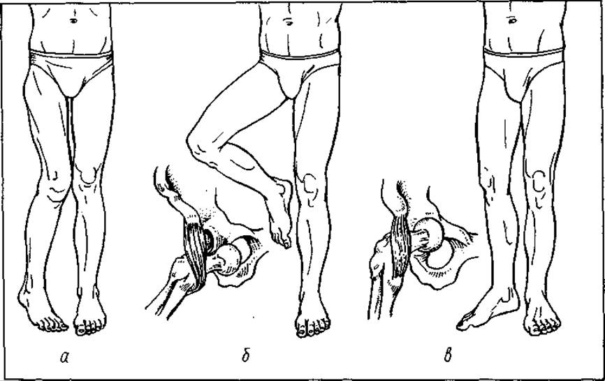
Fig. 33. Hip dislocation:
a — posterosuperior; b — anteroinferior (obturator); c — anterosuperior
Central hip dislocation. As a result of a fracture of the acetabular floor, the head is displaced into the pelvic cavity, depending on the magnitude of the traumatic force. Central dislocation most often arises from a direct force acting along the axis of the femoral neck and head. It is caused much less frequently by the indirect action of a traumatic factor.
Clinical manifestations of central dislocation: a slightly flexed, adducted, and moderately externally rotated thigh, slight relative shortening of the limb, limitation of flexion and extension in the hip joint, and the inability to perform abduction and rotational movements. Relative to the biomechanical axis of the lower limb, the axis of the thigh is displaced slightly inward, the contours of the greater trochanter are not defined, and the outer surface of the hip joint is less oval. Rectal examination reveals tenderness and a dome-shaped protrusion of the pelvic wall.
Clinical examination is followed by X-ray Diagnostics, after which a definitive diagnosis is established.
Complicated hip dislocations are those accompanied by a simultaneous fracture of the posterior rim of the acetabulum. The symptoms are identical to those of an uncomplicated posterior dislocation. To clarify the diagnosis, anamnesis data are utilized (the patient received an impact along the axis of the flexed thigh in the knee area, which is typical for this injury).
DISLOCATION OF THE LEG (KNEE DISLOCATION)
Dislocation of the leg is a rare injury, accounting for 1.5–2% of all dislocations. It occurs more frequently due to indirect force, when one of the articular ends (usually the femur) is stably fixed, while the other (the tibial end) is displaced forward, backward, or to the side. Dislocation of the leg occurs under the condition of a complete rupture of the cruciate ligaments and the joint capsule. In the case of a subluxation, one of them is ruptured (anterior or posterior, depending on the direction of the mechanical force). Anterior dislocation of the leg is characterized by a bayonet-like deformity of the knee joint. The leg is fixed in an extended position and appears shorter. The proximal articular end of the leg protrudes over the femur beneath overstretched skin. In a purely anterior dislocation, it is located along the axis of the lower limb; in anterolateral and lateral dislocations, it is displaced outwardly or inwardly. Hemarthrosis (bleeding into the joint) is observed.
In posterior dislocation, the articular end of the leg is displaced backward, forming a step-like deformity. On the anterior surface beneath the skin, the contours of the femoral condyles protrude prominently. While the Patella is mobile and its ligament is not taut in anterior dislocation, in posterior leg dislocation it is pressed against the femoral condyles, and its proper ligament is tense. The axis of the leg is displaced backward in the sagittal plane, whereas in anterior dislocation it is shifted forward. During a leg dislocation, especially a posterior one, the popliteal artery is traumatized, which manifests as foot swelling, cyanosis or skin pallor, sensory impairment, decreased temperature, and the absence of a pulse on the dorsal surface of the foot.
Traumatic dislocation of the patella (luxatio patellae) is rare (0.4–0.7%). Its diagnosis is straightforward. As a result of direct trauma, the patella is displaced laterally, less frequently medially. Hemarthrosis of the knee joint is observed. The leg is slightly flexed at the knee joint, which is deformed. The patella is palpable on the lateral surface of the knee joint. The direction of its proper ligament shifts in accordance with the dislocated patella. Patellar dislocation frequently occurs with minor trauma, which is explained by Dysplasia of one of the femoral condyles.
Isolated traumatic dislocations of the ankle joint do not occur. They always happen in combination with malleolar fractures and rupture of the distal tibiofibular syndesmosis.
Traumatic dislocation of the foot bones is likewise not an isolated injury. Such injuries include subastragalar (subtalar) dislocation of the foot (luxatio subtalo), dislocation at the Lisfranc joint, and isolated dislocations of the talus and navicular bones.
Vertebral dislocations are most frequently observed in the cervical spine (Fig. 34). They typically result from excessive flexion or, less commonly, extension of the head. This is accompanied by tears in the ligaments of the zygapophyseal joints. In such cases, the articular surfaces of the upper vertebra displace anteriorly over those of the lower vertebra; that is, one articular surface overrides the other, resulting in a bilateral vertebral dislocation.
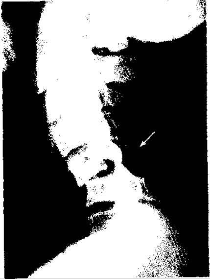
Fig. 34. Subluxation of the 5th cervical vertebra
Unilateral dislocations occur when an element of rotation is added to the flexion of the head.
Significantly more often, vertebral dislocations are accompanied by fractures of the articular processes, vertebral arches, or vertebral body.
In bilateral flexion dislocations of a vertebra, the head is tilted forward, with the chin almost touching the chest. Severe neck pain arises, which intensifies sharply at the slightest attempt to move the head, prompting the patient to support the head with their hands. The Neck Muscles are tense.
The spinous process of the dislocated vertebra appears more recessed, while the adjacent spinous process protrudes prominently beneath the skin above it. In unilateral vertebral dislocation, the head is turned away from the side of the dislocation, and the spinous process deviates toward the dislocation. A definitive diagnosis is established following a radiographic examination.

Fig. 35. Reduction of a shoulder dislocation using Kocher's method: a — Stage I; b — Stage II; c — Stage III; d — Stage IV
Treatment of traumatic dislocation. Since ancient times, it was believed that a dislocation that occurred before sunset should be reduced before sunset, and conversely, if it occurred before sunrise, it should be reduced before sunrise. To this day, the fundamental and urgent principle in the treatment of traumatic dislocations remains immediate reduction.
A dislocation must be reduced under anesthesia that achieves complete muscle relaxation. Only then will the reduction cause no additional trauma. Following reduction, immobilization of the injured extremity is mandatory (for at least 2–3 weeks) until the torn joint capsule heals.
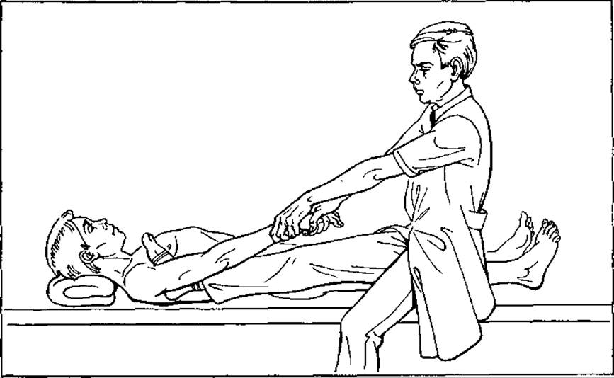
Fig. 36. Reduction of a shoulder dislocation using Hippocrates' method
Shoulder dislocations are reduced using the methods of Kocher, Hippocrates, Janelidze, Mota, and others. Anterior dislocations are best reduced using Kocher's method. Depending on the type of anesthesia, the reduction is performed with the patient lying supine or sitting. An assistant fixes the scapula to the table (or to the back of a chair if the patient is sitting). The surgeon grasps the patient's injured limb just above the elbow with one hand and the forearm with the other, bends the elbow to a right angle, and systematically and gently performs the following maneuvers (Fig. 35):
Stage I — smoothly and with progressively increasing force, the surgeon applies axial traction to the humerus downward, overcoming muscle contraction;
Stage II — externally rotates the humerus. In this position, the humeral head has its smallest diameter, and the angle between the head and the diaphysis is leveled out. This prevents the head from further traumatizing the muscles as it shifts toward the glenoid cavity;
Stage III — without reducing the axial traction on the humerus, the surgeon directs the humerus toward the body's midline so that it rests against the chest wall at the junction of the lower and middle thirds. The humerus thus acts as a two-armed lever. The upper and middle thirds form the long arm of the lever, while the lower third forms the short arm. Maintaining axial traction, the surgeon then presses on the outer surface of the elbow joint (the short arm) from top to bottom, generating a force at the end of the long arm that allows the head of the humerus to be brought to the level of the glenoid cavity of the scapula;
Stage IV — upon feeling that the humeral head has shifted and seeing that the contour of the shoulder joint has filled out, the surgeon executes a brisk internal Rotation of the humerus and, in a pronated position, places the hand and forearm across the chest at an acute angle. At this moment, the humeral head snaps back into place, accompanied by a characteristic click. As soon as the head is reduced, the contour of the shoulder joint immediately assumes an oval shape. Immobilization is performed using a Desault bandage, additionally secured with plaster bandages (for at least 3 weeks, until the joint capsule heals). Treatment without immobilization or its premature removal leads to a severe complication — recurrent shoulder dislocation.
Reduction of a dislocation using Hippocrates' method (Fig. 36) is also referred to as the military field method. The patient lies supine. The surgeon sits on the side of the dislocation, facing the patient, and grasps the injured arm with both hands by the forearm just above the wrist joint. Then, the midfoot of the foot corresponding to the dislocated arm (rather than the heel) is inserted as deeply as possible into the axillary fossa so that the instep covers it. In this position, the outer edge of the midfoot rests against the lateral chest wall, while the inner edge rests against the Medial surface of the upper third of the arm. This creates a two-armed lever, where the short arm consists of the humeral head and the upper third of the arm, and the long arm consists of the middle and lower thirds of the arm. The surgeon begins to gradually and smoothly increase the force of axial traction along the arm while adducting it toward the trunk. Operating on the lever principle, the head is thus gradually guided to the level of the glenoid cavity of the scapula and returns to its normal position. The shoulder joint acquires its usual shape, and passive movements become free and unrestricted. All of this indicates that the dislocation has been successfully reduced. Immobilization is then carried out using a Desault bandage.
Janelidze's method is an effective technique for reducing shoulder dislocations. The patient is placed on the table on the injured side so that the scapula does not extend beyond the edge of the table but is securely fixed to it. The patient's head is supported by an assistant or placed on an auxiliary stand. The arm must hang freely between the table and the stand. A mandatory condition is the fixation of the scapula to the table. Only under this condition can relaxation of the shoulder girdle muscles be achieved within 10–15 minutes. Once assured that the muscles have relaxed, the surgeon flexes the forearm at the elbow joint to a 90° angle and progressively, with increasing force, presses downward on the upper third of the forearm while making slight rotational movements. At this point, the reduction of the head takes place.
Reduction of posterior forearm dislocation. Posterior dislocations of the forearm are reduced using two methods: hyperextension and flexion. Anesthesia and muscle relaxation are mandatory.
Hyperextension method. An assistant grasps the patient's arm at the lower third, fixing it, while the surgeon hyperextends the forearm at the elbow joint. This maneuver achieves the disengagement of the coronoid process from the olecranon fossa of the humeral epiphysis. Axial traction is then applied along the arm until the olecranon approaches the level of the olecranon fossa. Without releasing the traction, the forearm is flexed to an acute angle. The disappearance of springy resistance and free passive movements in the elbow joint indicate that the forearm dislocation has been successfully reduced.
Reduction by flexion. An assistant grasps the forearm at the lower third and smoothly pulls it lengthwise with increasing force. The surgeon grasps the lower third of the arm so that both thumbs rest against the olecranon. Without reducing the traction force, the assistant gradually flexes the forearm, while the surgeon simultaneously guides the olecranon forward and pulls the distal end of the humerus backward. Reduction of the dislocation occurs, accompanied by an audible click. Immobilization of the elbow joint is performed using a posterior plaster splint extending from the upper third of the arm to the metacarpal heads, with the forearm flexed at an acute angle (up to 70–75°) for 3 weeks.
In posterolateral dislocation of the forearm, the lateral displacement must be corrected first. To do this (with the arm fixed), the surgeon shifts the forearm toward the axis of the limb, thereby converting the posterolateral dislocation into a posterior one, and then reduces it using the technique described above.
In anterior dislocation of the forearm, an assistant fixes the arm with both hands, grasping its lower third. Without extending the forearm, the surgeon applies axial traction downwards and, maintaining the traction, further flexes the forearm while smoothly pushing it backward. At the same time, the assistant pushes the distal third of the arm forward. Under these conditions, the dislocation is successfully reduced.
Reduction of hip dislocations. Given that powerful muscles attach to the femur, reduction must be performed under conditions of complete muscle relaxation.
Complete muscle relaxation can be achieved using general anesthesia or spinal anesthesia. The methods of Janelidze and Kocher are most commonly used.
Kocher's method (Fig. 37, a). The patient lies on their back. The assistant presses on the iliac crests to fix the pelvis to the table.

Fig. 37. Reduction of a hip dislocation using Kocher's method (a) and Janelidze's method (b)
The surgeon flexes the knee and hip to a 90° angle. Then, applying increasing force, pulls the femur upward while slightly abducting and externally rotating it. Once the femoral head is brought to the level of the acetabulum, the dislocation is reduced with energetic rotational movements and abduction, accompanied by a characteristic click. As soon as the dislocation is reduced, passive movements in the hip joint are fully restored, and the tip of the greater trochanter lies on the Roser–Nelaton line. After reduction, the limb is extended and immobilized (with a hip spica cast), or skeletal traction is applied to the lower third of the femur for 3 weeks.
Patients with traumatic hip dislocation must be monitored by a specialist for up to 2 years, because in more than 80% of cases, a severe complication occurs—aseptic Necrosis of the femoral head—which leads to disability.
Janelidze's method (Fig. 37, b). The patient is placed prone on the table so that the injured limb hangs freely, while the pelvis remains fully on the table. The patient lies in this position for 15–20 minutes. Under the weight of the limb, the muscles around the hip joint relax.
Having ensured that the muscles are relaxed, the assistant presses down on the pelvis toward the table to fix it. The surgeon stands between the table and the hanging limb of the patient, flexes the knee joint to a right angle, slightly abducts, and externally rotates the femur. Then, placing their knee against the posterior surface of the upper third of the lower leg, the surgeon pulls the femur downward along its axis until the femoral head reaches the level of the acetabulum. Afterward, without releasing the traction force, the surgeon performs rotational Movements of the femur, at which point the head slips into place. Passive movements become free, and the tip of the greater trochanter is positioned along the Roser–Nelaton line.
Dislocations of the lower leg and patella, if acute, are not difficult to reduce. Under deep anesthesia, with complete muscle relaxation, the assistant holds the lower third of the femur with both hands, while the surgeon applies axial traction to the lower leg. When the articular end of the lower leg reaches the level of the joint space, in cases of anterior dislocation, the articular end of the lower leg is displaced backward, and in cases of posterior dislocation, forward. The articular surfaces are easily aligned. Immobilization is performed using a long leg cast for 3 weeks.
Vertebral dislocations, if not neglected, are reduced by a closed, single-stage method. The assistant fixes the shoulders to the table. The surgeon smoothly and with increasing force pulls the patient's head, simultaneously performing slight rotational movements of the head, and tilts the head backward. The reduction of the vertebra is accompanied by a characteristic click. For long-standing dislocations, skeletal traction using Crutchfield tongs is effective. Two weeks after reduction, without removing the skeletal traction, a thoracolumbar cast is applied, or plastic orthoses are used for 1.5–2 months.
Treatment of neglected dislocations is surgical, meaning that open reduction of the dislocation or joint arthroplasty is performed.
PATHOLOGICAL DISLOCATIONS
Pathological subluxations and dislocations occur as a result of inflammatory, degenerative-dystrophic, and neurotrophic joint disorders, as well as (much less frequently) malignant and benign tumors.
In acute inflammatory processes, when a large amount of inflammatory fluid accumulates in the joint cavity (synovitis), intra-articular pressure rises so significantly—especially in the hip joint—that it causes rupture of the joint capsule and the ligamentum teres, pushing the femoral head out of the acetabulum. This is facilitated by stretched Muscles of the hip joint, which contract and displace the femoral head outward and upward. Such dislocations or subluxations are termed tangential.
Pathological dislocations or subluxations frequently occur as a result of the destruction of the articular ends of bones. This is observed in tuberculosis, osteomyelitis, aseptic necrosis, cystic restructuring, and arthropathies (syphilitic, syringomyelic, etc.).
Treatment of pathological dislocations is comprehensive. First and foremost, the underlying disease must be treated to halt the progression and eliminate (or achieve remission of) the pathological process in the joint. Only after this can reconstructive and restorative joint surgeries be performed. If inflammatory fluid rapidly accumulates in the joint during general treatment, joint puncture with aspiration of the inflammatory fluid should be performed in a timely manner. This helps reduce pressure within the joint cavity and pain response, and serves as a prophylaxis against tangential dislocation.
Specific therapy is administered depending on the Nature of the inflammatory process.
Congenital dislocations and subluxations result from exogenic and endogenic factors that cause developmental disorders and abnormal formation of all joint structures. Treatment is carried out from the first days of a child's life using orthopedic methods.
BONE FRACTURES
Among injuries to The Musculoskeletal System, bone fractures constitute one of the most severe types of trauma, accompanied by prolonged loss of working capacity and frequently leading to disability.
What does the term "bone fracture" mean? A fracture is defined as a complete disruption of bone integrity accompanied by the loss of its static and dynamic Functions. However, there are cases where bone integrity is not completely disrupted and function is partially preserved. Such injuries are referred to as fissures or cracks.
A bone fracture, fissure, or crack typically occurs as a result of a sudden, strong impact of external physical factors. If a fracture occurs at the site of the direct application of physical force, this mechanism is called direct; if the traumatic factor acts at a distance, the fracture mechanism is called indirect (excessive bending, extension, or twisting force applied to the bone).
In addition to traumatic fractures, fractures frequently occur as a result of bone Damage caused by various pathological processes, namely: osteomyelitis, benign or malignant tumors, metastases, dysplasia, and others. The bone is destroyed gradually, and a fracture occurs under the slightest stress, movement, or even during Sleep. Such fractures are called pathological.
Traumatic and pathological fractures are, in most cases, monofocal, meaning they occur in a single site. In cases of Polytrauma, tumor metastases, or tumors, fractures occur in two (bifocal), three, or more sites (polyfocal), and are localized in different areas and at various levels of the bone. Considering the Water/140.html">Anatomical structure of the bone, diaphyseal, metaphyseal, and epiphyseal fractures are distinguished. The most vulnerable zone of a tubular bone is the metaphysis. This is due to several reasons: first, the metaphysis serves as a transitional zone between the diaphysis and epiphysis, consisting of spongy tissue that lacks the dense cortical layer found in the diaphysis; second, although epiphyses share a structure similar to metaphysis, they are stronger due to a greater mass and a higher number of bone trabeculae. Furthermore, they receive additional reinforcement from the joint capsule. As for the diaphysis, in addition to its natural strength, it has extra protection provided by the muscles. Muscles not only cushion and absorb impacts, but through their contraction, they also present a significant barrier to traumatic forces.
In childhood, the metaphysis contains the growth plate, which is easily torn during trauma, resulting in partial or complete displacement of the epiphysis. Such traumatic injuries are referred to as epiphyseolysis (complete or partial). If a bone fragment breaks off simultaneously with the rupture of the growth zone, the injury is termed osteoepiphyseolysis.
Epiphysealis fractures are intra-articular in the vast majority of cases. In children, Bone tissue is more elastic and resilient, and the periosteum is significantly thicker and stronger; therefore, fractures with fragment displacement occur less frequently, while greenstick fractures are observed more often (Fig. 38).
Bone fractures may occur without displacement, or with minor or complete displacement of the fragments. Fragment displacement can take place: in a single plane—frontal, sagittal, or vertical; in two planes—frontal and sagittal; and in three planes—frontal, sagittal, and vertical. Depending on the nature of the displacement, the following are distinguished: displacement of fragments in width—ad latus, in length—ad longitudinem, at an angle—ad axim, and peripherally—ad periferiam (Fig. 39).
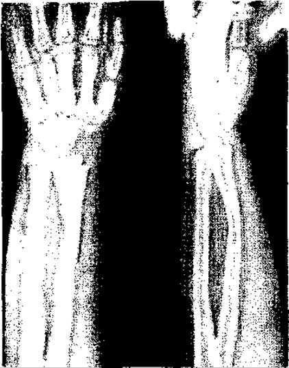
Fig. 38. Greenstick fracture of the radius
The direction of fragment displacement primarily depends on the strength and vector of the traumatic force, as well as on muscle biomechanics. In a direct fracture mechanism, when the traumatic force acts in an anteroposterior direction, the distal fragment is displaced posteriorly in the sagittal plane; conversely, if the force acts from back to front, the displacement is anterior. If the impact is directed at an angle from the outside forward, the distal fragment is displaced medially and anteriorly. When a limb segment is subjected to multiple forces involving an element of inward twisting (indirect mechanism), the distal fragment is displaced in length and width, and undergoes internal rotation. Such displacement is termed primary displacement. After the traumatic force subsides, the fragments are acted upon by the muscles attached to them. As a result of the primary displacement, the physiological balance of the muscles is disrupted, leading to their contraction and elastic retraction, whereby the evolutionarily stronger muscle group causes secondary displacement of the fragments.

Fig. 39. Displacement of long bone fragments in a fracture: a — in length; b — in width; c — impacted fracture; d — at an angle; e — peripherally
Thus, the degree of fragment displacement depends on the direction of the traumatic force and the muscle contraction force. Elastic retraction and reflex traumatic muscle hypertonus become persistent, resulting in the so-called muscular contracture of the fragments.
Based on the fact that fragment displacement is generally multi-planar, X-ray examinations should be performed in two projections. This allows for an objective Assessment of the fragments' alignment. Typical displacements occur depending on the fracture site and the attachment of muscles to the fragments. For instance, in a fracture of the distal metaphysis of the radius, the distal end is displaced upward (dorsally), while the proximal end is displaced downward in a volar direction, giving the bone a bayonet or dinner-fork shape. In fractures of the olecranon or the distal metaphysis of the humerus, the contraction of the triceps brachii muscle causes the proximal fragment to displace upward (Fig. 40). The proximal fragment of the patella is displaced proximally during a fracture due to the action of the quadriceps femoris muscle.
For a fracture of the lower third of the femur, posterior displacement of the distal fragment due to the contraction of m. gastrocnemius is typical. In a fracture of the upper third of the femur, the proximal fragment is displaced outward (retraction of the gluteal muscles) and forward (retraction of m. iliopsoas), while the distal fragment is displaced medially and upward (retraction of the adductor muscles). This results in a typical "jodhpurs" deformity. To select the correct tactics and treatment method, one must know not only the fracture mechanism and the type of fragment displacement, but also the nature of the fracture itself, namely the specific direction and surface planes of the fracture. According to the configuration of the fracture planes, diaphyseal, metaphyseal, and epiphyseal fractures are classified into transverse, transverse-oblique, oblique, spiral, comminuted (Fig. 41), crushed, and double fractures.
A transverse fracture occurs as a result of a sudden direct mechanical impact (a jolt or blow) perpendicular to the long axis of the bone. When the traumatic force acts at a slight angle, a transverse-oblique fracture occurs. If significant direct traumatic force acts over a prolonged period, a comminuted or crushed fracture develops.
If the traumatic force acts on a bend (moderate flexion or extension) while one end of the limb segment is fixed, or if multiple forces act simultaneously, an oblique fracture occurs. If an element of segmental torsion is also added, a spiral fracture results.
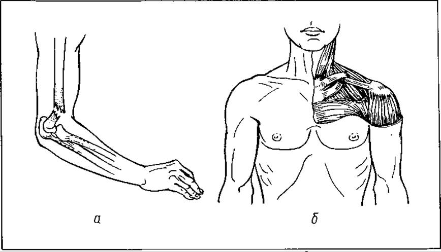
Fig. 40. Typical displacement in a fracture of the humerus (a) and clavicle (b)
If force is applied along the axis of the limb segment or spine, compression and impacted fractures occur.
Thus, the nature of a fracture resulting from direct or indirect mechanisms of traumatic force can be highly diverse and depends on the magnitude, shape, and duration of the traumatic impact, the Anatomical Features of the fracture site, and the biomechanics of the muscles and JOINTS OF THE injured limb.
Reparative regeneration of bone tissue. The outcomes of bone fracture treatment depend on the course of bone healing (reparative regeneration). Since ancient times, humanity has sought to understand this process and pursued methods to control reparative regeneration—the mechanism of bone fusion. The humoral theory (Hippocrates) was eventually succeeded by the cellular theory (Virchow). Specific Changes in the bone injury zone are investigated, and The Significance of various cellular and tissue elements, as well as bone tissue regenerate, is studied. The Role of the periosteum, Bone Marrow, endosteum, surrounding Connective Tissue, blood vessels, and the nervous and endocrine systems in fracture healing is being elucidated. Through meticulous analytical research, the concept was developed that all cellular elements of mesenchymal origin participate in bone regeneration, with the degree of their participation directly depending on the plastic capabilities of various Cells and environmental conditions. Researchers have examined how reparative regeneration depends on blood supply conditions, the state of the nervous and endocrine systems, and Homeostasis.
Scientists have studied the Dynamics of Biochemical changes during bone healing, the metabolism of Trace Elements (especially Calcium and phosphorus), and shifts in acid-base balance, Phosphatases, and other parameters. The Importance of stable fragment alignment throughout the entire healing period and the role of early functional restoration have been substantiated. In the closing decades of the 20th century, The Study of reparative regeneration processes advanced to the level of molecular biology and Electron Cell/15.html">Microscopy. These investigations have clarified the course of biological reactions. Numerous comprehensive studies have demonstrated that reparative regeneration and regenerate formation are staged processes that directly depend on the General condition of the body and local alterations in tissue metabolism. Three types of reparative regeneration are distinguished: desmogenous, chondrogenous, and angiogenous, all of which are characterized by a staged progression.

Fig. 41. Comminuted bone fracture with foot dislocation
The First stage of reparative regeneration is the stage of tissue structure Catabolism, dedifferentiation, and proliferation of bone elements. It begins at the moment of trauma. As a result of mechanical traumatic force, a bone fracture occurs. The trauma damages not only the bone tissue, but also the surrounding soft tissues, blood vessels, and nerve branches, resulting in hemorrhage (hematoma). The severity of the trauma is directly related to the magnitude and duration of the traumatic factor. Within the first minutes, the hematoma becomes filled with fragments of adjacent soft tissues, periosteum, bone marrow, endosteum, various cells and their constituent elements, fragments of nuclear membranes, nuclear and cytoplasmic contents, DNA, nuclear RNA fractions, lysosomal Enzymes, blood components, and other biological substances.
The epicenter of the injury is surrounded by a layer of soft tissues whose cells are in a state of parabiosis. The Fate of this paranecrotic layer depends on the severity of the paranecrotic process and The rate of microcirculatory bed restoration. Beyond the paranecrotic layer lie soft tissues with undamaged vessels and nerves that ensure normal metabolism. Trauma induces general and local protective, adaptive, specific, and non-specific neuro-reflex and humoral reactions within the body. In the fracture area—specifically within the hematoma, which transforms into a heterogeneous mass due to infiltration by tissue fragments, cellular elements, and Blood Cells—anaerobic processes (Glycolysis) develop. This leads to The formation of organic acids (pyruvates, lactates, etc.) and an increase in osmotic pressure, causing acidosis initially due to a depletion of reserve bases and later resulting from an increasing concentration of hydrogen ions. Leukocyte and protein emigration increases, a significant amount of degraded acidic muco- and Glycoproteins accumulates, and Collagen undergoes Denaturation, thereby increasing the hydrogen ion concentration. Collagen denaturation is also driven by proteases (Trypsin, fibrinolysin, Chymotrypsin, cathepsin, etc.). Tissue Water Metabolism is disrupted, cells lose potassium, and hyperkalemia develops at the injury site. Vascular stasis is observed along with fibrin precipitation, which has been mistakenly credited with The ability to transform into collagen-like fibers and form bone. It is now proven that collagen fibers in the body are formed exclusively through synthesis by connective tissue cells. Disintegration of the intercellular substance occurs in the injury zone, disrupting physical and mechanical bonds with collagen fibers, which undergo dismantling, breakdown, and degradation under the influence of proteases. Within the heterogeneous mass (hematoma), various chemical compounds of Polypeptides, oligopeptides, Amino Acids, and nitrogenous bases arise, which subsequently form histamine, bradykinin, serotonin, and acetylcholine (so-called tissue Hormones) through decarboxylation.
In catabolism, the Breakdown of Proteins, fats, and CARBOHYDRATES is accelerated, the secretion of glucocorticoids and thyroxine is intensified, Vitamin C reserves are depleted, and Mineral Metabolism is disrupted. A negative nitrogen balance occurs due to the combustion of free blood proteins as early as the first days following a fracture, accompanied by hypocreatininemia, dysproteinemia, and a surge in transaminase activity.
Energy Metabolism and the rhythm of biochemical processes are disrupted. The amount of ATP—the primary energy product—drops several-fold (down to 3). Calcium and phosphorus metabolism in both bone and Blood Plasma is impaired, resulting in a significant loss of calcium and phosphorus from the bone, not only at the fracture fragments but across adjacent skeletal segments.
Intact osteogenic and non-osteogenic cells in the interphase are stimulated by signals from the nervous and humoral systems, as well as directly by BIOLOGICALLY ACTIVE SUBSTANCES generated within the heterogeneous mass through catabolism. These biologically active substances (hormones) act not only as stimuli for interoceptors, but also as direct chemical Inducers of interphase cells regarding proliferation and dedifferentiation into young pluripotent cells—polyblasts.
Thus, on the one hand, these so-called tissue hormones play a crucial role in developing aseptic inflammation and triggering proliferation processes, and on the other hand, in the dedifferentiation of osteogenic cells into polyblasts.
The Second Stage is the stage of formation and differentiation of tissue structures.
The course of the second stage depends on general and local conditions, specifically: the patient's condition, age, the alignment of the fragments, the degree and quality of their reduction, and the period required to restore capillary Circulation. During this stage, capillaries actively sprout from all sides toward the ends of the fragments, supplying adequate nutrients and oxygen to the polyblasts. These polyblasts differentiate into osteoblasts, which in turn produce intermediate osteoid tissue. In such cases, reparative regeneration occurs via direct osteogenesis. However, if the fragments are not rigidly fixed, minor mobility between them is possible, capillaries are traumatized, and polyblasts fail to receive the required amount of nutrients. Under these conditions, they differentiate into a less "demanding" cell—the chondroblast, which has lower energy demands.
Chondroblasts produce chondroid intermediate tissue, which under favorable conditions transforms into osteoid tissue via metaplasia. This is the indirect pathway of reparative regeneration, and it takes significantly longer. However, under conditions unfavorable for metaplasia, Cartilage tissue forms, hindering bone union and resulting in a pseudoarthrosis (false joint) at the fracture site.
When fragments are unaligned or have undergone secondary displacement with mobility persisting between them, continuous trauma to the sprouting capillaries occurs. Consequently, polyblasts do not receive sufficient oxygen or nutrients, causing them to differentiate into fibroblasts, followed by The Development of Fibrous connective tissue between the fragments. This newly formed tissue typically transforms into a fibrous scar. A pseudoarthrosis then forms at the fracture site.
The process of capillary sprouting from the periphery toward the center intensifies, and osteoblasts align along their path. Their metabolic activity and Protein Synthesis increase, and the fracture zone becomes filled with a protein-polysaccharide matrix into which collagen fibrils—rather than whole collagen proteins—are embedded. As the capillary network is restored, aerobic processes increase, levels of histamine, bradykinin, serotonin, and other biologically active substances decline, vascular permeability decreases, oncotic pressure equalizes, the medium becomes more alkaline, and under the action of enzymes and hormones, the mineralization of collagen fibrils progresses. The reparative process transitions into the Third Stage—the formation of an angiogenic Bone Structure (Fig. 42).
The intensive growth of the capillary network from the periphery to the center, stretching from the proximal to the distal fragment, culminates in their merging into a single Vascular System. Osteoblasts and newly formed osteoid tissue reside between the loops of this capillary plexus. The angiogenic bone structure is well-supplied with oxygen and nutrients, supporting an intense metabolism under aerobic conditions. This angiogenic structure anchors the fragments, which begin to experience axial mechanical loads (physiological pressure) from muscles. Reparative regeneration then moves into the Fourth Stage—the formation of lamellar bone structure, periosteum, endosteum, cortical layer, bone structure in the epiphyses and metaphyses in accordance with biomechanical loads, and the bone marrow. The course of reparative regeneration directly depends on the patient's general physiological state and the local conditions at the fracture site. What specific general conditions can delay or disrupt this physiological process of reparative regeneration?
First, it is the environment in which the patient lives. For instance, residing in high-altitude regions with low partial pressure of oxygen, living through the polar night, experiencing starvation (chronic malnutrition, avitaminosis), or living in ecologically unfavorable areas; second, the patient's overall health status (presence of acute or chronic diseases, convalescence period, severity or multiplicity of trauma, or combined/polytrauma injuries).
Local factors that delay or disrupt reparative regeneration include the extent of damage to surrounding soft tissues, blood vessels, and nerves; incomplete or unstable fragment reduction; secondary displacement; unjustifiably frequent changes in treatment methods; unstable osteosynthesis; and premature static and dynamic loading of the angiogenic bone callus.
Delays and disruptions in reparative regeneration caused by unfavorable general and local factors can be observed at every stage. Homeostatic imbalance leads to suppressed catabolism, impaired proliferation and dedifferentiation, and hindered capillary sprouting, which in turn delays the differentiation of osteogenic cells and the formation of osteoid tissue. The lack of stable reduction or rigid osteosynthesis causes fragment mobility. Their sharp edges traumatize sprouting capillaries, meaning polyblasts fail to receive vital nutrients and oxygen, causing them to differentiate into chondroblasts or fibroblasts instead of osteoblasts. Energy metabolism in chondroblasts and fibroblasts is considerably lower than in osteoblasts, and they form chondroid or scar tissue between the fragments, blocking bone union and leading to pseudoarthrosis. Experimental research and clinical observations have demonstrated that early, excessive static and dynamic loading on the angiogenic bone callus damages the capillary network, impairs Blood supply to osteoblasts, and leads to pseudoarthrosis formation at sites of high mechanical stress concentration.

Fig. 42. Radiographs of a femoral shaft fracture before treatment (a) and 4 months post-treatment (b). A well-defined bone callus is visible.
Thus, knowledge and understanding of the staged nature of bone reparative regeneration provide the key to selecting appropriate treatment tactics and methods.
Based on etiological factors, bone fractures are classified into traumatic and pathological (Fig. 43). They can be closed or open. Closed fractures are those where the fracture zone does not communicate with the external environment (skin or mucous membranes remain intact). Open fractures involve at least a minor breach of skin integrity that connects the fracture site to the external environment. According to anatomical localization, fractures can be diaphyseal, metaphyseal, or epiphyseal. Epiphyseal and epimetaphyseal fractures are further divided into intra-articular and extra-articular. Depending on the orientation of the fracture line, they are classified as transverse, transverse-oblique, oblique, spiral, T-shaped, Y-shaped, or comminuted fractures.

Fig. 43. Pathological fracture of the radius due to hyperparathyroidism (a) and of the Tibia due to Paget's Disease (b).
Fracture-dislocations constitute a separate group. They are most commonly observed in injuries to the spine, the proximal humerus and femur, the forearm (Monteggia, Galeazzi injuries), and the hand (Bonnet injury).
Among traumatic fractures, avulsion fractures of apophyses—such as the greater and lesser trochanters, the tibial tuberosity, and epicondyles—deserve special mention. They are particularly frequent in children whose growth plates have not yet fused.
Basic principles of bone fracture treatment. A key feature of treating patients with bone fractures is urgency and promptness, paired with a strictly individualized approach depending on age, general health status, and injury severity. First aid is most effective and comprehensive when provided not by standard ambulance crews, but by specialized trauma teams. In such cases, depending on the patient's condition, timely and comprehensive care is administered, significantly reducing mortality rates and severe complications. THE PRINCIPLE OF transport immobilization must always be strictly observed. Joints above and below the fracture site must be immobilized. For femoral fractures, the Dietrichs splint is used, which immobilizes the foot, lower leg, knee, and hip joints, ensuring complete rest for the injured lower limb. This allows the patient to be transported to a medical facility without exacerbating pain or causing additional soft-tissue trauma around the fracture. Upper extremities are immobilized using a Kramer splint.
At the trauma or surgical department, the patient undergoes a comprehensive examination to determine the precise type, nature of the fracture, and degree of fragment displacement, taking into account the configuration of the fracture line. Afterward, treatment tactics and methods are justified based on the patient's general condition.
Basic principles of fracture treatment:
1) achieve fragment reduction in the shortest possible time;
2) alignment of the fragments is performed according to the following principle: the distal fragment is aligned with the proximal one;
3) the alignment of fragments must be stable throughout the entire consolidation period, meaning the aligned bone fragments must be securely fixed;
4) early restoration of the functional capacity of the injured limb.
Based on these principles, treatment tactics and methods are developed.
Both conservative and Surgical methods are used to treat bone fractures. These methods do not compete with each other; rather, they Complement one another and are applied according to clearly defined indications.
Conservative treatment methods include:
1) plaster immobilization;
2) closed reduction of fragments combined with limb immobilization using plaster casts;
3) skeletal traction;
4) external compression-distraction osteosynthesis using Kirschner wires and pin fixation devices.
Plaster immobilization of the injured limb is indicated for fractures without fragment displacement, cracks, infractions, and greenstick fractures in children. Single-stage reduction of fragments is effective in fractures with transverse, serrated-transverse, and oblique-transverse planes, where secondary displacement of bone fragments is impossible after reduction. Reduction is performed under complete anesthesia. For this purpose, 20–30 ml of a novocaine or trimecaine solution is injected directly into the hematoma at the fracture site using a syringe prior to reduction. Following anesthesia, the fragments are reduced, and a plaster cast is applied to the limb (Fig. 44). It should be noted that the plaster cast does not exert direct pressure on the fragments and does not hold them in place; it merely ensures limb immobilization and eliminates active muscle and joint function that could cause secondary displacement. If the fragments have an oblique fracture plane, then despite reduction and plaster immobilization, muscle tension (especially after the subsiding of reactive edema) may cause fragment displacement—meaning secondary displacement can occur under the plaster. Therefore, for oblique, spiral, and comminuted fractures, single-stage reduction with plaster immobilization is contraindicated. In such cases, closed reduction of fragments using skeletal traction (Fig. 45) or wire/pin fixation devices (Fig. 46) is effective.
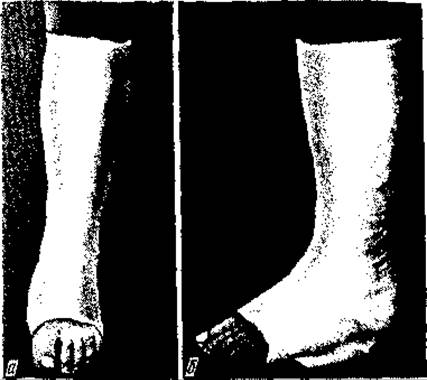
Fig. 44. Plaster immobilization for malleolar (a) and foot (b) fractures
Surgical treatment of fractures is indicated in the following cases:
1) damage to major blood vessels and nerves. For example, fractures of the lower third of the femur frequently damage the femoral or popliteal artery, while fractures of the middle third of the humerus often injure the radial nerve;
2) interposition of muscles and other soft tissues between the fragments;
3) intra-articular and juxta-articular fractures;
4) fracture-dislocations;
5) double fractures with displacement of all fragments;
6) patellar and olecranon fractures with displacement;
7) intolerance to skeletal traction or fixation devices, as well as mental disorders;
8) open Bone and joint fractures;
9) polytrauma and combined injuries.
Surgical treatment is contraindicated in the presence of pustular or inflammatory skin processes, wounds, severe general condition of the patient, or decompensation of vital organs and systems.


Fig. 45. Skeletal traction for a femoral fracture:
a — fracture diagram and fragment displacement; b, c — Braun and Braun-Böhler splints for limb positioning; d — diagram of the direction of traction force
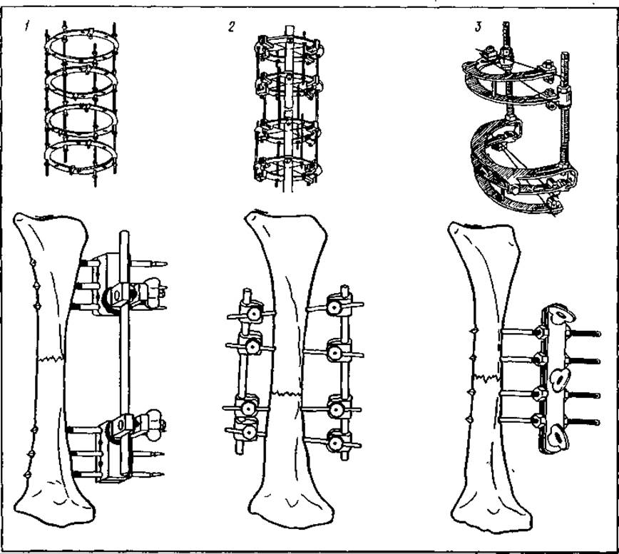
Fig. 46. Onlay fixation (osteosynthesis):
1 — Kalnberzs apparatus; 2 — Ilizarov apparatus; 3 — Sivash apparatus
The goal of surgical treatment is to restore the anatomical and functional parameters of the limb segment. Numerous methods have been proposed to achieve this. It all began because Conservative methods were insufficient to achieve fragment reduction. In such cases, open reduction was proposed. However, in most cases, the fracture line was oblique or spiral, or the fracture was comminuted, and secondary fragment displacement occurred after open reduction. Thus, clinical reality compelled physicians to adopt methods of securing fragments with osseous sutures. Surgeons began to secure fragments using bone sutures made of catgut, silk, fascial strips, etc. Nevertheless, bone sutures do not always provide stable fixation of bone fragments. Secondary displacement occurs, the healing period is significantly prolonged, and pseudoarthrosis frequently develops. All of this provided the impetus for the biomechanical substantiation of osteosynthesis methods (Fig. 47). These methods must ensure stable osteosynthesis of fragments with early functional mobilization of the injured limb. Plates (onlay fixators) proposed by Lane, Lambotte, K.M. Klimov, N.V. Novikov, S.S. Tkachenko, Polyakov, as well as screws, hooks, AO fixators, and hip joint prostheses, were introduced (Fig. 48). While fixators (plates) achieved firm, stable osteosynthesis, this required extensive surgical exposure, extensive skeletal stripping of the fragments, and, most importantly, removal of the fixation plate after fracture healing, which necessitated a second operation. Therefore, intramedullary osteosynthesis using rods (Küntscher, Dubrov, Klimov, Bogdanov, CITO, Sklyarenko-Voloshin, etc.) was proposed. However, its drawbacks became apparent over time. Firstly, the medullary canal is uneven in width; therefore, not only is an individualized rod size Selection required, but the canal must also be reamed in narrow areas. Only under these conditions can reliable fixation be achieved. Secondly, in oblique, spiral, and comminuted fractures, it is impossible to maintain the fragments in a reduced position.


Fig. 47. Intramedullary fracture fixation:
a — radius; b — malleolus; c — intertrochanteric hip fracture

Fig. 48. Hip joint replacement with a xenoprosthesis for a femoral neck fracture
In such cases, connecting plates of various designs, compression-distraction devices (Ilizarov, Kalnberzs, Tkachenko, Volkov-Oganesyan), and screws are used.
Ye.T. Sklyarenko and O.I. Voloshin developed a method for closed osteosynthesis of oblique and spiral diaphyseal fractures using screws. After fragment reduction, 3–4 pins are inserted through them, similar to skeletal traction. Next, a small incision of the soft tissues is made over the entry point of each pin, and a cannulated drill is placed over the pin. A tunnel is created in both fragments, mandatory through both cortical layers. The drill is removed from the pin, and cannulated screws are placed over the pin and driven into the tunnel, after which the pin is removed. The remaining screws are inserted in the same manner. The skin wounds are closed with 1–2 sutures and an aseptic dressing. This technique avoids trauma to adjacent soft tissues, the periosteum, and the vascular network, while providing stable fragment fixation that enables functional treatment.
Open fractures are fractures in which the fracture site communicates with the external environment.
In peacetime, open fractures account for 10–12% of all bone fractures. Any open fracture is accompanied by microbial contamination; therefore, the primary objective is the prevention of purulent complications. This prophylaxis is achieved through early primary surgical debridement of the wound (within the first 6–12 hours, or up to 24 hours post-injury if broad-spectrum antibiotics are available). The goal of primary surgical debridement is not only the prevention of purulent complications. After the removal of non-viable tissues, the open fracture is converted into a closed one and subsequently managed according to the principles of closed fracture treatment.
Osteosynthesis of open fractures should be performed only after wound healing (delayed). This helps to reduce the incidence of purulent complications.
Errors and Complications IN THE TREATMENT OF BONE FRACTURES
Errors in the treatment of bone fractures can be appropriately divided into three groups: organizational, tactical, and technical.
Organizational errors: 1) lack of Organization in the trauma service (trauma centers, departments, outpatient clinics); 2) inappropriate staffing of the trauma service (physicians, nurses, technicians); 3) insufficient equipment, instruments, apparatus, and fixators; 4) unsystematic continuing professional development for physicians.
Tactical errors: 1) unjustified choice of treatment tactics (conservative vs. operative); 2) single-stage fragment reduction without complete anesthesia and muscle relaxation; 3) application of single-stage closed reduction and a plaster cast for oblique and spiral fractures; 4) treatment of femoral neck fractures using skeletal traction; 5) early static and dynamic loading on the primary bone callus; 6) expansion of indications for surgical treatment; 7) untimely initiation of functional treatment, leading to joint stiffness and contractures; 8) diagnostic errors (untimely detected impacted fractures, interposition of soft tissues between fragments, combination of fracture and dislocation, rotational displacements); 9) overestimation of the compensatory capacity for angular and rotational displacements in pediatric fractures; 10) immobilization of an injured limb segment without capturing the joints above and below; 11) incomplete and unstable fragment reduction.
Technical errors:
1) a plaster cast applied too loosely or too tightly. In the first case, fragment instability occurs; In the second, BLOOD AND Lymph circulation is impaired, carrying the risk of ischemic contractures;
2) application of plaster casts without protective cotton-gauze padding over bony prominences of the limb, leading to pressure sores;
3) untimely "tightening" or revision of the plaster cast after the subsidence of reactive edema, resulting in secondary fragment displacement;
4) frequent changes of plaster casts, which disrupts reparative regeneration and leads to prolonged healing times or the development of pseudoarthrosis;
5) removal of casts without considering the time required for bone healing and the individual CHARACTERISTICS OF THE patient's body;
6) incorrect placement of the Kirschner wire during skeletal traction;
7) superficial insertion of the wire through the bone, leading to its cutout;
8) passing the wire through the joint capsule, which is accompanied by excruciating pain, reactive synovitis, and premature removal of skeletal traction;
9) inadequate weight selection and lack of monitoring over the dynamics of traction;
10) premature removal of skeletal traction;
11) improper surgical approach to the fracture site;
12) excessive skeletonization of bone fragments;
13) selection of fixation devices that fail to ensure fragment stability, thereby precluding the early application of functional loading;
14) use of bone sutures for the osteosynthesis of fragments.
Complications during fracture treatment: 1) pressure ulcers; 2) suppuration around the exit and entry sites of wires, pins, and the surgical wound; 3) embolism, thrombosis; 4) hypostatic Pneumonia; 5) cardiovascular decompensation; 6) delayed union; 7) development of a false joint (pseudarthrosis); 8) breakage of fixation devices; 9) ischemic contractures; 10) osteomyelitis; 11) joint stiffness.
The majority of complications are caused by violations of basic patient care requirements, failure to follow asepsis rules, insufficiently thorough patient examination, untimely response to adverse changes in the patient's condition and the fracture healing process, etc. Even in the times of Hippocrates, it was known that until "inflammation" in the fracture area (i.e., reactive edema) subsides, tight application of splint jaws (pads) is strictly prohibited, as it leads to necrosis of the limb tissues. This rule remains relevant in the treatment of fractures with circular plaster casts. As noted, a plaster cast does not hold fragments in place, but merely ensures limb immobilization. Therefore, applying a plaster cast as tightly as possible does not prevent secondary displacement of fragments, but only leads to complications such as pressure ulcers and ischemia. Tight application of a plaster cast in fresh fractures, when reactive edema is increasing, is the cause of such a severe complication as Volkmann's ischemic contracture. It frequently complicates forearm bone fractures in the upper (less frequently middle) third, as well as transcondylar and supracondylar humerus fractures. In the first case, due to significant hemorrhage in forearm bone fractures and an intact, strong deep forearm fascia, considerable hydraulic pressure is exerted on the surrounding tissues, blood vessels, and nerves, leading to their degeneration. In the second case, the fragments compress the artery in the cubital fossa region, resulting in ischemia, tissue and nerve degeneration, and the development of Volkmann's contracture. The clinical symptomatology of developing ischemia is quite typical: acute, burning pain, edema of the hand and fingers. Cyanosis of the distal extremity gradually diminishes, paresthesia in the fingers appears, along with numbness, decreased finger sensitivity, and limited movement. The rate of development of these symptoms directly depends on the progression speed of ischemia and edema. If the patient does not receive qualified care at this stage, an irreversible stage of contracture ensues. The pain gradually subsides, the skin becomes pale, subepidermal blisters appear, sensitivity becomes dull and eventually disappears, and finger movement becomes impossible as the fingers assume a semi-flexed position.
Thus, during limb immobilization with circular plaster casts or deep splints, constant monitoring and timely loosening are required upon the appearance of ischemia or local compression symptoms. If ischemia progresses due to significant subfascial hemorrhages, the deep Fascia of the forearm must be incised to prevent pressure buildup and, consequently, ischemia. If symptoms of ischemia appear in supracondylar fractures of the humerus, the compression of soft tissues by the plaster cast should be eliminated, the flexion angle of the forearm reduced, and thus optimal conditions created for the Blood vessels of the cubital fossa region. Volkmann's contracture causes severe disability due to profound degeneration and aseptic necrosis of the forearm muscles. The effectiveness of reconstructive and restorative surgical interventions depends on the extent and Location of muscle and nerve damage. Full functional recovery is impossible. Therefore, it is critically important to prevent this severe complication. A frequent complication resulting from a breach of fracture treatment principles is the formation of a false joint (pseudarthrosis) (Fig. 49). Pseudarthroses primarily arise due to violations of fracture treatment principles and the occurrence of complications. The first cause is improper reduction of fragments without ensuring stability throughout the entire healing period. The second cause is infection, which not only promotes the formation of a false joint but also leads to such a severe complication as osteomyelitis. The third cause is instability of osteosynthesis, and the fourth is early (at the stage of angiogenic callus) static and dynamic loading on the injured limb.
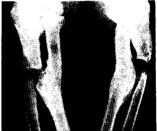
Fig. 49. Pseudarthrosis of the forearm
The Clinical symptoms of delayed union and pseudarthrosis are identical. These include pain during movement and static loading, and abnormal mobility at the fracture site. However, the patient's history must be taken into account. A pseudarthrosis forms over 6–12 months, whereas delayed union can be diagnosed 1.5–3 months after the fracture. Radiographic findings remain the primary differential criterion. In delayed union, the medullary canal in the fragments is always open, whereas in pseudarthrosis, it is always closed off by bone plates. Depending on their characteristics, pseudarthroses are classified into hypertrophied (with thickened fragment ends) and atrophic types. The ends of the fragments are osteoporotic and icicle-like in thickness. The type of pseudarthrosis determines the management strategy and treatment method.
Hypertrophic fragment ends in pseudarthrosis (unlike atrophic ones) unite well in the event of unstable osteosynthesis and opening of the medullary canals, and their resection is not required. The ends of atrophic pseudarthroses lack medullary canals; therefore, they must be resected and the canals opened, after which stable osteosynthesis and bone grafting should be performed. While apparatuses are used to treat atrophic pseudarthroses, distraction methods are applied for hypertrophic pseudarthroses, especially when there is limb segment shortening. Distraction methods are contraindicated in atrophic pseudarthroses, with compression being the method of choice. The treatment of pseudarthroses, much like bone fractures, must be comprehensive, taking into account the patient's general condition, age, profession, and living conditions.
Last update: 08/08/2026
Editorial and Educational Adaptation: This material has been compiled based on the primary/original source text. The project team performed an editorial review, corrected technical inaccuracies, structured sections, and adapted the content for an educational format.
What was processed:
- elimination of formatting defects (OCR errors, structural breaks, corrupted characters);
- editorial organization of content;
- standardization of terminology in accordance with academic sources;
- verification of factual statements against the original source text.
All mentions of the author, publication year, and origin of the primary text have been preserved in accordance with the source.