Biological Membranes - A. N. Ogurtsov 2012
Structure and Functions of Biomembranes
Membrane Lipids
Lateral Mobility in Membranes
The thermal motion of lipid molecules combines virtually free rotation about the molecular axis with lateral diffusion within the plane of the membrane. During these Two Types of motion, the hydrophobic fatty acid tails of the Lipids remain embedded in the Hydrophobic core of the membrane.
In both natural and artificial model membranes, the frequency of lateral lipid jumps at a physiological Temperature of 37°C is on the order of 107 s-1, and a molecule covers a distance of about a few micrometers per second. Such a rate of lateral diffusion indicates that the viscosity of a biomembrane is 100 times that of Water and roughly comparable to the viscosity of vegetable oil. Furthermore, although lipid molecules diffuse more slowly in a biomembrane than components in the surrounding aqueous solution, any membrane lipid molecule travels a distance comparable to the length of a bacterial Cell (~1 µm) in 1 second, and a distance on the order of an animal cell length in 20 seconds.
Absolute values for the lateral diffusion coefficients of lipids and Proteins in a biomembrane can be determined using fluorescence recovery after photobleaching (FRAP). The membrane objects under investigation—for example, a specific type of protein—are first labeled with fluorescent markers (Figure 20 (1)).
Class="center">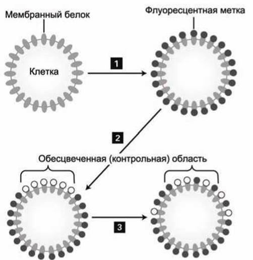
Figure 20 — Sequence of steps in the FRAP technique
A laser beam is then used to irreversibly bleach the fluorescent markers within a specific control region, resulting in a sharp drop in fluorescence intensity from this area (Figure 20(2)). Over time, mobile proteins diffuse out of this region, while unbleached proteins move in to take their place, leading to a recovery of fluorescence intensity in the control region of the membrane (Figure 20(3)).
If a fraction of these proteins is immovably anchored within the membrane, the fluorescence level after the curve reaches saturation will remain lower than it was before bleaching, making it possible to determine the relative proportions of mobile and immobile labeled molecules. Figure 21 shows an example of such a curve for the case where 50% of the labeled proteins were immobile.
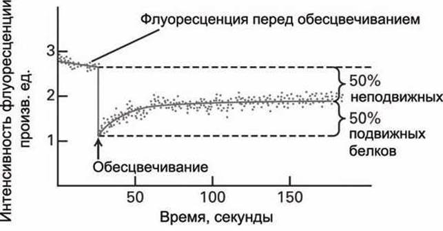
Figure 21 — Fluorescence recovery after photobleaching
Such a kinetic curve can be used to calculate the numerical value of the diffusion coefficient for the labeled protein in the membrane.
FRAP studies have demonstrated that in the Plasma Membranes of fibroblasts (the primary Connective Tissue Cells in vertebrates that synthesize Collagen), all Phospholipids can freely travel over a distance of only about 0.5 µm, but no further. Evidently, the fibroblast membrane is divided in a mosaic fashion into protein and lipid "islands" roughly 1 µm in size, and lipids can move freely only within their own island. In addition, the diffusion coefficient of lipids in The Plasma Membrane was found to be ten times lower than that in a "pure" lipid bilayer (10-8 cm2/s and 10-7 cm2/s, respectively). Based on these findings, it can be concluded that lipids may be associated (reversibly bound) with specific Membrane Proteins.
Last update: 13/08/2026
Editorial and Educational Adaptation: This material has been compiled based on the primary/original source text. The project team performed an editorial review, corrected technical inaccuracies, structured sections, and adapted the content for an educational format.
What was processed:
- elimination of formatting defects (OCR errors, structural breaks, corrupted characters);
- editorial organization of content;
- standardization of terminology in accordance with academic sources;
- verification of factual statements against the original source text.
All mentions of the author, publication year, and origin of the primary text have been preserved in accordance with the source.